Trichosporia
Historical document, translated for reference. It reflects medical knowledge of the 1920s–30s and is not medical advice.
Summary
Trichosporia is a fungal disease of the hair described in 1876, characterized by hard, stone-like nodules on the hair shaft. It is prevalent in the Americas and has a distinct European form affecting the beard and mustache. The prognosis is favorable, and treatment involves washing with hot water and mercuric chloride.
Encyclopedia article (1928–1936)
TRICHOSPORIA, trichosporia (syn. piedra, trichosporosis nodosa, trichosporosis tropica, trichomycosis nodularis), a peculiar fungal disease of the hair, first described by Osorio in 1876; it occurs mainly in Central and South America; in Colombia, Trichosporia is endemic. In Europe, a special form of Trichosporia is known—Tr. nostras, which occurs mainly in the Balkan Peninsula. Trichosporia manifests itself by the formation on the hair of hard, stone-like small nodules located along the length of the hair, the appearance of which changes little. These nodules are visible only under a magnifying glass, but when the hair is pulled between the fingers they are clear to the touch, forming complete or incomplete rings around the hair, sometimes in the form of continuous sheaths up to 2–5 mm long. The size of the nodules is therefore smaller than that of nits, their color ranging from whitish to brown. With long hair, the nodules are linked together, sometimes forming so-called Colombian knots. While in America Trichosporia occurs almost exclusively on the hairy part of the head, in the European form it is usually only the beard and mustache area that is affected. In America, Trichosporia mainly affects young women, which some authors connect with the practice, particularly in Colombia, of washing the head with a decoction of flaxseed, which is a good nutrient medium for the causative agent of Trichosporia—Trichosporon. The nodules on the hair consist of a large number of tightly glued together, mosaically arranged spores of this fungus. The spores are two-contoured, have a nucleus and a nucleolus. A large number of varieties of Trichosporon have been described, the most frequent of which is the one isolated by Osorio, Trichosporon giganteum. In cultures, in some cases white colonies grow first, and then greenish and brown colonies (on agar), while in other cases dark colonies grow on Sabouraud medium, the pigment of which corresponds to the pigment found in the nodules. The prognosis is always favorable. Treatment consists of washing with very hot water with the addition of sublimate (1:1,000–2,000); the most radical method of treatment is shaving the affected areas and subsequent washing with soap.
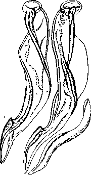
Figure 1. Trichostrongylus instabilis—spicules and gubernaculum.
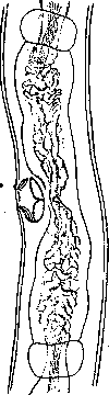
and so on, the exit of larvae from the egg and their molting occur at different times. Larvae enclosed in a sheath are invasive. When swallowed by a suitable host, they reach their localization site in the digestive tract, where they develop. Trichosporia of humans was considered extremely rare until recent years and was only established in isolated cases in some tropical countries, such as: Egypt, China, Japan, India. Being on the territory of Europe for the first time discovered in 1923 in the city of Moscow by Pod'yapol'skaya (Trichostrongylus instabilis), they are increasingly being established in various regions. In Moscow and the nearest localities, in 1 1/2 years, Trichosporia of humans was detected in 134 cases by methods of examining feces for helminth eggs (Davydov and Vinogradov). At present, Trichosporia are known in many regions of the USSR, and their distribution apparently is most widespread in Armenia (Kalantaryan's work).
The clinic and therapy of Trichosporia are almost not studied. The clinical picture apparently depends to a significant extent on the intensity of the invasion. With a weak invasion, clinical manifestations are weak (appetite disturbances and other disturbances of the digestive tract, weakness, irritability, etc.) or may even be absent altogether. With intense invasions, clinical manifestations can be very severe, resembling the clinical picture in ancylostomiasis—strongly expressed anemia, a sharp decline in strength up to cachectic states. With repeated courses of treatment with thymol (Kalantaryan), complete clinical recovery can be achieved. However, even after numerous courses, single eggs in the feces still remain. Since man is a facultative host, prevention should be directed along the line of fighting Trichosporia of domestic animals.
Related articles
Cite this page
“Trichosporia.” Soviet Medical Encyclopedia. English translation of Bolshaya Meditsinskaya Entsiklopediya, 1st ed. (Moscow, 1928–1936), ed. N. A. Semashko. https://sovietmedicalencyclopedia.pages.dev/article/trichosporia/