Epispadia
Historical document, translated for reference. It reflects medical knowledge of the 1920s–30s and is not medical advice.
Summary
Epispadia is a congenital malformation where the external urethral opening is located on the upper surface of the penis rather than at the tip. This article describes the condition's classification, symptoms, theories of origin, and surgical treatment methods from the 1930s perspective.
Encyclopedia article (1928–1936)
Epispadia (epispadia), or fissure of the upper wall of the urethra, represents a congenital malformation in which the external opening of the urethra is not located at the tip of the glans, but along the upper surface of the glans or shaft of the penis. E. occurs much less frequently than hypospadias. According to Baron (Wagon), for 300 cases of hypospadias there are 2 cases of E. Kaufmann in 1886 was able to collect 42 cases of E. from the literature, of which only in 3 cases was there E. of the glans. Burckhardt in 1906 collected 60 cases, with E. of the glans in 5 cases, and E. of the penis in 55 cases. In women, this malformation occurs less frequently than in men. Burckhardt cites only 26 cases of E. in women. -Regarding the origin of E., two theories deserve attention: 1) one explains the development of E. as a defect or delay in the intrauterine formation of the urethra (Thiersch), 2) the other suggests that here we are dealing with an intrauterine rupture of the urethra due to retention of urine after the complete formation of the bladder and urethra, but with the latter being closed in the glans of the penis (Kaufmann). There are 3 types of E. in males: 1) epispadia glan-dis, in which the urethra is split only in the area of the glans penis, with the external opening of the urethra lying either in the area of the glans or slightly above; 2) epispadia penis, in which the external opening of the urethra may be located at any point on the dorsal surface of the shaft of the penis up to the pubic symphysis; 3) epispadia totalis, or peno-pubica, when the entire urethra represents an upwardly open groove extending to the bladder, which may also be split to a greater or lesser extent. In these cases, a divergence of the pubic symphysis is usually observed. -In epispadia glan-dis, from the slit-like opening of the urethra to the tip of the glans there extends a deep groove lined with mucous membrane; the prepuce may be split or remain normal; the penis is in most cases of normal size or slightly shortened. In epispadia penis, the groove lined with mucous membrane extends along the dorsal surface of the penis from the abnormal opening of the urethra to the tip of the glans. The penis is usually reduced in size, often rudimentary developed and curved upward. The glans penis is sometimes only flattened, sometimes divided into two halves. The prepuce is split along the upper surface and hangs down in the form of an apron. In E. in women, the urethra passes over the split clitoris, may be partially split upward, or the groove extends from mons veneris downward under or even beyond the pubic symphysis and there directly opens into the bladder. Symptoms in E. depend on the degree of malformation. In mild forms, there is an abnormal direction of the urine stream, due to which surrounding parts of the body become wet. More severe forms are accompanied by partial or complete urinary incontinence, resulting in weeping eczematous surfaces on the skin of the scrotum and thighs. From constant soaking of clothing with urine, those suffering from incontinence give off an offensive odor, making them intolerable in society.

Fig. 1. Formation of the urethral canal according to Duplay.
Sexual function and the ability to generate offspring in mild forms (epispadia glandis) are usually preserved, but in severe forms the function of producing offspring is usually lost. The surgical treatment of E. aims to eliminate the malformation and, as far as possible, restore anatomical and functional relationships close to normal. It is most advantageous to perform the operation at the age of 6-8 years, since at this period the proportions of the parts are not too small, and erections that would hinder postoperative healing have not yet appeared. There are many methods of operations for E., of which only some deserve attention. In some methods, the edges of the urethral groove and adjacent tissues are freshened and sutured, in other methods, skin flaps on a pedicle are used to form the upper wall of the urethra. From the methods of the first group, the method of Duplay should be mentioned, which consists in that two incisions are made on both sides of the urethral fissure and the well-freshened edges of the wound are sutured over a catheter (fig. 1). From the methods of the second group, used for the formation of the urethra in E. of the penis, the method of Cantwell should be mentioned, in which the mucous membrane of the urethral groove is separated in the form of a flap with its base at the abnormal opening of the urethra. From this flap, a urinary tube is formed, which is moved in depth between the separated cavernous bodies, which are sutured over it. In the method of Nelaton-Dolbeau, a rectangular skin flap is outlined from the abdominal skin with its base above the abnormal opening of the urethra. The flap is turned downward and sutured to the freshened edges of the urethral groove in such a way that its epidermis-covered surface forms the upper wall of the urethra. Then, on the anterior surface of the scrotum, two parallel arcuate incisions outline a strip of skin, which is moved to the bloody surface of the abdominal flap. The advantage of the method is that the abnormal opening of the urethra is firmly and immediately closed. S71 The method of Thiersch consists of the following steps: 1) For urine diversion, a perineal urethrotomy is performed. 2) A urinary tube is created in the area of the glans penis according to the Duplay method. 3) For the purpose of forming a urinary tube on the penis, two quadrangular skin flaps are outlined, one of which has its base at the urethral fissure, the other on the side from it (fig. 2). Both flaps are separated to the required width. The flap with its base at the urethral fissure is turned over it and its free edge is sutured to the freshened edge of the fissure on the other side. The separated flap of the other side is stretched over the bloody surface of the turned-over flap and its edge is sutured to the lateral edge of the skin incision on the other side. 4) The urinary tube of the penis, formed in this way and healed, is connected with the urinary tube in the area of the glans. For this, a transverse incision is made in the prepuce, and through it the glans penis is passed. By freshening and suturing the tissues of the moved prepuce, the missing part of the urethra is created between the newly formed urinary tubes in the area of the penis and glans. 5) The abnormal opening of the urethra is closed, for which two triangular flaps from the abdominal skin are used. The main obstacle is the wetting of the wound with urine, as healing is greatly hindered due to soaking and incrustation.
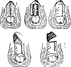
Fig. 2. Five consecutive moments of the operation according to Thiersch.
A rectangular flap is outlined from the abdominal skin with its base above the abnormal opening of the urethra. The flap is turned downward and sutured to the freshened edges of the urethral groove in such a way that its epidermis-covered surface forms the upper wall of the urethra. Then, on the anterior surface of the scrotum, two parallel arcuate incisions outline a strip of skin, which is moved to the bloody surface of the abdominal flap. The advantage of the method is that the abnormal opening of the urethra is firmly and immediately closed.
Related articles
Mentioned in
Cite this page
“Epispadia.” Soviet Medical Encyclopedia. English translation of Bolshaya Meditsinskaya Entsiklopediya, 1st ed. (Moscow, 1928–1936), ed. N. A. Semashko. https://sovietmedicalencyclopedia.pages.dev/article/epispadia/