Diaphysis
Historical document, translated for reference. It reflects medical knowledge of the 1920s–30s and is not medical advice.
Summary
The diaphysis refers to the main shaft of long bones, distinct from the epiphyses. It undergoes physiological twisting, bending, and thickening under functional conditions, and can develop deformations in pathological states like rickets or osteomyelitis.
Encyclopedia article (1928–1936)
DIAPHYSIS, diaphysis (from Greek dia- through and phyo- I grow), a term by Heister, denoting the body (corpus) or the middle, larger part of the so-called tubular bones; it is contrasted with the ends (extremities) or epiphyses (epiphysis). Under the influence of functional conditions, twisting along the axis of the diaphysis, physiological bending and thickening occur (fig. 1-4). In certain pathological conditions based on rickets, osteomyelitis, poliomyelitis, etc., deformations of bones develop; in these cases, various curvatures of the diaphyses in the plane or along their axis are often observed. Deformations of the diaphyses of long bones are more common in the lower extremities. Static conditions play an important role in the origin of these deformations. The structure of bone tissue in deformed diaphyses follows the general laws of bone tissue construction. The harmony between the form and structure of the bone, resp. diaphysis, remains unchanged.
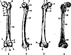
Figure 1-2. Femur (left-posterior, right-side): 1-fossa capit.; g-greater trochanter; z-lessertrochanter; 4-neck; S-popliteal line; b-medial epicondyle; 7-medial condyle; 8-lateral condyle; 9-pectineal line; 10 and 14-medial and lateral rough lines; 11 and 12-intercondylar line and fossa; 13-lateral epicondyle; 16-rough line. Figure 3-4. Femur (left-anterior, right-sawed): 1-fossa1 capit.; 2-neck; 3-greater trochanter; 4-intertrochanteric line; 5-lesser trochanter; 6 and 7-lateral and medial epicondyles; 8-patellar surface; 9 and 10-proximal and distal parts of epiphysis; 11-diaphysis; 12-spongy substance; 13-compact substance; 14-medullary cavity.
In pathological conditions based on rickets, osteomyelitis, poliomyelitis, etc., deformations of bones develop; in these cases, various curvatures of the diaphyses in the plane or along their axis are often observed. Deformations of the diaphyses of long bones are more common in the lower extremities. Static conditions play an important role in the origin of these deformations. The structure of bone tissue in deformed diaphyses follows the general laws of bone tissue construction. The harmony between the form and structure of the bone, resp. diaphysis, remains unchanged.
Related articles
Mentioned in
Cite this page
“Diaphysis.” Soviet Medical Encyclopedia. English translation of Bolshaya Meditsinskaya Entsiklopediya, 1st ed. (Moscow, 1928–1936), ed. N. A. Semashko. https://sovietmedicalencyclopedia.pages.dev/article/diaphysis/