Intestinal Suture
Historical document, translated for reference. It reflects medical knowledge of the 1920s–30s and is not medical advice.
Summary
This article provides a historical overview and technical description of intestinal suturing techniques as understood in the early 20th century. It details the evolution of suturing methods, from early historical practices to the development of the Lembert, Jobert, Czerny, and Schmieden sutures, and discusses the use of mechanical devices like the Murphy button.
Encyclopedia article (1928–1936)
Intestinal Suture serves: 1) to suture tears of the serous membrane on the surface of the intestines; 2) to suture openings in the intestines (in case of ruptures, wounds); 3) to suture the ends of the intestines during resection; 4) to form interintestinal anastomoses and anastomoses of the intestines with other organs (stomach, gallbladder, ductus choledochus). The first mention of the intestinal suture is in 431 B.C. by the Greek Praxagoras, who used it for intestinal injuries and laparotomies for obstruction. Later (40–20 B.C.), Meges and Celsus knew about the intestinal suture, although Celsus considered small intestine injuries fatal and believed suturing was possible only for large intestine wounds. Then, a thousand years later, Albucasis used sutures made from animal intestines. The first precise indications of the intestinal suture are found in Italian physicians (1200–1300). Italian physicians also introduced the continuous suture. After 1700, Ramdohr performed invagination of the damaged intestinal segment into the lower one and sutured it. Moreau Botards introduced an improvement, suggesting the removal of the mucous membrane in the lower limb of the intestine before invagination, thereby achieving a stronger adhesion. For small intestinal injuries, ligation was used—the damaged area was grasped with a clamp and tied. Until the 18th century, for complete transverse ruptures of the intestine, the only measure to save patients was considered the use of anus praeternaturalis, proposed by Paracelsus. Scientific justification and development of the intestinal suture began with the time of Jobert (1824) and Lembert (1826), who proposed using a suture to bring the serous surfaces of the intestines into contact, which best promotes adhesion. This position was confirmed even earlier by the experiments of Bichat. Since then, this suture bears the name of Lembert. The Lembert suture (incorrectly called Lembert's) consists of grasping the serous membrane of each of the sutured intestines in a fold. The needle is inserted 4 mm from the edge of the wound and passed obliquely 3 mm into the muscular tissue of the intestine, without piercing the mucous membrane, and brought out 1 mm from the edge of the same side. On the other side, the needle is inserted 1 mm from the edge of the wound, not piercing
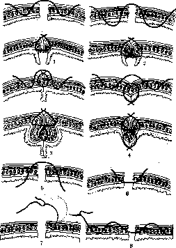
Figure 1-8.
the mucous membrane, and brought out again 4 mm outward. When the suture is tightened, the serous membranes are tightly applied to each other, thereby causing the approximation of the unsutured mucous membrane (Fig. 1). The suture is applied in one layer. The difference of the Jobert suture is only that it is applied through all layers (Fig. 2). The advantage of the first suture is that the thread does not come into contact with the mucous membrane and therefore the suture may not be infected, while in the second, the suture is infected from the mucous membrane. A further improvement was introduced by Czerny. The needle is inserted through the serous and muscular membranes 2–3 mm from the edge of the wound and brought out exactly in front of the mucous membrane at the edge; on the other side, the insertion is made at the edge above the mucous membrane and the exit is 2–3 mm from the edge of the serous membrane. When the knot is tied, all layers come into contact with each other (Fig. 6). The sutures are applied at a distance of 3–4 mm. The first row of sutures is reinforced for strength by a second row of Lembert sutures (Fig. 3). Given that sutures coming into contact with the mucous membrane or placed near it,

Figure 9.
Figure 10. are often infected and cut through into the intestinal lumen, to facilitate the passage of ligatures, which is hindered by the presence of a knot on the surface of the intestine, Albert proposed that the insertion and exit be made from the side of the mucous membrane. Thus, the knot will be in the intestinal lumen, and the suture can easily cut through (Figs. 4 and 5). Gradually, many surgeons switched from the interrupted suture to the continuous suture, which significantly reduces the time of suturing, prevents bleeding, and provides a more hermetic closure of the intestinal lumen (Fig. 9). For greater strength, the suture is applied in three layers: mucosa-mucosa, serous-muscular-serous-muscular, and the final Lembert suture. In this form, the suture is still used by some surgeons to this day. But the majority have already abandoned the intermediate suture, since good strength is achieved even when using a two-layer suture: the first suture through all layers and the second—Lembert. The Schmieden suture contributes to the greatest thoroughness of the contact of the serous covers. The needle insertion throughout the entire length of the suture is made from the side of the mucosa, the exit—from the side of the serous membrane. When the suture is tightened, the serous surfaces are turned into the intestinal lumen, and the serous surfaces come into contact (Fig. 10). Further observations showed that a suture grasping the mucous membrane not only does not promote its healing but even worsens it due to the formation of necrosis in the area of the tightened suture and subsequent inflammation around it. Besides this, a suture made of non-absorbable material on the mucous membrane becomes infected and cuts through into the intestinal lumen. As experimental data (Hilarowicz, Kopylov, Pokryshkin) and clinical observations have shown, a suture applied to the mucous membrane remained uncut for a long time: in Sokolov—2 years, in Schempp—3 years 3 months, in Pokryshkin—6.5 months, in Spasokukotsky—7 months and 2 years. Sometimes the thread hangs halfway into the lumen of the organ, and food constantly traumatizes the suture during passage. The presence of infection in the suture leads to the formation of inflammatory infiltration in the scar, to the disfigurement of the anastomosis, to narrowing. In the future, ulcers may form (Hilarowicz, Spasokukotsky). All this led to the fact that most surgeons are gradually abandoning the application of sutures to the mucous membrane, limiting themselves to a two-layer suture: serous-muscular and the Lembert suture. And in this case, the best contact is provided by the Schmieden suture, only without grasping the mucous membrane, or the Momburg suture (Fig. 8). The use of non-absorbable material in a two-row continuous suture often also leads to the cutting through of the suture and the formation of the same phenomena as with a three-row suture. Therefore, at present, a continuous suture made of absorbable material (catgut) or an interrupted silk suture is used, the cutting through of which is not so prolonged and does not cause narrowing of the lumen of the anastomosis. The outer suture is applied mainly as an interrupted silk one, since a catgut suture may be absorbed before the time of adhesion of the serous surfaces of the intestine and lead to the dehiscence of the suture. Besides this, according to some observations, with a catgut suture, the adhesion of serous surfaces does not occur quickly enough. The latest simplification of the intestinal suture was proposed by Bier and consists of a return to the initial proposal of Lembert to use a single-layer suture. Numerous clinical observations by Bier, Sokolovsky, and experimental studies by Kopylov speak for the sufficient strength of the single-layer suture. Instead of the intestinal suture, various prostheses were proposed for connecting the lumen of the intestines. Their use pursues two goals: 1) speed of connection and 2) asepsis. The Murphy button, proposed in 1892, enjoys the greatest popularity to this day. It consists of two halves, inside which there are two hollow cylinders, which, when connected, enter one into the other. On the outer cylinder, there are screw threads inside, and on the inner one, there are spring hooks on the outside and a spring ring on top of the cylinder (Fig. 11); when connecting the two halves, the spring hooks of the inner cylinder enter the threads of the outer cylinder and do not allow the button to open into two halves. The spring ring enhances the compression of the tissue located between the two halves. Opening the button into two halves can be done by unscrewing it. The technique of suturing by means of the button is as follows. The end of the afferent intestine is sutured with a continuous purse-string suture. One half with threads is taken with tweezers by the cylinder, inserted into the intestinal lumen, and the suture is tied on the cylinder (Fig. 12).

Figure 11.
Figure 12.
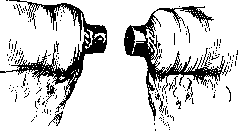
Figure 12.
The second half, which is heavier, is inserted in exactly the same way into the lumen of the efferent intestine, and the previously placed purse-string suture is tied onto the cylinder in the same manner. Then the forceps are removed, one cylinder is inserted into the other, and both halves are tightly closed. For greater strength, additional interrupted silk or catgut sutures are placed on the serous membranes of the contacting intestinal segments (Fig. 13). Usually, within the next two weeks, the button is passed per vias naturales. Observations on clinical material and in experiments have proven that the speed of creating an anastomosis is not worth the consequences. Trofimov points to the narrowing of the anastomosis, obstruction of the narrow lumen of the button by fecal masses, its long-term retention in the intestine, and perforation of the anastomosis, which he noted 36 times. In connection with this, modifications of the button made of absorbable material (calcified ivory, metallic magnesium, rutabaga, potato) appeared. At the present time, they are not used, and only the Murphy button is used in exceptionally rare cases when it is necessary to quickly finish the operation to preserve the patient's strength. The technique of applying an intestinal suture requires compliance with certain rules: 1) preparation of the intestinal segments for suturing; 2) strict asepsis; 3) correct placement and tying of the suture; 4) postoperative management of patients. Preparation of the intestinal segments when applying a suture consists of placing gentle Doyen intestinal clamps (see Vol. X, art. 449, Figs. 33 and 34) above and below the intended suture site to prevent the leakage of intestinal contents. For the same purpose, the intestine is emptied if the patient is not being operated on in an emergency, and before applying the clamps, the intestinal contents are pushed above and below the site of clamp application. In cases of incisions and wounds of the intestine, fecal masses are removed with moistened
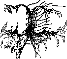
Figure 13.
napkins or cotton balls with physiological saline, and the mucous membrane is wiped with mercuric chloride cotton balls. When applying a suture to the large intestine, one must keep in mind the appendices epiploicae, which make it difficult to align the edges of the intestinal incision and to place the suture on the serous and muscular membranes, which may subsequently lead to dehiscence. Therefore, the suture site must be cleared of appendices epiploicae and the mesentery of the intestine, and then it is possible to apply a secure suture. Compliance with asepsis is achieved both by using clamps and wiping the suture area with mercuric chloride balls or iodine tincture, and by regularly changing instruments, gloves, or washing hands after each layer of sutures. Despite all precautions, it is still extremely difficult to achieve complete asepsis when opening the intestinal lumen, and a certain percentage of suture dehiscence is attributed to insufficient asepsis. Therefore, at all times there has been a desire both to improve the suture itself and to find methods that would allow for the application of a completely aseptic suture. For this purpose, in 1911, Rostovtsev proposed special narrow plates, compressed by strong clamps, which are applied to the site of the intended intestinal incision. The intestine is cut along these plates, they are joined together, and a Lembert suture is placed over them, leaving a small opening unstitched, through which the plates are removed. An additional suture closes this small opening. Thus, the suture is applied completely aseptically. For the same purpose, special instruments were proposed by Doyen, Braun, and a suturing machine (Cukor) (see Vol. X, art. 447-450, Figs. 19 and 24; art. 451, Fig. 48). Schumacher achieves this in the simplest way: he makes a circular incision of the serous and muscular membranes of the intestine down to the mucosa and applies Kocher clamps so that their tips exactly coincide with the edge of the intestine on the mesenteric side. Then the resected intestine is cut along the clamp, and this area is lubricated with iodine tincture. The clamps are brought together and sutures are placed over them, with the
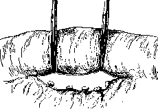
Figure 14.
exception of the place opposite the mesentery, where an opening remains through which the clamps are removed, and then this place is closed with an additional suture (Figs. 14, 15, and 16). The Schumacher method can be widely used in resection of the small intestine, but in resection of the large intestine it is of little
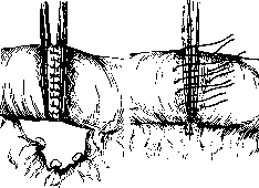
Figure 15.
Figure 16.
use, because it is difficult to dissect the serous and muscular membranes without opening the mucous membrane, and it is better to apply Kocher clamps to all layers of the intestine (Figs. 17, 18, 19, and 20), and to perform the suturing according to Lembert. Thus, by using Rostovtsev's clamps (see Vol. X, art. 447, Figs. 17 and 18) or Kocher's, one can achieve complete asepsis of the intestinal suture. A disadvantage of the aseptic suture is the formation of a significant fold in the intestinal lumen compared to a conventional suture, which, due to subsequent inflammation and edema, increases and can cause narrowing of the intestinal lumen (Fig. 21). And if in the small intestine such temporary narrowing does not cause suture dehiscence due to the liquid contents, which pass through the narrowed area without difficulty, then in the large intestine, in the presence of narrowing, even temporary, during the passage of stagnant and solidified contents, suture dehiscence may occur. To avoid this, it is recommended, simultaneously with the application of an aseptic suture to the large
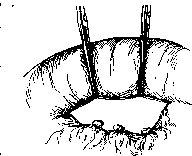
Figure 19.
intestine, to create a fecal fistula on the cecum according to Witzel, through which gases are easily removed and the dense contents of the intestine are liquefied by irrigation. To prevent the formation of internal strangulations, when applying an intestinal suture, the mesentery of the intestine must be well sutured (Figure 18). The asepsis of the suture is violated, in addition to opening the intestine, also by incorrect placement of the suture. Therefore, when applying a suture to an overly stretched intestine distended by gases, and especially to the large intestine in the area of the haustra, extreme caution must be exercised not to puncture the intestine. On the other hand, when suturing the muscular membrane, the puncture must be made not obliquely (indicated by a dotted line in Fig. 7), so as not to place the suture too superficially, but perpendicularly to the intestine, and only when the needle enters the muscular membrane should a turn be made.
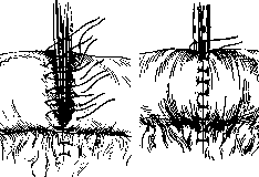
Figure 17.
Figure 18.
When applying interrupted sutures, each subsequent suture should be 3-4 mm from the previous one; the second layer of interrupted sutures should correspond to the gaps of the underlying one. Tying is performed tightly enough to bring the ends of the intestine together until the walls are in full contact, but not so tightly as to cause cutting through or subsequent necrosis of the intestinal wall in the area of the suture. The knot is tied as a simple, non-surgical one; the second knot, for greater strength, should be a sailor's knot, especially when tying catgut, which easily unties when swelling. Tightening of the suture should be strong enough so that dehiscence of the wound edges does not occur. To avoid unnecessary trauma to the intestinal wall, when applying an intestinal suture, round intestinal (straight or curved) or thin cutting needles and thin catgut and silk threads are used. With a continuous suture, the stitches should not be too far apart, so as not to cause narrowing of the intestinal lumen when tightening (Haberer). For suturing the duodenal stump during Billroth II gastric resection, the stump of the appendix, or the resected intestine, previously
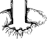
Figure 20.
clamped with strong clamps, ligated, and transected, instead of conventional sutures, a purse-string suture is used, which fully guarantees asepsis (Figs. 22 and 23). In the postoperative period, the intestinal suture requires special care for patients. With a suture
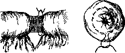
Figure 22.
Figure 23.
on the small intestine, especially in the upper section, the patient's nutrition is initially provided by means of enemas. With a suture on the large intestine, the patient can be fed liquid food from the very first days, strictly observing the stool, and from the 3rd-4th day, drip or small (100.0) oil enemas can be given, which facilitate the liquefaction of feces and easier bowel movements.
Related articles
Mentioned in
Cite this page
“Intestinal Suture.” Soviet Medical Encyclopedia. English translation of Bolshaya Meditsinskaya Entsiklopediya, 1st ed. (Moscow, 1928–1936), ed. N. A. Semashko. https://sovietmedicalencyclopedia.pages.dev/article/intestinal-suture/