Strongyloidiasis
Historical document, translated for reference. It reflects medical knowledge of the 1920s–30s and is not medical advice.
Summary
This article from the 1928–1936 Soviet medical encyclopedia details Strongyloidiasis, a parasitic disease caused by the nematode Strongyloides stercoralis. It covers the parasite's life cycle, including direct and indirect development, the clinical symptoms of infection, and diagnostic and therapeutic approaches of the era.
Encyclopedia article (1928–1936)
STRONGYLOIDIASIS (anguilluliasis, angiostomiasis), a helminthic disease of humans and some other mammals, as well as birds, caused by the nematode of the genus Strongyloides Grassi, 1879, belonging to the suborder Rhabdiasata and the family Rhabdiasidae. The genus Strongyloides includes a whole series of species that are parasites of mammals and less frequently birds; although many of these species are poorly differentiated from each other morphologically, nevertheless it is considered that they are independent species, more or less stenoadaptive, i.e., specific with respect to their hosts. It is believed that there are their own species in monkeys (Strongyloides cerjus Darling, 1911; Strongyloides lulleborni Linstow, 1905), as well as in a whole series of other animal species [in the dog - S. cards Brumpt, 1921; in cattle - S. vituli Brumpt, 1921; in sheep, goats, rabbits, etc. - S. papillosus (Wedl, 1856); in horses - S. wes-teri Ihle, 1917, and others]. The human form Strongyloides stercoralis can infect a dog, cat, monkey, in which the infection usually lasts no more than several months, after which the parasites die out. Other authors deny the possibility of such infection.
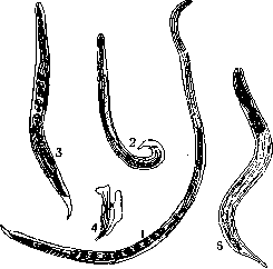
Figure 4. Strongyloides stercoralis: 1 - mature female of the parasitic stage; 2 - male of the free-living stage; 3 - female of the free-living stage; 4 - spicula and gubernaculum of the male; S - rhabditiform larva.
--Strongyloides stercoralis [synonyms: Anguillula stercoralis and Anguillula intesti-nalis Bavay, 1877; Strongyloides intestinalis (Bavay, 1877); Rhabdonema strongyloides Leuck, 1883; Rhabdonema intestinale Blanchard, 1886, and others]. It was first discovered by Norman in 1876 in the feces of French soldiers returning from Cochinchina and suffering from diarrhea. Bavay (1877) described these nematodes under the name Anguillula stercoralis. Some of these soldiers died, and upon autopsy parasites were found in them, which bore no resemblance to those previously found in the feces, so they were described as a special form Ang. intestinalis Bavay, 1877; it was suggested that both species were the cause of the observed disease, which Bavay named "Cochinchinese diarrhea". Later Leuckart (1882) proved that both forms described under different names are merely different stages of development of one species, whose biology is characterized by the so-called heterogony (see). Thus it was established that the parasitic stage is represented only by one hermaphrodite female inhabiting the upper parts of the small intestine (Fig. 1 and 2). This female is 2.2 mm long and 0.030-0.075 mm wide and is characterized by a cylindrical esophagus reaching 1/3 of the length of the body; the vulva is at the boundary of the middle and posterior thirds of the body. Females penetrate into the thickness of the mucosa, into the Lieberkühn glands (Fig. 3), where they lay eggs, which are 0.050-0.058 mm long and 0.030-0.34 mm wide; they ripen here as well, so that a larva (Fig. 4) hatches already in the human organism, emerging into the lumen of the intestine and then being thrown out with the feces outside; less often the larvae emerge in the eggs (Fig. 5) still unhatched from the latter. Larvae reaching 0.200-0.250 mm in length and 0.016 mm in thickness upon hatching and growing rapidly are characterized by the so-called "rhabditiform" esophagus, i.e., equipped with an anterior elongated and posterior bulbous expansion (bulbus). Further development can proceed in two ways: in direct development (homogony) rhabditiform larvae by further transformation pass into the so-called "filariform" larvae, characterized by a cylindrical esophagus. These larvae, reaching their larval maturity ("invasiveness"), can cause a new infection of humans or other animals. In indirect development, larvae in the external environment transform within 30 hours into a separate-sex generation of males and females (heterogony), morphologically sharply differing from the females of the parasitic stage: both the male and the female have a rhabditiform esophagus; the male has an unpaired spicula and gubernaculum. The female lays eggs in the external environment, measuring 0.070x0.040 mm. Larvae hatching from these eggs with a rhabditiform esophagus molt and in 3-4 days turn into filariform invasive larvae. Invasive larvae infect humans (and other animals) percutaneously, and upon oral penetration they perform a migratory cycle, penetrate into the mucosa of the upper part of the intestinal tract, enter the venous system, the right heart, and the small circle of circulation, from whence, disrupting the integrity of vessels and alveoli, they pass into the lumen of the lungs; from here the larvae rise upward along the respiratory tract, enter the oral cavity, are swallowed and settle in the small intestines, where, penetrating into the thickness of the intestinal wall, they grow into mature females. Upon swallowing of invasive larvae, the latter can also get directly into the small intestine, where they reach maturity without migration. From the moment of infection until the achievement of maturity and appearance in the feces of rhabditiform larvae, about 17 days pass. Many authors believe that in some cases rhabditiform larvae, hatching in the organism of an animal from eggs, may, without leaving the intestine, turn into invasive filariform and thus give rise to the so-called autoinvasion (Skryabin and Wagner, Nishigori, etc.). Apparently this explains the cases of finding larvae of Strongyloides in the blood, urine, sputum (Fr6es, 1931). S. stereoralis, inhabiting in the thickness of the intestinal walls, penetrate to the submucosa, invade the chyle vessel and affect the Lieberkühn glands; at the same time a catarrhal state of the mucosa, swelling of follicles, ulceration is observed, and apparently intoxication also takes place. The main complaints in strongyloidiasis consist of painful phenomena on the part of the gastrointestinal tract: pains in the abdomen, nausea, meteorism, tenesmus; diarrhea is most often observed, often with admixture of blood. Among general phenomena it is necessary to note headaches, dizziness, fainting states, anemia, exhaustion up to cachetic states. Eosinophilia can be of varying intensity: from 2.5% to 76%. Upon penetration of parasites from outside through the skin they cause various skin symptoms: itching, urticaria, papules, edema, which can last up to a month.--Diagnosis is based on the finding of a typical rhabditiform larva or eggs; it is easier to detect these elements after vigorous laxatives. In milder cases of infection, the so-called Berman method is applied, thanks to which larvae are concentrated in greater numbers. Therapy is medicamentous unreliable; it is considered that good results are given by thymol, male fern extract, chenopodium oil, gentian violet, etc.--Preventive measures in main points coincide with those in ancylostomiasis (see Ankylostoma, ancylostomiasis). The geographical distribution largely coincides with that of ancylostomiasis, since strongyloidiasis is encountered primarily in tropical and subtropical countries, where the percentage of infection reaches up to 35. Sporadic cases of strongyloidiasis have been repeatedly detected on the territory of the USSR - in Central Asia, in Armenia, Azerbaijan, Ukraine.
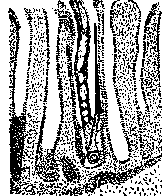
Figure 3. Strongyloides stercoralis in the small intestine of a human. Adult female in the intestinal gland.
They are characterized by the so-called "rhabditiform" esophagus, i.e., equipped with an anterior elongated and posterior bulbous expansion (bulbus). Further development can proceed in two ways: in direct development (homogony) rhabditiform larvae by further transformation pass into the so-called "filariform" larvae, characterized by a cylindrical esophagus. These larvae, reaching their larval maturity ("invasiveness"), can cause a new infection of humans or other animals. In indirect development, larvae in the external environment transform within 30 hours into a separate-sex generation of males and females (heterogony), morphologically sharply differing from the females of the parasitic stage: both the male and the female have a rhabditiform esophagus; the male has an unpaired spicula and gubernaculum. The female lays eggs in the external environment, measuring 0.070x0.040 mm. Larvae hatching from these eggs with a rhabditiform esophagus molt and in 3-4 days turn into filariform invasive larvae. Invasive larvae infect humans (and other animals) percutaneously, and upon oral penetration they perform a migratory cycle, penetrate into the mucosa of the upper part of the intestinal tract, enter the venous system, the right heart, and the small circle of circulation, from whence, disrupting the integrity of vessels and alveoli, they pass into the lumen of the lungs; from here the larvae rise upward along the respiratory tract, enter the oral cavity, are swallowed and settle in the small intestines, where, penetrating into the thickness of the intestinal wall, they grow into mature females. Upon swallowing of invasive larvae, the latter can also get directly into the small intestine, where they reach maturity without migration. From the moment of infection until the achievement of maturity and appearance in the feces of rhabditiform larvae, about 17 days pass. Many authors believe that in some cases rhabditiform larvae, hatching in the organism of an animal from eggs, may, without leaving the intestine, turn into invasive filariform and thus give rise to the so-called autoinvasion (Skryabin and Wagner, Nishigori, etc.). Apparently this explains the cases of finding larvae of Strongyloides in the blood, urine, sputum (Fr6es, 1931). S. stereoralis, inhabiting in the thickness of the intestinal walls, penetrate to the submucosa, invade the chyle vessel and affect the Lieberkühn glands; at the same time a catarrhal state of the mucosa, swelling of follicles, ulceration is observed, and apparently intoxication also takes place. The main complaints in strongyloidiasis consist of painful phenomena on the part of the gastrointestinal tract: pains in the abdomen, nausea, meteorism, tenesmus; diarrhea is most often observed, often with admixture of blood. Among general phenomena it is necessary to note headaches, dizziness, fainting states, anemia, exhaustion up to cachetic states. Eosinophilia can be of varying intensity: from 2.5% to 76%. Upon penetration of parasites from outside through the skin they cause various skin symptoms: itching, urticaria, papules, edema, which can last up to a month.--Diagnosis is based on the finding of a typical rhabditiform larva or eggs; it is easier to detect these elements after vigorous laxatives. In milder cases of infection, the so-called Berman method is applied, thanks to which larvae are concentrated in greater numbers. Therapy is medicamentous unreliable; it is considered that good results are given by thymol, male fern extract, chenopodium oil, gentian violet, etc.--Preventive measures in main points coincide with those in ancylostomiasis (see Ankylostoma, ancylostomiasis). The geographical distribution largely coincides with that of ancylostomiasis, since strongyloidiasis is encountered primarily in tropical and subtropical countries, where the percentage of infection reaches up to 35. Sporadic cases of strongyloidiasis have been repeatedly detected on the territory of the USSR - in Central Asia, in Armenia, Azerbaijan, Ukraine.
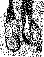
Figure 5. Eggs of Strongyloides stereoralis in the glands of the small intestine.
strongyloidiasis is encountered primarily in tropical and subtropical countries, where the percentage of infection reaches up to 35. Sporadic cases of strongyloidiasis have been repeatedly detected on the territory of the USSR - in Central Asia, in Armenia, Azerbaijan, Ukraine.
Related articles
Mentioned in
Cite this page
“Strongyloidiasis.” Soviet Medical Encyclopedia. English translation of Bolshaya Meditsinskaya Entsiklopediya, 1st ed. (Moscow, 1928–1936), ed. N. A. Semashko. https://sovietmedicalencyclopedia.pages.dev/article/strongyloidiasis/