Larva (Ml GRANS, "bluzhdayushchaya lichinka",)
Historical document, translated for reference. It reflects medical knowledge of the 1920s–30s and is not medical advice.
Summary
This article from the 1928–1936 Soviet medical encyclopedia details 'larva migrans,' a skin disease characterized by a creeping red ridge. It covers the etiology, caused by various parasites including horse botfly larvae and nematodes, and describes the clinical signs, diagnosis, and treatment methods of the time.
Encyclopedia article (1928–1936)
LARVA Ml GRANS, "bluzhdayushchaya lichinka", designation of a skin disease characterized by the appearance of a creeping reddish ridge moving over the skin and giving various course figures. Synonyms for L. t. are the designations: creeping disease, creeping eruption, Hautmaulwurf, hypenomoderma, linea migrans, dermatitis herpetiformis migrans, epidermiditis linearis migrans, gastrophilosis cutis and others; the Russian folk name is volosatik, hair- is explained by the belief that horse hair comes to life in water (confusion with the hairworm Gordius aquaticus) and, when a person bathes, penetrates into his skin. The etiology of this disease is different; in some cases the skin lesion depends on the penetration into the thickness of its epidermis of the larvae of botflies of the genus Gastrophilus; in others, larvae of other insects figure (these cases are studied much weaker), in the third, the cause of skin changes are the larvae of certain round worms. Cases of L. m. oestrosa, conditioned by the parasitism in the epidermis of larvae of botflies of the genus Gastrophilus, which are parasites of the stomach or intestine of the horse, have been described in different countries (including in the USSR) quite a lot. The duration of the stay of L. t. in the skin in different cases fluctuates from a week to two years. The lesion can be in any part of the body. The larva of the first phase of transformation of Gastrophilus (see vol. VI, p. 330, figure 2) bores into the Malpighian layer of the epidermis, where it moves forward. The course usually does not branch. As the larva moves forward, the old course disappears; with ordinary processes of movement of epidermal cells to the periphery of the skin as they are replaced by young cells, the course itself also moves into the horny layer of the epidermis and then disappears completely from the skin. It is noted that courses in the skin can be laid by the mite Rhizoglyphus, the ant, and even the larva of a cochineal, but all these indications are doubtful. The larvae of the skin botfly Hypoderma lie in the connective tissue part of the skin and do not cause the formation of linear stripes on the skin. Larva migrans Larva migrans oestrosa Infection is often in people who have dealt with horses, on pastures or near stables. Slight exudation, urticarial erythema. Mostly one course. The course is long corresponding to the duration of the disease, since the larva does not spontaneously leave the epidermis. Larva migrans nematosa No definite cause of infection is noted in the anamnesis. The disease is associated with lying on damp sand, soil, during peat works, etc. Strong exudation; formation on the skin of blisters or crusts. Courses are usually multiple. Courses are short; often spontaneous recovery after a short period of time (when the larvae leave the epidermis deeper into the vessels). At the same time, only the larvae of the dog nematode Uncinaria stenocephala, recently described in the south of the USA, Agamonematodum migrans (as well as Strongyloides, Gnathostoma and some others), give phenomena of creeping disease, depending on the wandering of the implanted larvae in the thickness of the epidermis itself. The courses of nematodes in the skin are usually multiple. Differential diagnosis of various forms of L. t. is performed by the signs indicated in the table given.
E. Pavlovsky. Larva migrans is a relatively rare skin disease described for the first time in 1874 by R. Lee; it occurs more often in young and childhood ages. The clinical picture is characterized by the appearance on the skin of edematous whitish-pink and red slightly elevated stripes, which continue to grow with their active end until the larva lives in the skin (fig. 1). L. t.. nematosa is conditioned by the penetration into the skin of larvae of nematodes. For some species of round worms (Ankylostoma duodenale, Necator americanus, Strongyloides and others), the skin is the usual place of penetration-
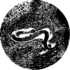
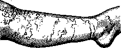
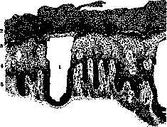
Figure 1. Section of human skin with a nematode larva in the thickness of the epidermis. it begins with itching and the formation of a blister, and then a pustule at the site of implantation of the larva; after a few days an edematous ridge appears at this place. The movable end of the course is recognized by a pale-red, then dark-red wheal, from which through 1/2-2 hours a linear continuation of the course is formed. This wheal is not the true final point of the course, since the larva, during the time necessary for the occurrence of the wheal, manages to penetrate further and is usually located in a clinically still non-reactive zone, at a distance of 1/2-3 cm from the visible final point of the course; this explains the difficulty of finding the larva in L. t. The lines of L. t. in appearance resemble stripes of urticaria factitia; the width of the stripes depends on the intensity of inflammation; the closer to the active end, the wider, higher and redder the stripes; above fresh courses sometimes blisters appear. The lines of L. t. are usually zigzag and go in the most various directions, giving often bizarre drawings (figure 2). Their length depends on the duration of the disease (known 379 cases with a duration of more than two years) and on the speed of movement of the larva in the skin. The periods of movement of the larva, which, according to Rille, occur mainly at night, alternate with periods of rest. In the case of Caposi, the larva moved with a speed of 30 cm per day. Subjectively - slight itching and burning, in children often the general condition is disturbed, sometimes even convulsions. It is rarely possible to detect the larva histologically (Melczer, Selissky); the course is usually (with larvae of the genus Gastrophilus) in the upper layers of the epidermis, sometimes deeper; the horny layer above it is fibrillated. Epithelial cells surrounding the course are edematous, vacuolated; slight acanthosis; papillae are edematous, in the papillary and subpapillary layers vessels are dilated and a greater or lesser round-cell infiltrate (fig. 3). The detection and extraction of the larva have naturally not only diagnostic, but also therapeutic significance. Examining carefully under a lens control the clarified (aniline or vaseline) skin in the area of the movable end of the course, it is often possible to trace the movement of the larva and remove
Figure 2.
it with a needle. In case of failure, one of the many other recommended means can be tried: cauterization of the area of the active end with CO2, excision of the same area, injection of ether, carbolic acid (3-5%), cocaine, electrolysis, etc.; some recommend making a superficial incision at the active end and rubbing in iodine tincture.
Figure 3. Section of human skin with a course (1),
performed in the Malpighian layer by larvae of Gastrophilus: 2-str. corneum; 3-str. lucidum; 4-str. Malpighi; 5-corium.
Related articles
Mentioned in
Cite this page
“Larva (Ml GRANS, "bluzhdayushchaya lichinka",).” Soviet Medical Encyclopedia. English translation of Bolshaya Meditsinskaya Entsiklopediya, 1st ed. (Moscow, 1928–1936), ed. N. A. Semashko. https://sovietmedicalencyclopedia.pages.dev/article/larva/