Operating Tables
Historical document, translated for reference. It reflects medical knowledge of the 1920s–30s and is not medical advice.
Summary
This article describes the historical development of operating tables from simple benches to specialized, adjustable metal tables with various attachments for different surgical procedures. It details various models, their features, and accessories like leg holders used in gynecological and rectal surgeries.
Encyclopedia article (1928–1936)
Operating tables, which are the center of every operating room (see), have undergone several stages in their development, connected with the rapid and brilliant development of surgery during its antiseptic and especially aseptic periods. In the pre-antiseptic era, any bench, table, bed, etc. could serve as an operating table, whereas the modern O. t. must satisfy a number of special conditions. The most important of these are the stability of the table, the ability to quickly disinfect it and give it one position or another, facilitating the surgeon's access to the operated part of the body or organ. At the beginning of the aseptic era, O. t. were mostly white-painted wooden tables with an elevated head end. After Trendelenburg proposed operating on pelvic organs with the head end of the table lowered, wooden tables of the Delageniere type (Fig. 1 and 2) became widespread, which can still be found in small surgical (especially gynecological) departments, mainly in rural hospitals. At that time, these tables were enthusiastically recommended by Snegirev and Gubarev, who pointed to their low cost and simple construction, allowing them to be built independently in any conditions. The rapid development of surgery in general and instrumental technique in particular soon required further improvements, and O. t., according to the proposal of de Quervain, and then Hahn, began to be made of metal, equipping them with various auxiliary devices that allow easy changing of the position of the patient lying on them (Fig. 3, 4 and 5). Further modifications are only a development of the original model of de Quervain (Fig. 6). At present, there are numerous models of O. t. on the market, but most of them differ from each other only by insignificant, often insignificant technical details. The ideal of a modern O. t. should be considered a table whose top board consists of several separate parts (joints); each of them can be given the necessary position for a given operation by means of one or another device. The more of these separate parts, the more convenient the table, but the higher its cost. The most perfect at present is considered O. t., the upper (steel, polished) board of which consists of 6 separate joints (Fig. 7), corresponding to 1) the head, 2) the neck, 3) the chest, 4) the abdominal cavity and pelvis, and 5) and 6) the lower extremities of the patient. The last 2 parts are usually made removable. For operations on the skull (Fig. 8 and 9), the head part of the board can be replaced by a special device - a hoop that tightly fixes the head (Fig. 10). On the same figure, a bow is depicted, on which a sheet is placed, isolating the anesthetist and the patient's face from the operating surgeon. For operations in the oral cavity and on the chest, the O. t. can be given the appearance of a chair (Fig. 7), giving it one or another inclination by means of a side lever (wheel). Such a lateral (Fig. 11) position of the table is also very convenient for emptying the abdominal cavity of large accumulations in it. In this case, the patient's torso is fixed by various removable devices (Fig. 12 and 13), screwed to the existing splints along the edges of the table with special screws (Fig. 14). For operations on the kidney, spleen, liver, a special roller covered with oilcloth is placed under the patient (Fig. 15), bringing the operated organ closer to the body surface. In the latest models of O. t. made in Germany, this roller is replaced by a corresponding device (Fig. 16) (Beckenbank, Beckenstutze), which allows, with the help of a rotating handle, to easily give the patient's body the necessary bend. For applying a bandage to a patient under anesthesia, corresponding supports are placed under the sacral region (Fig. 17 and 18), and in the latest O. t., a device similar to that shown in Fig. 16. Raising or lowering the O. t. in old models was done by rotating a handle located under the upper board of the O. t. In the latest tables, an oil pump is used for this, easily operated by a foot pedal (similar to a dental chair). Usually next to the pedal there is a second one, allowing to rotate the upper board of the O. t. around its axis by 360°, without moving its legs. To give the O. t. the Trendelenburg position or the opposite elevated position of the upper part of the torso, a very ingenious device (a lever with a weight at the end) (Fig. 19) exists in new models, allowing one person without difficulty to give the O. t. the desired position, without removing the patient from the table.
Figure 1. Delageniere table.
Figure 6. Table for urological manipulations.
For operations on the perineum and anus, leg holders (see below) are used, screwed to the foot end of the O. t. The cost of O. t. with the listed improvements is very high (about 2,500-3,000 German marks). It should be noted that many surgeons, while giving due credit to the improvements in the latest O. t., prefer to operate on simpler O. t. of the Hahn type (Fig. 4), the advantage of which is the absence of complex mechanical devices, facilitating their easier disinfection. Not a small advantage of the Hahn table is also its low cost. In the USSR at present, operating tables of the standard type according to the Hahn model (Fig. 3) with manual or hydraulic lift are manufactured. In wartime conditions in mobile field hospitals, various folding operating tables (Fig. 20) of the Tilman's and Biaumenthal type can be used.
Figure 2. Delageniere table. The position of the lid in Trendelenburg position is indicated by a dotted line. Dimensions are given in centimeters.
Figure 5.
For operations on the perineum and anus, leg holders (see below) are used, screwed to the foot end of the O. t. The cost of O. t. with the listed improvements is very high (about 2,500-3,000 German marks). It should be noted that many surgeons, while giving due credit to the improvements in the latest O. t., prefer to operate on simpler O. t. of the Hahn type (Fig. 4), the advantage of which is the absence of complex mechanical devices, facilitating their easier disinfection. Not a small advantage of the Hahn table is also its low cost. In the USSR at present, operating tables of the standard type according to the Hahn model (Fig. 3) with manual or hydraulic lift are manufactured. In wartime conditions in mobile field hospitals, various folding operating tables (Fig. 20) of the Tilman's and Biaumenthal type can be used.
Figure 7.
Figure J Martel table.
Figure 8. Martel table.
Figure 10.
Figure 12. Figure 13. Leg holder-device for holding the lower extremities of patients during gynecological, obstetric and rectal operations. Sanger as early as 1884 at the congress of German naturalists and physicians in Magdeburg proposed his model of leg holder. Since then, a number of new models have been proposed, of which the most widespread are leg holders of Zweifel, Fritsch, Dieffenbach, Ott (Zweifel, Fritsch, Dieffenbach). The use of leg holder is based on the need in the above operations to fix the lower extremities of patients in a strictly defined position. The purpose of this is to reduce the number of assistants during the operation. The simplest and most convenient is the Ott leg holder, first described by Blumberg in 1886 (Fig. 21 and 22). It consists of a hollow metal rod 0.25 m long, into which a second metal rod of the same length is inserted (with notches). The second rod can be extended to a greater or lesser distance and fixed in this position by a screw located near the central end of the hollow rod. Wide belt rings are attached to the outer ends of both rods, which are covered with curved metal plates fastened with narrow straps and metal buckles. The maximum bend
Figure 16.
Figure 17.
Figure 19. is calculated in such a way that the lower extremities, bent at the knee and hip joints and raised at the same time to the required height, are sufficiently close to the anterior abdominal wall and spread apart. In addition, by using the leg holder-
bending of the lower extremities at the hip joints and bringing the thighs to the abdominal wall is achieved by means of a narrow strap thrown over the shoulder girdle and connected to the other end of the leg holder with a metal buckle located on a second short strap. As a result of the adduction of the thighs, there is relaxation of the abdominal muscles, which is very important both during examination, and especially during vaginal operations and on the rectum. The Ott leg holder is not only simple in design, but also inexpensive, and most importantly it allows
Figure 22.
the necessary spreading of the lower extremities, bent at the hip and knee joints. Among its shortcomings, one should note the incomplete fixation of the entire patient's body, which is especially important during obstetric operations (forceps, perforation), during which displacement of the entire patient's body from the table is possible. In such cases, the use of metal, vertically arranged rods fixed at a distance of 10 cm from the foot end of the table with metal clamps is most appropriate. Three screws make it possible to fix the lower extremities, bent at the knee joints, at the required height (36-38 cm). The Zweifel leg holder (Fig. 23) also allows wide spreading of the lower extremities apart. Martov (1930) proposed a new model of leg holder (Fig. 24), built on the principle of maximum use by means of spherical joints of the movements that normally occur in the hip and knee joints. This leg holder can be attached
Figure 23.
Figure 24. new model of leg holder (Fig. 24), built on the principle of maximum use by means of spherical joints of the movements that normally occur in the hip and knee joints. This leg holder can be attached
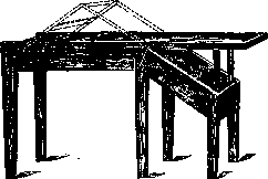
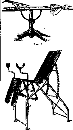
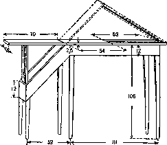
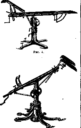
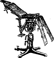
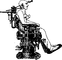
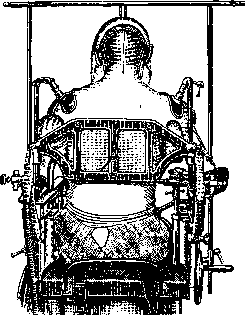
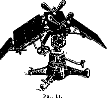
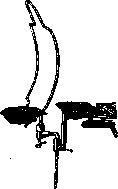
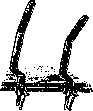

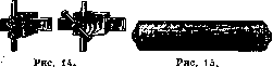
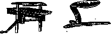
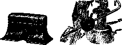
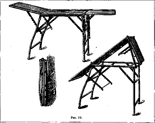
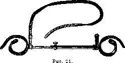
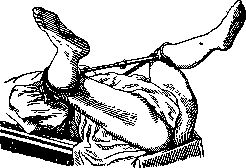
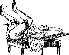
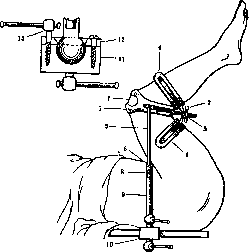
Figure 24. Martov's leg holder: two side bars (6 and 8) made of hollow tubes. One tube (3) is inserted into another (9) as needed. On both sides, the bars with tubes are attached at the top and bottom in the manner of a spherical joint fixation, which makes it possible, thanks to the crater-shaped cover (12) and by means of the clamp (13), to give any direction to the bar (9) and the ring (4) in all planes. Clamp for fixing the leg holder to the table (10 and 11), wooden and metal, which is one of the essential advantages of the leg holder for work in the conditions of district practice. Leather socket for the kneecap (1 and 7). Device (4), with the help of which, by acting on the nut (2 and 5), any position can be given to the knee bend (to prevent blood stagnation).
fit to any table. This last model will apparently also find application in practical life (fig. 25). Of the simple leg holders that allow fixing
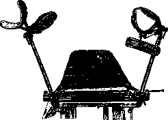
Figure 25.
the lower extremities of patients in the required position directly on the bed, one can mention the leg holder -30; the opium of Zangemeister (fig. 26).- In addition to the complex metal models mentioned, improvised leg holders from an ordinary sheet are widely used in practice (especially in the district), with the help of which the lower extremities are held in the necessary position. Nevertheless
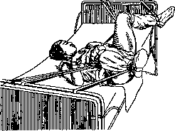
Figure 28.
the most convenient, when there is a sufficient number of assistants, is for assistants to hold the patient's legs. The patient's pelvis is placed slightly in front of the table, and the assistants hold the bent LEGS.
Related articles
Mentioned in
Cite this page
“Operating Tables.” Soviet Medical Encyclopedia. English translation of Bolshaya Meditsinskaya Entsiklopediya, 1st ed. (Moscow, 1928–1936), ed. N. A. Semashko. https://sovietmedicalencyclopedia.pages.dev/article/operating-tables/