Bone Suture
Historical document, translated for reference. It reflects medical knowledge of the 1920s–30s and is not medical advice.
Summary
This article from the 1928-1936 Soviet Great Medical Encyclopedia discusses the surgical procedure of bone suture (osteosynthesis), detailing indications, operative techniques, material choices such as wire and bone pegs, and potential complications.
Encyclopedia article (1928–1936)
BONE SUTURE. To restore the integrity of a bone, it is often necessary to resort to osteosynthesis—a bone suture (Lister, 1873), e.g., in irreducible, periarticular fractures, etc. A properly applied bone suture eliminates pain in the case of a fresh fracture, makes it possible to begin treatment early with massage and passive gymnastics without fear of displacing the fragments, shortens treatment time, and provides good restoration of the function and anatomy of the bone. The improvement in the results of bone sutures in connection with better asepsis and the development of technique has significantly expanded the indications for its use. A necessary condition for the success of a bone suture in closed injuries is the most scrupulous observance of asepsis during the operation. In open injuries, a bone suture can be used if sufficiently reliable disinfection of the wound is feasible by excision of the infected wound surface followed by tight closure. If the wound is unreliable, it is better to drain it and postpone the suture for 8–14 days. In infected granulating wounds, a suture can be used if proper alignment of the fragments cannot be achieved by other means. The best time for a bone suture in fresh injuries is considered to be the 2nd week. By this time, part of the hemorrhage has been resorbed, and tissue regeneration is enhanced under the influence of irritation by the remnants of the hemorrhage. In general, it is desirable to operate before the onset of bone atrophy, while the bone is still dense enough not to resorb quickly from the pressure of the applied sutures. During the operation, wide access to the suture site is necessary so as to handle the soft tissues less and to more easily freshen and properly fit the ends of the bone being sutured. To avoid working in the wound with fingers, Lane recommended long instruments for these operations. Careful cleaning of the operative field from everything that could interfere with the placement of the bone suture and subsequent healing is necessary: small fragments must be removed, especially those not connected with the periosteum, embedded in the soft tissues, tissue fragments, and blood clots; larger fragments are recommended to be preserved, since with proper placement they contribute to bone restoration, growing together with the main fragments and serving as a kind of bridge between them. The fitting of the exposed ends of the fragments in fresh cases is relatively successful until full contact and restoration of the bone shape are achieved. In older cases, it is necessary to properly freshen the fracture surfaces for the widest and most precise contact of the fragment ends. Scars, overgrown bone callus, roughnesses are removed, or the ends of the fragments are sawn off subperiosteally so that the sawed surfaces closely adhere to each other. Depending on the shape of the fracture, the ends are sawn transversely, obliquely, in the form of the so-called Russian lock (Sklifosovsky, Nosilov) (Fig. 1), etc. During processing, the bone is held with bone forceps (see Fig. in the article Surgical Instruments). After this, the fitted ends of the fragments are reduced using bone forceps or special elevators, e.g., Kocher's (Fig. 2). If the fragments are severely displaced lengthwise, considerable traction must be applied, and in older cases, if it is impossible to overcome the resistance of the remaining scar tissue after excision, the ends of the fragments are sawn off to the necessary length. Mendler's forceps may be useful for bone reposition: screws with loops are screwed into the fragments, into which the ends of the forceps are inserted (see Fig. in the article Surgical Instruments), and by compressing the handles of the forceps, the bone is shifted in the desired direction. After fitting and reduction, the fragments are held in the correct position using bone clamps and fastened in one way or another.
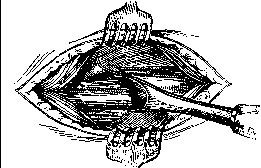
Figure 2.
In some forms of fracture, sufficient fixation is sometimes obtained by driving the end of one fragment into the medullary cavity of the other. Bone sutures are most often placed subperiosteally to minimize disruption of the periosteal blood supply and obtain a better bone callus. When applying metal sutures, it is best to use a homogeneous metal, since the presence of different metals in the wound (e.g., gold-plated steel screws, aluminum plates, copper screws, etc.) creates a galvanic cell with a constant current, which entails electrolysis, impaired healing, and aseptic inflammation in the soft tissues. For binding (cerclage) and suturing the bone, bronze-aluminum wire, silver wire (worse), wire cable, the so-called Wiener Draht, as well as thick silk, silkworm gut, catgut, and broad fascia are most often used. All these materials are not strong enough to always provide tight approximation of the bone ends and eliminate the possibility of displacement. Recently, piano steel wire (0.8 mm), Krupp stainless steel wire, and steel strip have been proposed for sutures. They withstand high tension, allow the use of strong tightening forceps, and thus provide stable fixation of the fragments. Kirschner, Borchardt, Demel, and other forceps are used for tightening (see Fig. in the article Surgical Instruments), or heavy pliers as a last resort. Kirschner solders the ends of the wire tightened by the forceps, protecting the bone from burns with a fiber plate, which does not always save from more or less deep necrosis. Demel twists the ends of the wire,
e
/
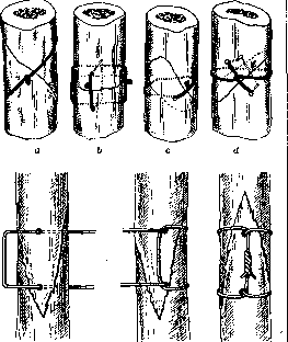
Figure 3. a - bone binding; b - d - various methods of wire suturing. Borchardt passes the end through a loop on the other end and bends it sharply. Magnus, in order not to leave non-absorbable material in the tissues, brings the ends of the wire out through a stainless steel tube and bends the ends, removing the wire and tube upon bone healing. Leaving foreign bodies (metal sutures, silk, silkworm gut) is not always indifferently tolerated by tissues. Sometimes even stainless material has to be removed. Some surgeons therefore use thick, strong catgut, but a sufficient bone callus does not always have time to form before it is resorbed. The simplest method of bone suture is binding the fragments, mainly in oblique fractures. For proper strength, it is recommended to bind perpendicularly to the fracture line, and grooves should be made (e.g., with a hacksaw) on the bone surface so that the wire does not slip (Fig. 3a). Binding is done in various ways, depending on the shape of the fracture, the presence of fragments, etc. If the shape of the fragment ends does not allow secure binding, a suture is used: the fragments held in the correct relationship are drilled in 1-2 places and various sutures are passed. Here, too, various methods of passing the suture are possible, depending on the shape of the fracture (Figures 3 b-d and 4). In addition to binding and suturing the bone, nailing (less often) and screwing are used. Metal and bone nails of various sizes are used. On diaphyses and dense areas, it is necessary to drill a hole for the nail slightly smaller than the diameter of the nail so that it sits tightly. With a very small diameter of the hole, the driven nail can split the bone. Nails are more applicable for fractures in the epiphyseal region, where pre-drilling is often not required for metal or sharp bone nails. Wiemers proposes elastic ("glue") nails made of decalcified, dried, and nail-shaped bull bone. They are strong, flexible, swell rapidly after insertion into the prepared hole, firmly hold the fragments, and eventually resorb. Stronger connections are provided by Gussenbauer staples driven into the bones (Fig. 5). Attempts were made to make absorbable nails and staples from magnesium, but they are not strong enough. Direct screwing of the diaphysis is almost never used, but it is very suitable for epiphyses and short bones. Screws (Fig. 6) hold stronger than nails. The correct shape of the bone and a very strong connection of the fragments are achieved by screwing on metal plates [see separate
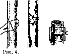
Wire suturing.
Figure 6 (see pp. 159-160, Figures 12 and 13). These plates are made of hardened steel, vanadium, or stainless steel, of various sizes for different bones, and are provided with holes for screws. For epiphyses, Lane's plates with three ends of various lengths converging at different angles are convenient. The plate is placed subperiosteally (usually) on the bone held in the correct position; through its holes, holes for the screws are made in the bone with a twist drill. The diameter of the drill must not be thicker than the screw shaft. It is better to make the holes sequentially, securing the plate with screws. It is possible to first screw the end of the plate to one fragment, then reduce the bone and screw the other part of the plate, but it is not always easy with this method to position the fragments correctly. In some cases, the plates do not irritate the tissues and can be left permanently, while in other cases they have to be removed after the fracture has healed, which prevents more significant bone changes around the screws. A plate screwed to the bone directly under the skin easily causes irritation and must be removed in a timely manner. Lambotte therefore proposed (1902) replacing the plates with long screws that are screwed into the fragments in a straight line. The ends of the screws protrude from the wound and are connected outside the sutured wound by a special holder. The cumbersomeness of the method and complications in wound healing in the presence of screws passing through them prevented the widespread adoption of this method. Schanz screws for the correct positioning of the bone during osteotomy and Rosen's osteostat are constructed on approximately the same principle. The long screws of this device, after preliminary closed reduction of the fragments,
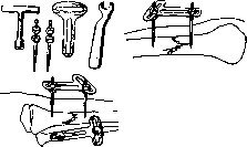
Figure 7. Rosen's osteostat and its application.
are introduced into the cortical layer of the bone without a skin incision, into a depression made in the bone with an awl. The outer ends are held by a T-shaped holder allowing fixation of the screws in different planes after final bone positioning (which can be done under X-ray control) (Fig. 7). In all cases of using screws, nails, and other metal sutures, after some time they loosen due to greater or lesser significant resorption of the bone around them; but by this time a sufficient callus has usually already developed. Plates with screws, nails, etc., are inapplicable in cases of severe comminution and increased bone fragility. In addition to the above-mentioned methods, fragments can be joined using a bone peg inserted into their medullary cavities (so-called Bolzung), or by attaching a bone plate to the fragments externally (Schienung). Trojan proposed metal plates with ears attached to the bone with piano wire. Metal pins proposed earlier are of little use because they are too bulky a foreign body. Resorbable ivory (or bovine bone) is more convenient, and the patient's own bone is even better. Horn (Mysz, Rehn) and magnesium pins were also proposed for this purpose. For pinning, the patient's fibula without periosteum is most often used, as well as other bones. A piece is selected or cut that matches in thickness the diameter of the medullary canal of the bone being joined. The length of the piece corresponds to the size of the bone being repaired
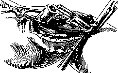
Figure 8. Pinning - Bolzung.
(up to 8-10 cm for the femur). If the medullary canal is narrow, it is widened without disturbing the integrity of the cortical layer using a narrow chisel or burr to the thickness of the pin, which is carefully driven in with a hammer to half its length (Fig. 8). Having prepared the canal in the other fragment, the free end of the pin is inserted into it and pushed in. It is useful to fix the pin with forceps or by wrapping it tightly with silk so that during reduction and advancement it does not slip deeper into the cavity of the first fragment. If it is not possible to pass the end into the cavity of the second fragment, part of its wall can be knocked off to facilitate the introduction of the pin, the pin introduced, and the knocked-off piece attached by tying with catgut or wire. Pinning is more applicable in fresh fractures when the medullary cavity is not altered by scarring. In old injuries, the bone must be drilled out if possible down to healthy bone marrow in order to involve the endosteum in the formation of bone callus. It must be pointed out that significant destruction of the bone marrow during pinning sometimes very strongly slows down bone healing. - In cases with severely damaged or scar-altered
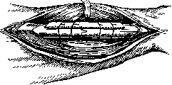
Figure 9. Plating - Schienung.
periosteum, the fragments are fastened externally with a bone plate taken with the periosteum, usually from the tibia. The plate must be long enough so that it can be firmly tied to the fragments with wire and make them immobile (Fig. 9). It is useful to make small notches on the edges of the plate and on the fragments so that the wire does not slip, but the ends of the plate should not be drilled for this purpose. The side of the plate covered by the periosteum should face the soft tissues so that the nutrition of the periosteum is established better and faster. After applying the bone suture, carefully adapting the periosteum with the restoration, as far as possible, of its continuity in the area of the junction of the ends of the fragments, and stopping the bleeding, the wound is sutured tightly. With strong bone and a strong metal suture, one can be limited to a usual splint bandage, and light massage, gymnastics, and baths can be started early, which significantly accelerates bone healing. With other sutures, it is safer to apply a good plaster modeling bandage (with a window in the area of the incision if possible) until sufficient callus has formed, and then leave an open plaster bandage or splint and conduct treatment with massage, etc. If possible, it is useful to check the position of the bone with X-rays after applying the plaster bandage, since displacement sometimes occurs during plastering, and it is better to correct the position in the first days. X-rays also help monitor the development of bone fusion. It must be kept in mind that the bone used for the pin or splint becomes looser after some time, which is why fractures and disruption of the synostosis are possible with careless handling before the development of callus. The use of a pin and splints made of living bone from the same patient has become widespread thanks to the ever-increasing development of bone-plastic operations.
N. Terebinsky. Lit.—see lit. to article Bone.
Related articles
Mentioned in
Cite this page
“Bone Suture.” Soviet Medical Encyclopedia. English translation of Bolshaya Meditsinskaya Entsiklopediya, 1st ed. (Moscow, 1928–1936), ed. N. A. Semashko. https://sovietmedicalencyclopedia.pages.dev/article/bone-suture/