Thrombus
Historical document, translated for reference. It reflects medical knowledge of the 1920s–30s and is not medical advice.
Summary
A thrombus is a mass of blood clots that forms within blood vessels during life, potentially obstructing blood flow. The article details the formation process, distinguishing between primary white thrombi and red thrombi, and discusses the pathogenesis involving blood flow slowdown, vessel wall damage, and changes in blood properties.
Encyclopedia article (1928–1936)
THROMBUS, n. (from Greek thromboō - I clot). Thrombosis - the process of the in vivo formation of dense masses from blood that may to a greater or lesser degree occlude the vessel lumen. A thrombus - a mass of blood clots (a dense mass, "plug") forming from blood as a result of thrombosis. The terms "thrombosis, thrombus" are also used in a broader sense, denoting similar phenomena of the formation of dense masses from blood that sometimes occur in tissues outside vessels during slow bleeding (extravascular thrombi). Furthermore, thrombi (essentially incorrectly) are sometimes used to denote dense masses that do not originate from blood as a whole (hyaline thrombi), and some that even form within the lumen of other tubular systems (bile thrombi in the lumen of bile capillaries, thrombi from clotted lymph in lymphatic vessels), thrombi from tumor cells, from bacteria. The generally accepted definition of thrombosis given above is somewhat formal and descriptive. A definition that would better correspond to the essence of the process would be one that highlights the main moment of thrombosis - the disruption of normal relationships between the vessel wall and blood on the basis of changes in both. The development of thrombosis in its simplest form has been studied under the microscope on living objects (transparent membranes) in both cold-blooded (Mantegazza, Zahn, Klemensiewiez) and warm-blooded (Eberth, Schimmelbusch) animals. Thrombosis was caused by the action of predominantly mechanical or chemical irritants on the vessel wall (mainly veins). In the damaged vessel, blood flow slows down, the peripheral plasma layer of the blood column becomes narrower, its demarcation from the central stream is blurred, and leukocytes and especially thrombocytes (blood plates, Bizzozero's plates) appear in it in greater numbers than normal. The latter accumulate in large numbers on the damaged area of the vessel wall, adhere to each other (agglutination of plates) and merge into a single continuous, colorless, homogeneous or slightly granular mass (conglutination of plates). To the latter, leukocytes also adhere to a greater or lesser degree along the periphery. The resulting colorless mass on the inner surface of the vessel wall is called the primary white thrombus of Cohn. Cohn, the first to observe this phenomenon in frogs, considered the primary thrombus to consist of leukocytes, since thrombocytes - spindle-shaped cells in cold-blooded animals - do not have the typical appearance as in warm-blooded animals, and only later observations on warm-blooded animals, especially by Eberth and Schimmelbusch, allowed to establish the important role of thrombocytes in thrombosis. More precise observations on thrombosis in vivo, mainly by Laker and Klemensiewicz, showed that even before the deposition of plates on the damaged area of the vascular wall, a thin, structureless film precipitates from the blood plasma (primary fibrin membrane), to which blood plates already adhere. The observations cited show that the process of thrombosis cannot be equated with simple clotting of blood in vessels, since the primary event is the deposition of blood plates, and clotting is only secondary; hence it is clear that blood plates play a particularly important role in thrombus formation. Pathohistological studies on post-mortem and experimental material have fully confirmed these findings. Often at sites of vessel damage, one can clearly see the deposition of a granular or homogeneous mass along the wall, consisting of altered thrombocytes with an admixture of leukocytes and the fibrin threads forming around them. The further development of the thrombus depends on the ratio of the processes of plate sedimentation and blood clotting. Blood clotting in thrombosis is still considered mainly from the point of view of the fermentative theory (see Blood). In accordance with the latter, it is assumed that thrombokinase is released from the blood plates (partly also leukocytes) accumulated on the vessel wall when they are destroyed, and, activating thrombogen present in the plasma, leads to the conversion of fibrinogen to fibrin. This is facilitated by the stagnation of blood that occurs in the spaces between the accumulations of plates on the vessel wall. Experiments by L. Léba showed that damage to vessels in animals whose blood has been artificially deprived of the ability to clot (introduction of hirudin, peptone) does not completely cause thrombosis, although small deposits of blood plates on the walls do form. In general, in the formation of certain parts of complex thrombi (the tail part, red layers of the body) and in the development of so-called red thrombi (see below), blood clotting plays the greatest role. It also comes to the forefront in the formation of thrombi as a result of substances entering the blood that cause its rapid clotting (for example sometimes during blood transfusion, introduction of pepsin, ether, salvarsan into the blood, in burns, etc.). However, in these cases, we actually have not thrombosis, but rather in vivo clotting of blood in vessels. In the pathogenesis of thrombosis, three main factors are emphasized: slowing of blood flow, damage to vessel walls, and changes in blood properties. The following arguments speak in favor of the important role of slowing of blood flow in the development of thrombi: a) slowing of blood flow, observable ad oculos when observing the development of a thrombus under the microscope; b) the much greater frequency of thrombosis in veins compared to arteries: according to the data of Lyubarshev, thrombi are found in the right heart and veins in 30.1% of all autopsy cases, in the left heart and arteries in 7.6%; c) the predominant development of thrombi in such areas of the venous system where blood flow is particularly slow (venous plexuses of the pelvis, veins of the lower extremities); d) frequent development of thrombi in areas of pathological dilatation of vessels (in aneurysms, varicose veins); e) frequent formation of thrombi with weakening of cardiac activity (especially in old age), when blood flow is slowed (see separate table, figure 3); f) usually smaller thrombi in arteries than in veins. However, in all the cases of thrombus development mentioned now, damage to the vessel wall cannot be excluded. If such damage is not always detectable even microscopically, its presence in the sense of changes in physicochemical and biological properties of the vessel endothelium is quite conceivable, especially in cases of thrombosis in infections. Experimentally, it is also not possible to cause thrombosis with only slowing of blood circulation. In clarifying this question, experiments with double ligation of vessels played a major role. If the ligation is performed carefully, blood in such vessels remains in liquid form for several months. Consequently, stagnation of blood alone is not sufficient for thrombus formation. The following arguments speak in favor of the important role of vessel wall damage in the development of thrombi: a) in experiments, vessel damage easily causes thrombosis, which is also observable when observing blood circulation in vivo; b) thrombi are usually observed at damaged sites of vessels, e.g., on atherosclerotic ulcers of arteries, at sites of vessel ligations, with inflammatory changes in vessels, with infectious-toxic damage to the inner layer of the vessel wall (or heart valves in endocarditis), etc.; c) cauterization of a vessel ligated at both ends quickly causes the transformation of the liquid blood contained in it into clots. However, the arguments presented raise doubts. The main importance in vessel damage was always attributed to changes in the vascular endothelium. According to these views, the integrity of the latter is necessary for keeping blood in a liquid state, either due to the smoothness of the normal endothelial lining or to the content in endothelial cells of a substance that prevents blood clotting (antithrombin Brücke). However, the presence of antithrombin in the vessel endothelium has not been proven; the positive data of Gutschy in this regard have not been confirmed by L. Léba; moreover, at the beginning of thrombosis, it is not blood clotting but the deposition of thrombocytes that is important. The smoothness of the inner surface of vessels as a factor preventing thrombosis should not be given exceptional importance; thus Cohn, Eberth and Schimmelbusch, Arnold, etc. did not always observe thrombosis after introducing foreign bodies into vessels; in humans, cases of finding foreign bodies inside vessels or in heart cavities without any significant thrombosis have also been described. Cauterization of a vessel ligated at both ends leads not to the formation of a proper thrombus in it, but simply to clotting of blood. Finally, the formation of thrombi on atherosclerotic ulcers is observed far from always; in this case, as in others, we have, along with damage to the vessel wall, conditions leading to mechanical disruption of blood flow (formation of vortices, areas of blood stagnation, etc.). The views presented essentially emphasize only mechanical factors in the pathogenesis of thrombosis. The latter acquired particular importance after the research of Aschoff on the role of vortex movements and standing waves in blood in thrombus formation. On the surface of mural thrombi, a peculiar arrangement of their constituent elements in the form of parallel, transversely directed ridges is often observed.
Such a structure of thrombi is easily explained by the mechanical conditions created in a flowing fluid when there is any obstacle on the vessel wall. Observations by Aschoff and Rehbock on the flow of water in a channel to which sawdust was added showed that near obstacles, vortex movements and standing waves occur due to the reflection of water from the obstacle. In the spaces between these, areas of stagnation form and correspondingly, sawdust not carried away by the current settles to the bottom in the form of parallel ridges. The slower the water flow, the easier the settling of sawdust occurs in such places. Similar phenomena can be observed near obstacles, e.g., on the bottom of a stream carrying fine sand particles, or when sand particles are carried by the wind. By analogy with these phenomena, the formation of layered, ridge-like structures in thrombi can also be explained. The plates, being the lightest of the elements suspended in blood plasma, precipitate from the blood near obstacles, i.e., various irregularities on the vessel wall, due to the formation of vortex movements and standing waves here, especially with slow blood flow. It is impossible to explain thrombosis by mechanical factors alone. In particular, the important role played by platelets in thrombi requires special explanation, which relates to the question of the significance of changes in blood in the pathogenesis of thrombosis. Previously, in explaining the agglutination of plates, their special 'stickiness' was mentioned, however, an explanation of these phenomena from a physicochemical point of view has only been given in recent years, mainly by Lampert. The surface activity of platelets and leukocytes is particularly important for understanding thrombosis; in addition, fibrin is also a surface-active substance (Guchi). The surface activity of platelets is counteracted by their electrical charge, thanks to which platelets, repelling each other, remain in suspension. The stability of platelets, depending on their electrical (negative) charge, can change depending on a number of conditions. In particular, it decreases when weakly negatively charged globulins or almost uncharged fibrinogen increase in the blood, and when the acid-base balance shifts in the acidic direction. In almost all conditions, especially those frequently accompanied by thrombosis (infections, puerperium, weakening of blood circulation, postoperative period), the indicated shifts (Starlinger) are usually observed, leading to a decrease in platelet stability, which is an important factor in the pathogenesis of thrombosis. To this must be added that with slowing of blood flow, as well as in many infections, the number of platelets in the blood increases. The second physicochemical phenomenon, which apparently has particularly great significance in the pathogenesis of thrombosis, is the phenomenon of wettability of the vessel wall by blood plasma. The wettability of vessel walls, i.e., the force of attraction by them of the components of blood, increases primarily depending on the increase in wetting time, which is observed, for example, with slowing of the current and blood stasis. Furthermore, wettability increases with an increase in the roughness of the vessel wall. At the same time, a thin film of fibrin (Guchi's model experiments) first forms on the wall, similar to the primary fibrin membrane in thrombosis. When the vessel wall is damaged, its wettability increases, i.e., the surface-active forces of attraction between it and the blood increase, resulting in the precipitation of platelets, which among blood elements have the greatest surface activity. Finally, acidoses of the body also lead to an increase in the wettability of the vessel wall, i.e., they predispose to thrombosis. However, consideration of thrombosis from a physicochemical point of view does not exhaust its biological nature. This question has been particularly emphasized in the works of Dietrich and Siegmund. They consider thrombosis as a 'reactive' phenomenon on the part of the vascular endothelium, leading to the retention of blood elements, bacteria, etc. Dietrich also speaks of the increase in resorptive properties of the endothelium, caused by the 'adaptation' or sensitization of the latter to proteins of bacterial bodies or generally to foreign proteins. Sensitization of the endothelium leads to the precipitation of homogeneous protein masses from plasma onto it, to which platelets and leukocytes attach; thus a primary white thrombus is formed, and then mechanical conditions (vortex movements of blood) develop around it, contributing to further growth of the thrombus. Thus, in both these views and the physicochemical views, the factor of changes in the relationship between blood and the vessel wall is particularly emphasized in the pathogenesis of thrombosis. In the occurrence of these changes, all three factors discussed above are of importance, i.e., slowing of blood flow, damage to the vessel walls, and changes in the blood itself. All these factors to some degree participate in thrombosis of various etiologies. Thus, in thrombosis on the basis of atherosclerosis, along with damage to the wall, formation of vortex movements of blood, areas of slowed blood flow, and changes in physicochemical relationships in the boundary layer of the blood stream are always present near atherosclerotic ulcers and plaques. In the formation of so-called dilational thrombi (in dilated vessels), along with slowing of blood flow, changes in vessel walls and changes in blood properties are of importance. We have similar phenomena in stasis thrombi (thrombosis in venous stasis). So-called marantic thrombi, i.e., thrombi developing with general nutritional decline (marasmus), mainly in veins, auricles of the heart
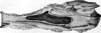
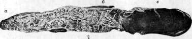
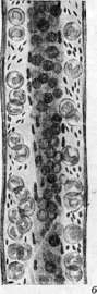
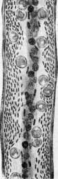
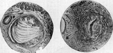
Figure 1. Lamellar thrombus of the femoral vein. Fig. 2. Longitudinal section of a femoral vein thrombus: a-head; b-layered part (body); c-tail part. Fig. 3. Scheme of blood flow (according to Kbertu-Schimmelbusch): a-normal circulation (axial flow, plasma peripheral zone with individual platelets); b-slowed circulation (erythrocytes visible in the axial flow, peripheral standing of leukocytes and beginning peripheral standing of plates); c-strongly slowed circulation (peripheral standing of plates and leukocytes, decrease of axial flow). Fig. 4. Organization of the thrombus (beginning). Fig. 5. Organization of the thrombus, formation of vessels. Fig. 6. Thrombus of a vein. Fig. 7. Organization and vascularization of the thrombus (late period).
and at its apex between the crossbars (cardiac 'polyps'), arise on the basis of general slowing of blood circulation from the decline of cardiac activity; the same changes in the walls of blood vessels and properties of blood have a certain significance in the pathogenesis of these thrombi as in stasis. The significance of mechanical moments in thrombosis is especially prominent in stasis and marantic thrombi: they often form at the valves of veins and at the points where lateral branches flow into veins; here vortex movements of blood especially easily arise - at the valves [see separate table (art. 823-824), fig. 1], due to their insufficient opening during stasis, at the points of entry of veins - due to the meeting of two blood streams. In thrombosis of inflammatory origin, all pathogenetic moments of thrombosis are expressed especially sharply: changes in the endothelium of blood vessels, slowing of blood flow, shifts in physicochemical constants of blood and tissues, accumulation of leukocytes and thrombocytes. Especially favorable conditions for thrombosis are created in infectious-inflammatory changes located directly in the walls of arteries (thromboarteritis) and veins (thrombophlebitis). These changes, especially thrombophlebitis, occur as a cause of thrombosis much more frequently than was previously thought: only in 13% of all cases of thrombosis could Lubarsch exclude the participation of infectious processes, and in 55% he confirmed them with localization mostly in the roots of the thrombosed veins. There are also indications of frequent findings of bacteria in apparently non-infected thrombi (Rosenow). Thrombosis, often accompanying various general infectious diseases, is apparently of complex origin. It is especially often observed in prolonged, sluggishly proceeding infections. Along with changes in the properties of blood (decrease in stability of thrombocytes, often their increased number and number of leukocytes, increase in globulins, often increase in fibrinogen content, acidic shifts in the body), here the change in properties of the endothelium ('activation' of it in the sense of Dietrich, see above) and finally slowing of blood flow due to weakening of the heart or decrease in vascular tone are also of significance. The observed in some patients as if special tendency to thrombosis (chronic thrombopathy - thrombopathia chron. migrans) can be explained by the presence of hidden infection or intoxication, for example by products of tissue breakdown (especially in cancer). In addition, recently constitutional factors have been given significance in this respect, especially highlighting the picnic type; according to Lampert, in individuals of this type, an increased number of thrombocytes and a tendency to acidic shift of acid-base balance are often observed, which explains the predisposition to thrombosis. Thrombosis, often observed after surgical operations, also has a complex origin. Here, apparently, both infectious factors and changes in blood (acidic shift of acid-base balance, etc.) as well as weakening of cardiac activity, slowing of blood flow in veins due to recumbent position, sometimes application of bandages compressing veins, etc. are of significance. Distinctive features of thrombi from postmortem blood clots, which have the greatest significance in patho-anatomical and forensic-medical practice: a) thrombi are almost always drier, denser, more brittle and less elastic than blood clots; b) the surface of thrombi is almost never completely smooth and shiny, like a clot; it is usually uneven, often showing ridge- or rib-like elevations; c) on cross-section, thrombi often reveal an uneven layered structure with alternating white and red layers in the middle (body) part of the thrombus; d) thrombi are firmly connected with the wall of the vessel, especially in their initial (head) part, whereas clots lie freely in the lumen of the vessels. Types of thrombi and their structure: a) White or colorless thrombi may consist of a collection of only thrombocytes, especially in the early stages of thrombosis or with its insignificant development, as well in test cases when rapid blood flow prevents further growth of the thrombus, for example in the aorta (in atherosclerosis). Such thrombi often look like glassy, translucent deposits (thrombocyte thrombi, platelet thrombi). Predominantly larger white thrombi have the appearance of grayish-white masses; they consist of a coarse spongy framework formed by conglutinated thrombocytes, under the microscope almost homogeneous or fine-grained in appearance. To the crossbars of the framework are attached in greater or lesser number collections of leukocytes and fibrin threads. In the loops of the framework are present in different amounts leukocytes, fibrin, and also sometimes erythrocytes. With a large predominance of leukocytes in white (usually infected) thrombi, they are called leukocyte thrombi, although thrombocytes are always also present in them. The so-called leukemic thrombi, occurring in small vessels in leukemia, do not actually represent thrombi, but only masses of accumulated leukocytes. White thrombi form mainly parietally on altered areas of the vascular wall under the condition of relatively rapid blood flow in the vessel, due to which the process of blood coagulation does not reach any significant size. b) Red thrombi have the appearance of dark-red masses and often completely fill the lumen of the vessel (mainly veins). They consist of a dense network of fibrin, in the loops of which are enclosed erythrocytes and leukocytes. The ratio of these and others with rapid formation of the thrombus is approximately the same as in the blood, with slower formation they are distributed less uniformly. Red thrombi are structurally very close to blood clots, the process of blood coagulation in them sharply predominates over the process of agglutination of thrombocytes (hence the name coagulation thrombi). However, they always arise on the basis of a primary parietal thrombocyte thrombus, which however can be very small and is detected with difficulty. Red thrombi usually form with sharp slowing of blood flow and stasis. Purely red thrombi also occur when substances are introduced into vessels that very quickly cause blood coagulation (pepsin, ether, ferric chloride). In humans they are sometimes observed in the lungs after ether-chloroform anesthesia, in small vessels of the brain and other organs after infusions of salvarsan, blood transfusions, introduction of extracts from organs (toxic and enzyme thrombi), further in eclampsia, burns (autotoxic thrombi). However, in all these cases, it is rather about in vivo coagulation of blood in vessels than about thrombosis. Red thrombi, as weakly connected with the vessel wall with their entire mass and relatively loose, especially easily detach and give rise to embolism. In older red thrombi, erythrocytes hemolyze, break down, the thrombus thus gradually discolors. c) Mixed or layered thrombi, representing a combination of white and red thrombi, consist of three parts: the white head end (head of the thrombus), firmly attached to the vessel wall, the intermediate layered part containing alternating layers of white and red color, and the red tail end [see separate table (art. 823-824), fig. 2]. The head of the thrombus, corresponding to the place of its primary origin, consists of a solid mass or separate collections of thrombocytes. In the middle layered part, having an uneven surface with elevations in the form of ridges or ribs, collections of conglutinated thrombocytes (white layers) form a complexly constructed branched framework of the thrombus, resembling coral or sponge. On the beams of this framework there are layers of leukocytes, and the spaces between the beams are occupied by clotted blood, i.e. consist of a network of fibrin with captured erythrocytes (red layers of the thrombus). The tail part is constructed on the type of red thrombus. Individual parts of the mixed thrombus are expressed to varying degrees: both the middle, layered part and the tail part can be either very short or of considerable length. The structure of the mixed thrombus is an expression of the complex process of its development. The latter begins with the formation of a white parietal thrombus from thrombocytes (head of the thrombus). At some distance from it and from each other corresponding to the intervals between areas of vortex movements and standing waves occurring around the primary thrombus, new masses of thrombocytes are deposited. Accumulating in ever greater quantities and conglutinating with each other, thrombocytes form branched beams of the white framework of the thrombus. On them leukocytes are deposited, and the blood remaining in the spaces between the beams clots under the influence of thrombokinase, released during the breakdown of thrombocytes. The fibers of the precipitating fibrin here due to vortex movements of blood have the appearance of curls or garlands. On the surface of the thrombus continues the settling of thrombocytes, arranged in ridges under the influence of vortex movements caused by the blood flow. Finally when the layered thrombus reaches a considerable size and, more or less closing the lumen of the vessel, causes sharp slowing or stopping of blood flow, the remaining mass clots, due to which the red tail part of the thrombus is formed.
Blood clotting usually continues to the mouth of one of the collateral branches. The continuing blood flow through it prevents further development of thrombosis. Thus, depending on the location of the thrombus in relation to the mouths of the lateral branches of a given vessel, the tail part of the thrombus can grow in the direction of blood flow (in veins toward the heart) or against it. Thrombi in veins are especially typical, with the tail part directed toward the heart (greatest danger of detachment and formation of an embolus). Depending on the relationship of thrombi to the lumen of the vessel, they are distinguished as mural and occluding (obstructing). The first are found mainly in the cavities of the heart, especially in its auricles and between the trabeculae of the ventricles (cardiac "polyps", spherical heart thrombi), as well as in the aorta at the site of atheromatous ulcers and in large veins; the second occur in smaller vessels. Occluding thrombi form either by gradual growth from mural ones or during rapidly progressing thrombosis, immediately obstructing the vessel. Depending on the place of formation, thrombi are distinguished as autochthonous, i.e., located at the site of primary development (root of the thrombus), and continued, corresponding to further layers developing after the primary formation of the thrombus (body of the thrombus). Thus, in mixed thrombi, their head corresponds to the primary autochthonous thrombus, while the middle layered and red tail parts correspond to the continued thrombus. Hyaline thrombi are found in capillaries and small veins, especially in the brain, kidney glomeruli (see separate plate for article on Turk's cells, fig. 2), intestines, and lungs. They appear as solid homogeneous masses or separate spherical homogeneous formations that give a positive reaction for fibrin. They occur mainly in certain intoxications (by enzymes, snake and mushroom venom, also in burns, frostbite, eclampsia) and in infectious diseases. The origin of hyaline thrombi is not fully elucidated. This is either a special form of fibrin coagulation or the transformation of blood plasma as a whole into a gel. With stasis, hemolysis, and toxic destruction of erythrocytes, hyaline masses can also form in capillaries and small veins in the form of uniform shiny deposits that stain intensely with eosin. Some authors equate hyaline thrombi with so-called spodogenic thrombi. The latter are accumulations in small vessels of various breakdown products of blood elements (platelets, erythrocytes), forming especially in certain intoxications (lead, aniline) and burns, as well as in rapidly developing hemolysis; these also include thrombi from the "shadows" of erythrocytes (e.g., after unsuccessful blood transfusion). Further transformations of thrombi. Soon after formation, thrombi begin to undergo some shrinkage, decrease in volume, loss of weight; they become as if drier and more brittle. This phenomenon, which is of great importance in terms of the possibility of thrombus detachment and formation of an embolus, is compared to the process of syneresis of a jelly, i.e., the contraction of a jelly with the release in free form of a certain amount of colloidal water. The same process occurs with a blood clot in vitro, and the degree of retraction of the clot depends on the number of platelets in the blood and certain physicochemical factors (state of acid-base equilibrium, etc.). Apparently the same conditions are also important in the shrinkage of thrombi. Subsequently, with prolonged stay of a thrombus in a vessel, swelling of the fibrin and platelet masses may occur, and thrombi sometimes transform into a uniform glassy hyaline mass. Subsequent changes in thrombi come down to their softening and organization through young connective tissue growing into them from the vessel wall. In different cases, one or the other of these processes may predominate. Softening of the thrombus proceeds either as aseptic or as bacterial (septic) softening. In the first case, enzymatic dissolution occurs mainly of the platelet and leukocyte masses, apparently by enzymes released from these. Small mural platelet thrombi may completely disappear due to dissolution and washing away by the blood. In larger thrombi, softening usually begins in the central parts, where semi-liquid pus-like masses form, containing fatty and protein granules, especially for example in round thrombi (polyps) of the heart. White thrombi, containing many leukocytes, soften especially quickly. Septic (yellow) softening occurs in thrombi formed in thrombophlebitis, but secondary infection of initially aseptic thrombi also occurs. In the disintegrating thrombus masses, which take on a greenish-yellow color, large accumulations of bacteria and secondarily penetrating leukocytes are found in septic softening. Finally, the thrombus turns entirely into a mass of pus filling the vessel and containing crumbly remnants of thrombotic masses. In rarer cases, putrefactive softening of thrombi is observed due to the penetration of putrefactive microbes (mainly bacilli and spirochetes) into them. In these cases, thrombi are melted down, forming a greasy, dirty-gray mass. Organization of the thrombus [see separate plate for article on Turk's cells, fig. 1 and separate plate (art. 823-824), figs. 4-7] consists in the fact that from the inner coat of the vessel, on the 2nd-3rd day, cellular elements of the fibroblast type grow into it. The possibility that the endothelium of the vessel also participates in this process is not excluded. Poliblastic wandering cells and capillaries also penetrate into the thrombus from the vessel wall, representing offshoots of vasa vasorum (vascularization of the thrombus). Erythrocytes in the thrombus disintegrate, leaving accumulations of hemosiderin phagocytized by macrophages; all other elements of the thrombus also disintegrate, are resorbed, and are replaced first by loose, cell-rich, and then by dense fibrous hyalinized connective tissue. Organization of mural thrombi ends with the formation of dense connective tissue thickenings or strands. Capillaries that have grown into the thrombus partly transform into fairly large vessels. When the thrombus shrinks, fairly wide cavities sometimes form inside it and between it and the vessel wall. They are lined with endothelium, filled with blood, and blood circulation can be restored in this vessel through them - canalization of the thrombus. Sometimes with pronounced canalization, the entire thrombus is penetrated by cavernous spaces containing blood (cavernous transformation of the thrombus, especially frequent for example in the portal vein). Sometimes organization of the thrombus leads to abundant growth of cell-rich connective tissue in it with swollen mucoid-like interstitial substance. Such thrombi, usually observed in the left atrium, are by some considered tumors - myxomas. In the absence of organization, thrombi that have long lain in vessels undergo shrinkage and become impregnated with lime salts (ossification, petrification of the thrombus), which especially often occurs in dilated veins and venous plexuses. Then detaching from the vessel wall, such thrombi remain lying freely in their lumen as vein stones (phleboliths). Statistical data on thrombosis. a) General frequency of thrombosis. According to Lubarsch (1905), out of 1,932 autopsies, thrombi were found in the heart or vessels 733 times (37.7%), of which in 584 cases thrombi were found in the right heart and veins (30.2%) and 149 times in the left heart and arteries (7.6%). Older autopsy materials, collected for example by Mannaberg (Mannaberg, 1899), give much lower figures (96 cases of thrombosis out of 1,800 autopsies, i.e., 6%), which Lubarsch explains by insufficient thoroughness of research, due to which small thrombi that did not cause phenomena during life were not noted. Therefore, clinical statistics of thrombosis also give lower figures than pathological anatomical ones. Thus, according to Albanus (Albanus, 1903), out of 1,140 cases of laparotomies, venous thrombosis was observed only 53 times. Figures given by authors for the pre-war and first post-war years fluctuate for autopsy material roughly within the range of 1% to 4.2% (Fahr, Oberndorfer, K. Schmlz, etc.), and only cases of sharply expressed thrombosis were taken into account. Clinical statistics of the same period give figures of 0.88% (Kiibler), 1.1% (Fehling and Rost for postoperative thromboses), 1.5% (Hering), 0.97-1.56% (Singer), etc. Great attention was drawn to data on a sharp increase in the number of thrombosis cases in recent years. Autopsy statistics in Germany indicate an increase in such cases from 2% to 5% and even to 20% (Oberndorfer, 1928). Fahr and Schultz speak of an increase in the number of thrombosis cases by 10-11 times. Clinical statistics also indicate an increase in the number of thrombosis cases by approximately 3-4 times, and an increase in cases of both postoperative thrombosis and thrombosis on the basis of circulatory weakness (mainly in heart defects) has been noted. The increase in thrombosis cases observed in recent years is explained by some as the fact that clinics began to frequently use the method of intravenous injections of medicinal substances.
However, the data of Singer, Schultz, and others showed that the increase in thrombosis occurs equally in cases not treated with intravenous injections. Other authors explain the increase in cases of thrombosis by the growing frequency of infectious diseases (influenza epidemics), however, convincing data in this regard is not provided. Oberndorfer, Morawitz, Schultz, and others tend to explain the increase in cases of thrombosis in recent years by the widespread use of means that support the dying in a state of agony, creating favorable conditions (slowing of blood circulation) for the development of thrombosis. Indeed, with a general increase in the number of cases of thrombosis, it, as before, most often develops in those parts of the vascular system where blood flow is particularly slow (in veins, mainly of the lower extremities). It is interesting to note Singer's observation that the number of cases of thrombosis in tuberculosis and cancer has not increased, and the increase mainly affected cases of thrombosis in cardiac insufficiency. The latter circumstance some explain by the fact that in modern therapy for edema on the basis of cardiac insufficiency, means are widely used that cause an increased loss of water by the body, associated with changes in the physicochemical constants of the blood (Nordmann, Schonbauer). Schloffer and Vente found that thrombosis indeed occurs particularly often in cardiac patients in the absence of edema, and in the presence of the latter it is not observed. The loss of water is also used by some to explain the origin of postoperative thrombosis (Gordon-Watson), pointing at the same time to the influence of laxatives given before surgery, vomiting, restriction of drinking in the postoperative period, etc. b) Distribution of thrombosis cases by sex. Usually, a predominance of thrombosis cases in women is noted, according to some data almost twice as many (Singer). This is apparently explained by the influence of pregnancy and childbirth, in particular postpartum infections. The observed increase in cases of thrombosis in recent years affects both sexes, but is expressed to a relatively greater degree in men (Singer). c) Distribution of thrombosis cases by age. The largest number of thrombosis cases according to Far, Oberndorfer, Singer, and others falls on the age of 60-70 years, which is easily explained by the weakening of blood circulation, changes in blood vessels, and the frequency of marantic conditions at this age. The consequences and significance of thrombosis in terms of harm to the organism consist partly in the mechanical obstruction of blood circulation caused by thrombi, and partly in creating conditions favorable for the development and spread of infectious agents in the body. Obstruction of blood circulation in arteries in cases of thrombosis leads to local anemia, infarctions, gangrene; in thrombosis of veins, stasis, edema, necrosis of the extremities are observed; in thrombosis of the portal vein - stasis in the organs of the abdominal cavity, ascites (only in rare cases of slowly developing thrombosis of the portal vein is the development of collaterals sufficient for the restoration of blood circulation possible). Thanks to the development of collaterals, and partly to the canalization of the thrombus, blood circulation can be restored after some time. The organization of the thrombus is favorable in terms of fixing it and reducing the danger of embolism; however, the organization of thrombi on the heart valves in endocarditis leads to the disfigurement of the valves, the fusion of their leaflets with each other, etc., leading to phenomena of valvular defect. Very dangerous consequences are the detachment of parts of the thrombus with the further development of embolism phenomena, especially in the case of septic thrombosis. Relatively favorable is the significance of thrombosis in the closing of a bleeding vessel by a thrombus and in the development of thrombi in dilated vessels (reduction of the dilated cavity of the vessel). As a physiological phenomenon, thrombosis is widespread in humans and mammals, having important significance in stopping postpartum bleeding after the separation of the placenta, and further in the obliteration of some vessels that function in the fetal circulation (Botallo's duct, Aranzio's duct, etc.).
Related articles
Mentioned in
Cite this page
“Thrombus.” Soviet Medical Encyclopedia. English translation of Bolshaya Meditsinskaya Entsiklopediya, 1st ed. (Moscow, 1928–1936), ed. N. A. Semashko. https://sovietmedicalencyclopedia.pages.dev/article/thrombus/