Bizzozero's Plaques
Historical document, translated for reference. It reflects medical knowledge of the 1920s–30s and is not medical advice.
Summary
This article from the 1928–1936 Great Medical Encyclopedia discusses Bizzozero's plaques (blood platelets or thrombocytes), detailing their morphology, staining properties, and role in blood coagulation. It also reviews historical theories of their origin, clinical variations in disease, and methods for counting and studying them.
Encyclopedia article (1928–1936)
BIZZOZERO'S PLAQUES (Bizzozero), blood platelets, thrombocytes, represent the smallest formed elements of the blood. In a blood smear prepared in the usual manner and stained with Giemsa, they appear as round or oval disks, with a diameter equal to 1/7 to 1/3 of the erythrocyte's diameter, lying between red blood cells in isolation (or, more often, forming groups). The narrow, peripheral, hyaline-structureless part of Bizzozero's plaques takes on a light blue hue, while the center appears reddish-purple from the accumulation of a large number of small granules that stain with azur (azurophilic granulation; see table, Fig. 1). In a preparation of fresh blood protected from coagulation by the addition of some anticoagulation fluid (a solution of sodium oxalate or sodium citrate, Witte's peptone), Bizzozero's plaques appear as colorless disks, the center of which possesses a much greater light-refractive property than the periphery (see table, Fig. 2). This light-refractiveness is associated with the presence of granules that intensely stain upon the application of vital staining (Methylviolett, Brillantkresylblau), whereas the hyaline peripheral belt weakly takes up the dye. In preparations of fresh blood not protected from coagulation, Bizzozero's plaques undergo changes very rapidly, sometimes in less than a minute, during which agglutination and sticking of the plaques occur. They lose their regular rounded form, acquire a spiky, stellate appearance and, joining together, form larger or smaller granular conglomerates in which individual elements are no longer distinguishable. With the simultaneously occurring blood coagulation, these granular masses serve as the site of attachment for fibrin threads. The agglutination of Bizzozero's plaques must be related to physico-chemical changes in the medium during the blood coagulation process. Such changes in blood platelets can also occur with insufficiently rapid mixing of the blood with the preserving fluid, with slow outflow of blood from a wound, and other defects in sampling material and preparing the smear. In view of the small size of the object and its property of easily changing its shape, the fine structure of Bizzozero's plaques has been poorly studied; in connection with this, the question of the essence and origin of Bizzozero's plaques is distinguished by an abundance of contradictory opinions. Some believe that the central granulation of Bizzozero's plaques represents the disintegration of the nucleus of a blood platelet degenerating during the preparation of the smear. By applying special precautionary measures, they succeeded in obtaining Bizzozero's plaques in the form of true cell-«thrombocytes» (Deetjen, Kopsch, Eminet). However, the majority of investigators do not confirm this and consider Bizzozero's plaques to be protoplasmic formations connected in their origin with other formed elements of the blood or hematopoietic apparatus. Some derive Bizzozero's plaques from the protoplasm of leukocytes, linking their origin either with the process of disintegration of white blood cells or with phenomena of vital detachment of protoplasmic particles (Al. Schmidt, Dominici, Decastello, and Kryukov); others from erythrocytes (Arnold, Pappenheim, Schilling, Shchastny, Tatarinov); thirdly, others believe that Bizzozero's plaques originate through detachment from the cytoplasm of bone marrow giant cells—megakaryocytes, whose protoplasmic processes penetrate into the bone marrow sinuses. These processes, breaking down into individual segments, enter the bloodstream in the form of Bizzozero's plaques (Wright, Ogata, Downey, Maksimov). The latter view is currently the most widespread [although the works of A. Perroncito (1920, 1921), G. Pianese (1920), and Cesaris Demel (1919) make one strongly doubt its correctness]. Regarding the physiological significance of Bizzozero's plaques, their participation in the formation of certain types of thrombi can be considered proven. The platelets, possessing the property of easily adhering to the damaged vascular intima, accumulate in these places, then agglutinate and provide the basis for the development and growth of a thrombus. A prominent role in blood coagulation processes is ascribed to Bizzozero's plaques by many researchers, specifically the role of producers of enzymatic substances necessary for the coagulation phenomenon (Deetjen, Noli). In connection with this view of platelets, pathology of the latter explains the origin of certain types of hemorrhagic diatheses, such as morbus mac. Werlhofii (Frank; see below). There is a tendency to look upon Bizzozero's plaques also as elements playing a role in the production of defensive substances against infection (N. Ya. Chistovich, O'Holenghi, Eminet). This view is based mainly on the fact that during the infectious process, a regular decrease in the number of Bizzozero's plaques is noted at the height of fever, replaced (after the temperature drops) by an increase in their number, sometimes by 3-4 times above the norm. The number of blood platelets in human blood normally fluctuates within wide limits (from 130,000 to 750,000); on average, it equals 200,000 - 300,000 per 1 cubic mm. In various pathological conditions, the count of Bizzozero's plaques can sharply deviate from normal figures, as, for example, in the blood of febrile patients. Diseases of the blood and hematopoietic organs are often accompanied by a reaction on the part of the number of Bizzozero's plaques; in secondary anemias, a small increase in the number of Bizzozero's plaques is observed, pernicious anemia is accompanied by a sharp drop in the number of platelets, myeloid leukemia proceeds with «thrombocytosis», lymphatic leukemia with a low content of Bizzozero's plaques in the blood, purpura haemorrhagica and morbus mac. Werlhofii are characterized by a drop in the number of platelets and even their absence in the blood (so-called thrombocytopenia). Experimentally, one can easily cause a decrease in the amount of Bizzozero's plaques in the blood by parenteral (intravenous) administration of a small amount of serum or Witte's peptone; in this case, agglutination of Bizzozero's plaques rapidly occurs in the blood, and they collect in the vessels of internal organs. Probably, the mechanism of the drop in the number of Bizzozero's plaques during the infectious process is also explained by this same phenomenon, since Bernhardt found that a very large number of platelets accumulate in the spleen of those who died from acute infections, undergoing phagocytosis by the macrophages of the pulp. In the blood of lower vertebrates, whose erythrocytes contain permanent nuclei, there are spindle-shaped cells which Bizzozero also called blood platelets. But since these elements possess a true nucleus and centrosome, most researchers currently consider these cells to be formations completely distinct from the blood plaques of mammals. - Methods of study. In blood discharged into physiological saline with the addition of 0.4% sodium oxalate, Bizzozero's plaques remain undamaged and unagglutinated for several hours. The same occurs in Afanasyev's fluid containing 0.6% Witte's peptone in physiological saline. For vital staining of the platelets, Methylviolett or Brillantkresylblau (1:5,000) is added to these fluids. Counting of Bizzozero's plaques is performed either in a preserving fluid or in a dry smear preparation. In the first case, the blood under study is discharged into a watch glass with Afanasyev's fluid and thoroughly mixed; then the ratio of the number of Bizzozero's plaques to the number of erythrocytes in the counting chamber is determined (at least 300 erythrocytes and the corresponding amount of Bizzozero's plaques are counted). Parallel determination of the number of erythrocytes in 1 cubic mm of the given blood makes it possible to calculate the absolute number of Bizzozero's plaques. When counting in a dry preparation, blood is obtained by pricking the skin through a large drop of a 14% MgSO4 solution. After mixing, a smear is made on a glass slide, stained according to Giemsa, and the ratio of Bizzozero's plaques to erythrocytes is determined while counting under an immersion system. Knowing the number of erythrocytes in 1 cubic mm of blood, the absolute number of Bizzozero's plaques is calculated (Schilling, Fonio). When centrifuging blood preserved against coagulation, the platelets settle very slowly and remain suspended in the plasma, while other formed elements of the blood have already completely settled to the bottom of the test tube. This property of Bizzozero's plaques makes it possible to obtain them isolated from erythrocytes and leukocytes (Shchastny, Tatarinov).
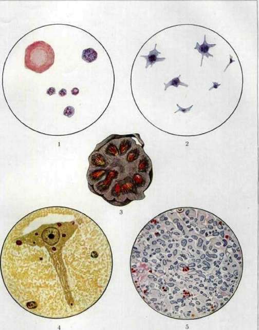
Fig. 1. Blood platelets (Bizzozero's plaques). Above on the left - an erythrocyte. Fig. 2. Preserved Bizzozero's plaques, swollen upon prolonged storage in plasma with sodium oxalate. Fig. 3. Wedge-shaped kidney infarct of a dog. [...] Fig. 4. [...] Fig. 5. Large-cell proliferation [...] Fig. 6. [...]
Related articles
Mentioned in
Cite this page
“Bizzozero's Plaques.” Soviet Medical Encyclopedia. English translation of Bolshaya Meditsinskaya Entsiklopediya, 1st ed. (Moscow, 1928–1936), ed. N. A. Semashko. https://sovietmedicalencyclopedia.pages.dev/article/bizzozeros-platelets/