Osteomalacia
Historical document, translated for reference. It reflects medical knowledge of the 1920s–30s and is not medical advice.
Summary
Osteomalacia is a general metabolic disease primarily affecting bone tissue, known since ancient times but not properly distinguished until the 18th century. The article discusses its historical recognition, epidemiology, predisposing factors including nutritional and social conditions, and demographic patterns of occurrence.
Encyclopedia article (1928–1936)
OSTEOMALACIA (from Greek osteon-bone and malakos-soft) (syn. mollities ossium, ostitis malacissans-softening of bones), a general metabolic disease with predominant affection of bone tissue. O. was known even in deep antiquity, but exact descriptions of it were not available; apparently for many centuries it was identified with other pathological processes of the bone system, incorrectly or not diagnosed at all. Diseases of bones were known in the school of Hippocrates, there are indications of them in Arabic medicine as well, where apparently in the 7th century the Arabic physician Gschuzius described the first confirmed case of O. in a man. Individual descriptions also appear later: worldwide fame was acquired by cases of O. in the Marquise d'Armagnac, who died at 22 years of age from softening of all bones of the skeleton and face, and in the Parisian Supiot (her skeleton is preserved in the Dupuytren museum). The beginning of clinical study of O. was laid only at the end of the 18th century, when the osteomalacic pelvis (Cooper, 1776), postpartum O. (Stein senior, 1787) and the significance of pregnancy in its occurrence (Conradi, 1797) were first described in England. The question of O. attracted the greatest attention of researchers in the first decades of the 19th century, when works appeared devoted to the pathological anatomy of O. (Lobstein, Weidmann and others), its connection with rickets (Cohnheim, Kassowitz, Pommer, Virchow and others); in the second half of the 19th century the first classifications of O. appear (Kschap, 1857), the difference in changes in puerperal and non-puerperal forms is indicated (Volkmann, 1865, Hennig, 1873), the clinical symptomatology is clarified, additional changes from the psyche, nervous and muscular systems are described (Friedberg, 1858; Friedreich, 1873; Hosslin, Trousseau, Charcot and others), more attention is paid to obstetric therapy and generally medicinal treatment, and finally in 1887 (Fehling) the endocrine theory arises, the development of which is also devoted to the first decades of the current century. Frequency of O. Osteomalacia occurs not infrequently and has a tendency to manifest in certain localities, especially along the course and in the valleys of large rivers. Thus, many data have established that it endemically occurs along the banks of the Rhine, Danube, Po, is observed in some cantons of Switzerland, in Bavaria, Flanders, in northern Italy, in China and others; in the USSR foci were also noted in the Volga region (especially the Kazan district) and in Transcaucasia. Recently, the relative frequency of O. is also indicated in Azerbaijan. It is difficult to judge the frequency of O., since apparently not all cases are described, and many initial forms are not diagnosed at all. For this reason, even published statistics cannot give an exact idea of the prevalence of the disease. In the USSR O. occurs rarely; the first case belongs to Bredov (1862), while the first description of the osteomalacic pelvis is by Pirogov (cited in the operative obstetrics of Krassovsky). In 1895 Eig indicated only 14 cases; the first Russian statistics (1902, Pobedinsky) contains 21 cases, in 1908 Skrobansky already brings 50 cases, and in 1914 Selytsky collected 71 cases. During the years of war and revolution individual cases of O. were also described and material from the Kazan clinic was published, while in 1930 O. was discussed at the 1st Transcaucasian Congress of Gynecologists and Obstetricians. Predisposing factors. The focal distribution of O. served as the reason that among predisposing factors geographical conditions, climate, composition of soil, water began to be considered. Along with this, the significance of social and domestic conditions was indicated. Thus, many authors point out that O. most often occurs in the poorest classes of the population and that along with insufficient nutrition, housing conditions (crowding, dampness, lack of air, light) and heavy physical labor may play a certain role. Although these factors, indicated even by Winckel, are disputed by some, they can undoubtedly play a certain additional role, not constituting the main cause of the disease. The significance of insufficient nutrition is also confirmed by observations during wartime in Germany and Austria; the increase in frequency of O. noted during the war is explained by Dieffenbach as due to the small intake of phosphates with food (Fromme, Zondek also believe that hungry O. is the result of food poor in lime). There is also a view of O. as an avitaminosis (Funk), caused by the lack of a certain substance for regulating metabolism. Recently, the influence of social conditions is also indicated (Klyuchevsky); Grusdev also comes to the conclusion that O. occurs predominantly among poor Tatar women living in difficult conditions and leading a sedentary, closed way of life. The significance of nationality was also taken into account earlier; apparently the predominant percentage of morbidity among Tatar women depends not only on nationality but also on living conditions-religious customs not yet completely overcome in our time, the subordinate position of the eastern woman and the associated secluded way of life. Hutchison and Patel on the basis of observations of Mohammedan women in Bombay also speak of the fact that among the factors contributing to the appearance of O. is their way of life, limited physical movements and lack of lig
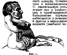
it occurs at 30-40 years (fig. 1); in Skrobansky's statistics of 50 cases, only one woman under 20 years of age is noted]. In earlier (childhood and adolescent) and in senile age O. occurs extremely rarely. In men individual cases are described (thus, in Litzmann's statistics of 131 cases it is noted only in 11, in Tikanadze of 50 cases-once).
Pregnancy and lactation also play a significant role. Thus, observations show that O. primarily affects women who have given birth, most often develops during pregnancy or shortly after childbirth, during prolonged breastfeeding; cases of O. in nullipara are according to some authors as rare as in young girls, old women, and men (in the latest Russian statistics, the puerperal form occurred 64 times out of 71 cases). Cases of O. during first pregnancy are not common; on the contrary, most believe that women who have had multiple births are most predisposed to it, and that osteomalacic women even exhibit increased fertility (the average number of births in osteomalacic women according to various authors ranges from 4.0 to 8.2). Although some dispute the increased fertility in O. and this question in general requires further investigation, the fact of the influence of the number of pregnancies undergone is undeniable, and the opposite viewpoint (held by a few authors) that a large number of pregnancies somehow protects a woman from the disease is probably incorrect. In this regard, frequently consecutive pregnancies, which are also prolonged and exhausting to the body, may also play a significant role. Consequently, it can also be said (Guggisberg) that O. is not a cause but a consequence of numerous births, and the question of excessive fertility should be comprehensively reconsidered, since statistics relate to an earlier period when only exceptional cases came under observation. The fact noted by Eisenhart of greater frequency in O. during multiple pregnancy (1:15) has not been further confirmed, nor is it mentioned in Russian statistics (Skrobansky noted it only once in 50 cases).- Besides pregnancy, other biological phases of the female organism and certain diseases of the sexual sphere (menstruation, menopause, abnormalities of the ovarian-uterine cycle, uterine bleeding) can in some rarer cases also influence the first appearance of O. as well as its course (see below). The previously expressed opinion about the great predisposing influence of syphilis has not been confirmed by later data. Constitutional factors are noted among predisposing factors. Thus, O. is more often observed in persons prone to obesity, with dark hair and swarthy skin. Theories of O. The acid theory (Schmidt) was the most widespread for a certain time. Its basis was the finding of lactic acid only in the affected areas of bone tissue (as well as observations of the identity of osteomalacic changes in bone with those resulting from acid exposure). Despite this theory being later confirmed by new research (e.g., the finding of lactic acid in the urine of patients), it was soon refuted, since lactic acid was also found in persons who had never had O., and moreover was not always found in bone affected by O. From the modern point of view, the acid theory, at least in relation to cases of the predominant puerperal form, can receive a somewhat different interpretation thanks to more clearly defined predisposing factors from pregnancy itself and all processes associated with it. The physiological hyperfunction of the entire organism, of individual systems and organs, and the altered metabolism observed during pregnancy always stand on the borderline between physiology and pathology. This hyperfunction of the organism and the observed state of acidosis in almost every pregnancy is accompanied by a series of characteristic symptoms (e.g., nausea, vomiting, chloasma gravidarum, etc.); they can of course directly affect bone tissue, the demands on which from the fetus are particularly high. In other words, along with other physiological changes in individual organs caused by pregnancy, similar changes can also occur in the bones. This gives reason to speak not only of pathological changes and disorders of bone tissue during pregnancy but also of physiological changes in it, that is, of 'physiological osteomalacia.' The possibility of such a condition is confirmed by observed clinical symptoms as well as by research conducted. Thus, microscopic examination has revealed changes in bones similar to osteomalacic ones in pregnant and parturient women who had no pathological manifestations during life; in clinical practice, pains in the pelvis, in the pubic joint, increased sensitivity on pressure, as well as transitional physiological mobility of the pelvic joints and their ligamentous apparatus are fairly often noted. In such cases, altered gait, changes in the inclination of the trunk and pelvis can also be observed. Guggisberg believes that on the basis of existing research, both these and deeper changes can be explained by the internal secretory influence of the placenta; for cases of non-puerperal origin, a similar state of acidosis can be explained by increased ovarian activity. Fowweather (1927) considers this acidosis as 'hunger' acidosis and believes that it arises on the basis of chronic malnutrition; he also explains the special tendency of pregnant women to O. by this acidosis. The theory of nutrition deficient in substances necessary for bone construction was also quite widespread. This theory of calcium starvation arose from the established deficiency of salts in affected bones as well as observations of O. in animals (see below). This theory, which caused great objections based on experiments and research on water in corresponding areas (the water contained the usual amount of lime, experiments showed not osteomalacic changes but simple osteoporosis, it was also pointed out that the deficiency of lime cannot be the main factor since its administration does not cure the disease), has recently again attracted attention; many authors believe that for certain forms of O. it cannot be definitively rejected. Particularly demonstrative in this respect are the experiments of Korenchevsky (1922) with rats; they, on the one hand, confirmed the great influence of vitamins on calcium metabolism and their role in the etiology of rickets and O., on the other hand, allowed the conclusion that there is no significant difference between rickets and O. and that some difference between them is due to age. Many studies have also been devoted to clarifying whether O. is caused by any microbe. Initially, on the basis of its endemic distribution, some (Kehrer) equated it with chronic infections, but later so-called specific causative agents of O. were even discovered, which were not confirmed by further research. The infectious theory was mainly supported by the Italian school. At present, this theory has been abandoned by almost everyone, while the sometimes observed rise in temperature and sudden onset of the disease with fever in O. can be easily explained not by infection, as some believe (Hutchison, Patel), but by the same causes that sometimes cause a rise in temperature in other disorders of pregnancy. Endocrine theories. In recent decades, the study of the pathogenesis of O. has most extensively involved the endocrine glands, especially those that participate in calcium metabolism to some extent. The so-called ovarian theory initially proposed by Fehling (1887) was the most widespread from the moment of its inception. In the form proposed by the author, it is now accepted by almost no one (according to Fehling, O. represents a trophoneurosis caused by irritation originating from the ovaries being reflexively transmitted via the sympathetic nerve to the bone vasodilators, resulting in their hyperemia and resorption). This theory soon came under criticism. Thus, from the very beginning, observations with ovarian transplantation showed that the matter does not lie in reflex irritation and nervous influence but in the entry into the blood of some substances produced by the ovary. Although the opinion about the exclusive significance of increased ovarian activity (hyperovaria) was held almost until the very last time, it could not but evoke criticism due to newly published clinical observations. Thus, alongside a certain percentage of cases in which castration had no therapeutic effect, cases of O. (though occurring much less frequently) in the climacteric period, in old women, and finally osteomalacia virginalis could not but attract attention. The data from microscopic research were also contradictory (see below). For this reason, it is more correct to adhere to the view that the ovary indeed plays a major role in the development of O., but still other endocrine glands are also involved in the process, and that in view of our insufficient knowledge about the ovarian hormone, it is better to consider the occurring changes from the point of view of ovarian dysfunction. Histological studies of removed ovarials also do not yet allow us to definitively state that hyperovaria is always present in O., and moreover, a uniform characteristic picture is not found in every case.
Thus, atrophic processes are noted, a decrease in the number of follicles, degeneration of the vessel walls, strong development of the interstitial gland, an increased number of atretic follicles with strong proliferation of the thecae internae, sharply expressed hyaline degeneration of the vessel walls, marked proliferation of the elements of the thecae internae with insignificant atrophy of the follicle, a large number of Call-Exner bodies in various stages of development, hypersecretion of the follicular apparatus, enhanced development of follicular atresia (Kushnir, Selitsky), etc. There are also reverse data. For example, some authors could not find an increase in the interstitial gland in O., while others, on the contrary, observed its strong development during normal pregnancy, which made it difficult to establish its development in a causal relationship with O., all the more so since it was not always present (besides, the general significance of the interstitial gland as an endocrine organ is still considered debatable). Moreover, some researchers have indicated that no characteristic changes are found in those observed, that they occur under physiological conditions as well, and that when considering them, one must also take into account the age of the patients. In view of all the above regarding the phenomena of hyperovaria, as well as in view of cases where reverse changes were observed (e.g., Curschmann's with aplasia of the ovaries) or where other endocrine glands may have predominant importance, it is better to interpret O. from a more acceptable point of view for all cases, namely from the point of view of ovarian dysfunction. The view concerning the connection of O. with the parathyroid glands, first expressed by Erdheim and later confirmed in experiments with rats, appears sufficiently justified. These experiments speak of the enhanced activity of the epithelial bodies in O. and their great influence on calcium metabolism. Their increase and hyperplasia are noted. The participation and even the leading role in O. of the thyroid gland (Hoenicke) has also been indicated, but the question of whether there is hyper- or hypothyroidism here and whether the observed changes are primary or secondary has not yet been resolved; apparently, for the present moment, it is more acceptable to speak of their secondary origin and to consider them one of the manifestations of the pluriglandular osteomalacic syndrome. Predominant importance in the pathogenesis of O. was also attributed to other endocrine glands—the adrenal glands, the appendage of the brain, and the thyroid gland, but even in relation to them, no data has been obtained on the basis of which the disease could be attributed to the lesion of any single gland. For example, the adrenal theory, proposed by Bossi, who believed that the primary cause of O. lies in the decreased activity of the adrenal glands (in experiments with partial or complete removal of them, changes similar to osteomalacic ones occurred in the bones), was not fully accepted. The research of other authors showed that there is indeed a decreased function of the entire chromaffin system (this is confirmed, among other things, by the fact that osteomalacic patients are less sensitive to adrenaline and that adrenal glycosuria is absent in them), but this is caused by the inhibitory ovarian secretory influence, which disrupts the existing relationships between the chromaffin system and other glands regulating calcium metabolism and bone growth. (Varaldo, by administering adrenaline to animals, found that under the influence of injections, the ovary increases in volume and degenerative changes occur in it.) The pituitary theory also found few supporters, and few shared the opinion about the greater participation of the thyroid gland (this theory was based on the decrease in calcium content in the bones after removal of the thyroid gland and on the similarity of bone changes in thymectomized young dogs with O., etc.). The entire sum of the data obtained regarding the endocrine glands allows one to see in the osteomalacic process not an isolated lesion of any single gland, but a violation of the chemical correlation in the entire endocrine system during pregnancy, probably with the participation of a new gland—the placenta, which plays a large role in metabolism. Classifications of O. Naturally, with the beginning of the scientific study of O., there also appeared a desire to classify the observed cases of it, but as before, and now, due to the unclear etiology, none of the classifications can be considered satisfactory. Just as incorrect was the initial classification of Killian (1857), which distinguished O. according to the occurring changes (O. fragilis, s. fracturosa, characterized by bone fragility, and O. segrea with waxy bone flexibility), so unsatisfactory are the subsequent, as well as modern classifications, based on the etiological factor, clinical symptoms, or severity of the disease. Ligmav? (1861) distinguished 4 forms: 1) rheumatic, 2) syphilitic, 3) senile, and 4) nervous; at the present time Zondek divides O. into 1) puerperal (strongly predominant), 2) rheumatic-marantic senile, 3) juvenile, and 4) hungry. Aschner accepts two main forms—juvenile or maiden and O. of multiparous women—and believes that rheumatic-marantic is rarely encountered. Some still divide O. into atrophic and hypertrophic, into vagosympathicotonic, etc. Only the isolation of puerperal O. as a separate group is most correct, which Pashutin considered the main type (he also proposed the special name 'genitogenous' O. due to the close connection with the sex glands); while Skrobansky considers it a sui generis form, completely independent in character, in the gradualness of the occurring changes, as well as in the course of the process and outcomes (he also distinguishes three types of puerperal O.—malignant, severe, and mild, occurring covertly, without skeletal deformities and standing on the border of 'physiological' changes during pregnancy). Pathological anatomy. The most significant changes occur in O. in the skeletal system. The bones decrease in size, lose in weight, become soft and very pliable. The basis of this change in the skeletal system is the disappearance of the old dense bone substance and its replacement with soft, lime-free osteoid tissue. However, the question of how this happens, i.e., what pathological process takes place in the bones in O., has not yet been finally resolved. The opinion, quite common earlier, not entirely abandoned even now, that the main process here is halisteresis (see), has been significantly shaken by very weighty research showing that the softening of bones consists not only in the simple disappearance of lime from the finally formed bone, but also in the new formation of bone tissue devoid of lime (osteoid tissue), which then remains in this state (Schmorl and others). As is known, such an explanation of the essence of the process of formation of osteoid tissue in O. has brought the change in the bones in O. much closer to those characteristic of rickets. This observation, however, does not exclude the possibility that along with the new formation of osteoid tissue, halisteresis may also occur, in other words, the process of disappearance of calcium salts from the bones may be based on atrophic and hyperplastic changes [some, on the basis of the predominance of one or the other of these processes, even distinguish forms—atrophic (porotic) and hypertrophic (hyperplastic)]. Nikiforov believed that the new formation of osteoid tissue occurs unevenly and predominantly where the bone tissue is subjected to increased pressure. All the above changes occur with a certain gradualness and are not equally expressed in the individual components of the bone; the trabeculae of the spongy bone are affected first, while the bone marrow, as well as the substantia compacta, are affected later, the periosteum usually does not change or thickens slightly; in severe, advanced cases, the picture becomes more uniform, and almost everywhere instead of normal bone, osteoid tissue is noted. The most characteristic appearance is that of the bone trabeculae of the spongy bone (thinning, layering, violation of the correct arrangement), from which the loss of lime actually begins. They seem to be composed of two completely different parts, separated by a sharp boundary and clearly distinguishable in their structure; one part, facing the Haversian canals and the bone marrow cavity, appears lime-free and shows traces of an atrophic process (atrophy of bone corpuscles), while the other, remote from the bone marrow tissue, consists of bone of completely normal structure. With the further development of the process, decalcification spreads further, the trabeculae become increasingly thinner, while the bone marrow cavities expand (Fig. 2 and 3). In parallel with this, the substantia compacta also undergoes changes, which, depending on the degree and stage of the process, may thin to one degree or another. In far advanced cases, the diaphyses of the long bones may appear as thin-walled, flexible tubes.
In the bone marrow itself, apart from congestion, there are no noticeable changes, and in general the bone marrow, in appearance and structure, corresponds to the age. Only in areas of strong bone remodeling does the bone marrow acquire a connective tissue nature (German Fasermark). Osteoclasts and osteoblasts are found extremely rarely in the affected bones in O. Along with changes in the bone system in severe forms, changes in the muscular system and in the nerves are noted (the latter, however, are rarely changed). Thus, Schlesinger (1893), upon examination of the peripheral nerves in one case of O., noted lesions of the nn. ischiadici, radialis and ulnaris and considered them as degenerative neuritis. Changes in the muscles are more frequently observed; they consist of atrophy of muscle fibers and fatty degeneration (Friedreich, Chambeis, Hosslin and others); sometimes complete disappearance of muscle tendons also occurs. Clinic, symptomatology, diagnosis, course, prognosis. The pathological symptom complex of O. is quite complex and diverse; the nature and intensity of pain, all individual symptoms generally vary depending on the stage and degree of the process. In the changes in the skeleton, a certain systematicity is also observed, which depends, among other things, on the form of O. (puerperal and non-puerperal). The predominant symptom is pain. They appear at the very beginning of the disease, being one of the earliest symptoms. The pains either localize predominantly in one place (in the sacrum, in the pelvic area - a feeling of heaviness) or are more widespread (in the chest, spine, lower limbs and even throughout the body) and depend not only on changes in the bone system but also on changes in the nerves and muscles, which can be affected earlier. The pains are pulling, of a rheumatic nature, occurring even in a state of rest. Early symptoms also include easy fatigue, difficulties in movement, muscle weakness (decreased tone, trembling, sub-tetanic state) and associated changes in gait (so-called duck-like, unsteady, "rolling" gait). An early characteristic sign is also contracture of the adductors (patients cannot lift their legs and spread them) (fig. 4 and 5). Quite early, paresthesias, pareses, and even in moderately severe cases appear; marked tenderness is noted with even slight pressure, increased galvanic and mechanical excitability, increased knee reflexes develop. In the bone system in the puerperal form, the pelvis is first affected, then the spine and chest; the lower extremities are affected less frequently (and rarely the bones of the skull). With further development of the disease, individual symptoms progress - pains intensify, movements become completely impossible, deformations in the skeleton begin (kyphoses, scolioses), decrease in height due to shortening of the spine; asymmetry of the pelvis and the entire torso is not uncommon (fig. 6 and 7); bones become brittle, lax - curvatures, cracks and fractures of individual bones are not uncommon (in puerperal O. with chronic course, repeated fractures have been described only with newly occurring pregnancy - Savulescu, 1921, at the 11th, 12th and 13th pregnancies). The phenomena from the side of the nerves progress (osteomalacic paralyses), the muscular system is more deeply affected (atrophy, fatty degeneration, cases of lipomatosis have also been described). Disorders from the side of the psyche are observed, general swelling of tissues, nephropathy and other general disorders. The general condition in mild forms is not disturbed, in more severe forms - dyspeptic phenomena are noted, loss of appetite, constipation, breathing is difficult, stagnant phenomena in the lungs, disorders of blood circulation and cardiac activity are not uncommon. Sexual life is possible only in initial forms, when apparently the sexual sense is not disturbed. Earlier, when almost only advanced cases came under observation, tuberculosis of the lungs, chronic bronchitis, nephritis, general marasmus were not uncommon. From the side of the blood in puerperal O., nothing characteristic has been noted; for example, a moderate decrease in Hb, leukocytosis, marked eosinophilia, lymphopenia, and in severe cases - anemia have been indicated. A decreased alkalinity of the blood is also noted. The data regarding metabolism are extremely contradictory: both an increase and a decrease in the excretion of phosphorus and calcium with urine have been noted; observations about increased content
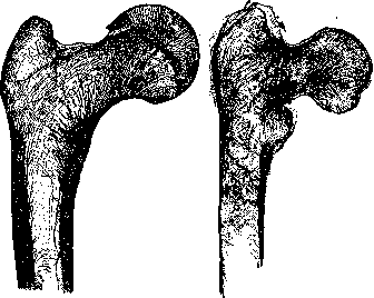
Figure 2. Saw cut of a normal femur.
Figure 3. Saw cut of a femur in osteomalacia.
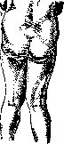
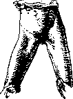
in the bone system in the puerperal form first the pelvis is affected, then the spine and chest; the lower extremities are affected less frequently (and rarely the bones of the skull). With further development of the disease, individual symptoms progress - pains intensify, movements become completely impossible, deformations in the skeleton begin (kyphoses, scolioses), decrease in height due to shortening of the spine; asymmetry of the pelvis and the entire torso is not uncommon (fig. 6 and 7); bones become brittle, lax - curvatures, cracks and fractures of individual bones are not uncommon (in puerperal O. with chronic course, repeated fractures have been described only with newly occurring pregnancy - Savulescu, 1921, at the 11th, 12th and 13th pregnancies). The phenomena from the side of the nerves progress (osteomalacic paralyses), the muscular system is more deeply affected (atrophy, fatty degeneration, cases of lipomatosis have also been described). Disorders from the side of the psyche are observed, general swelling of tissues, nephropathy and other general disorders. The general condition in mild forms is not disturbed, in more severe forms - dyspeptic phenomena are noted, loss of appetite, constipation, breathing is difficult, stagnant phenomena in the lungs, disorders of blood circulation and cardiac activity are not uncommon. Sexual life is possible only in initial forms, when apparently the sexual sense is not disturbed. Earlier, when almost only advanced cases came under observation, tuberculosis of the lungs, chronic bronchitis, nephritis, general marasmus were not uncommon. From the side of the blood in puerperal O., nothing characteristic has been noted; for example, a moderate decrease in Hb, leukocytosis, marked eosinophilia, lymphopenia, and in severe cases - anemia have been indicated. A decreased alkalinity of the blood is also noted. The data regarding metabolism are extremely contradictory: both an increase and a decrease in the excretion of phosphorus and calcium with urine have been noted; observations about increased content
Figure 4.
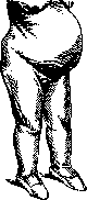
Figure 5.
of phosphorus and calcium in the blood and urine have not yet been proven. The data regarding the nervous system are also contradictory: some authors point to the toxic nature of the nervous lesions, others consider them as secondary to bone changes. The diagnosis of O. is not always easy, especially in the initial stages. The clinical picture, X-ray examination (which reveals the characteristic decalcification and the so-called pseudofractures), and the determination of the calcium content in the blood and urine are of importance. Differential diagnosis must be made with rheumatism, multiple myeloma, leukemia, bone tuberculosis, syphilitic periostitis, etc. Treatment. The treatment of O. should be comprehensive and prolonged. The main thing is diet therapy: abundant nutrition with sufficient protein and fats, restriction of carbohydrates, especially sugar. Of great importance is the administration of vitamin D (in the form of irradiated ergosterol, cod liver oil, etc.). Calcium preparations are also prescribed (calcium lactate, etc.). Sun baths and electric light baths are indicated. In case of severe pain, rest in bed is necessary. In advanced cases with severe deformities, orthopedic treatment is necessary. The prognosis depends on the form and stage of the disease; in puerperal O., the prognosis is worse, especially if the disease develops during pregnancy. Prevention consists in proper nutrition, especially during pregnancy and lactation, and in sufficient exposure to sunlight. In endemic areas, prophylactic administration of vitamin D is indicated. state in mild forms is not disturbed, in more severe forms - dyspeptic phenomena are noted, loss of appetite, constipation, breathing is difficult, stagnant phenomena in the lungs, disorders of blood circulation and cardiac activity are not uncommon. Sexual life is possible only in initial forms, when apparently the sexual sense is not disturbed. Earlier, when almost only advanced cases came under observation, tuberculosis of the lungs, chronic bronchitis, nephritis, general marasmus were not uncommon. From the side of the blood in puerperal O., nothing characteristic has been noted; for example, a moderate decrease in Hb, leukocytosis, marked eosinophilia, lymphopenia, and in severe cases - anemia have been indicated. A decreased alkalinity of the blood is also noted. The data regarding metabolism are extremely contradictory: both an increase and a decrease in the excretion of phosphorus and calcium with urine have been noted; observations about increased content
Figure 6.
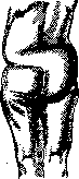
Figure 7.
calcium in the blood. Recent research by Klyuchevsky shows that in puerperal osteomalacia there is a significant loss of calcium (increased excretion of Ca through the intestines). Diagnosis of O. in advanced cases presents no difficulty; in initial forms, however, with a slowly and gradually developing process, with slightly expressed symptoms and absence of clinical manifestations from the bones, O. can easily be overlooked or confused with another disease. In differential diagnosis in cases of puerperal O., one must mainly consider rheumatism, neuralgia (especially of the sciatic nerve), as well as general fibrous osteitis and multiple myeloma (hysteria is also considered by some); in the senile form-osteoporosis (see.), arthritis deformans, Paget's disease, progressive muscular dystrophy, malignant diseases of the bone marrow. In diagnosis, X-rays have recently been of great assistance. The course of O. is clearly dependent on a number of accompanying conditions; of these, the onset of a new pregnancy is of greatest importance, which as a rule exacerbates and worsens the process. Generally, the course of O. is wave-like. The prognosis is generally serious [in early statistics (Litzmann)-mortality from puerperal O. was noted at 80%, in recent ones, on the contrary, recovery is confirmed in 85-90%], however, a number of authors consider the prognosis favorable. Cases of self-healing have been described even with continuing pregnancy. After recovery, of course, deviations and deformities of the skeleton remain. Non-puerperal forms of O. differ considerably from puerperal forms both in the nature and gradualness of changes, and in the course and prognosis (the spine and thorax are affected first, later the pelvis; in male O., the latter is very rarely affected). These forms also differ in the localization of pains, which in such cases are predominantly observed in the spine and lower extremities. The pathological symptom complex is more sharply expressed, and additional dystrophic disorders are observed, which are an expression of the general insufficiency of the organism. Thus, for example, combination with general obesity, tetany, with severe mental disorders, dementia praecox; cases of Charcot's disease, trophic disorders (severe hair loss, marked changes in the skin), multiple neurofibromatosis, etc., have been described; thyrotoxicosis is also frequently observed. In general, frequent cases of O. are noted in the mentally ill. Some difference has also been noted in relation to the blood (for example, Chistovich and others described a sharp lymphocytosis-up to 60%). Malignant forms with a more rapid course are often observed; these forms are more difficult to treat, especially male O., which many consider incurable. Their course is usually chronic (also wave-like), it depends on the form, external conditions, and the observed combined symptoms; they can last for several years, sometimes exacerbating, sometimes weakening in their course. Of cases of non-puerperal O., the prognosis is most favorable apparently in O. infantilis. This latter form may be associated with the period of puberty (according to Grace Stapleton-in India usually); on the other hand, cases have been described where it occurred with the onset of menstruation. The most unfavorable prognosis is in male O. (rapid course, high mortality rate). Prevention and therapy (obstetrical, see Pelvis). All those, as yet only tentative, data that exist regarding the etiological factors of O., as well as the insufficiency (far from in isolated cases) of both medicinal treatment and more radical intervention, force one to pay attention to prevention. In view of the fact that in O. symptoms are often observed that characterize the osteomalacia patient as an inadequate subject, this prevention must consist not only in the proper upbringing of youth and hygienic environment, but must also be carried out antenatally. Prevention should have special significance in areas where O. is noted as an 'endemic' phenomenon; here, along with special attention to social conditions (the housing question, nutrition, in particular on increasing vitamins in food), a systematic struggle must be conducted against harmful habits, customs, and prejudices in the life of the eastern woman. Prevention should be particularly deepened at the first symptoms of the disease, as well as in its further development. The favorable effect of climatic conditions, the importance of improving hygienic, preventive factors and nutrition are noted in recent times by many authors. Thus, for example, Miles and Feng (Miles, C. T. Feng, Peking) saw good results only with appropriate modification of diet in Chinese women; this is pointed out by Stone (Emerson Stone, 1924) and Stapleton (Grace Stapleton, India), who considers vegetarian diet (milk, butter) as one of the main therapeutic measures. The basic principles of therapy depend directly on the type and character of O. In puerperal O. in mild and moderately severe cases, preliminary conservative treatment is permissible under strict control; if unsuccessful, pregnancy must be interrupted without delay, followed by (depending on the case) removal of the ovaries. The principle of gradualness in relation to conservatism or radicalism in therapy must also be observed in cases of non-puerperal O. Of therapeutic means, preparations of phosphorus (Kassowitz, Latzko) have deserved success, which are recommended in combination with cod liver oil (Vogt also used cod liver oil alone with success). Thus, Latzko advises using the following composition: 0.06 g of phosphorus per 100 g of cod liver oil daily by the teaspoonful; in cases difficult to treat, the dose is increased to 0.1 g; these same doses are also adhered to by Gruzdev. Kurshman believes that it is better to introduce relatively large doses, for example Phosphor 0.1, Succus et Pulv. liq. q. s. ut i. pil. № 150 two to three times a day by 1 pill, and to use it for many weeks; according to Kurshman, phosphorus is better tolerated by osteomalacia patients than any other means. Mykertch'yants A. M. (1905) pointed out that phosphorus is a reliable and trustworthy remedy and that it should be taken 'in proper doses and for a sufficiently long time'. Other authors (Seitz) also indicate the advisability of using large doses of phosphorus; among others, Zondek considers its action in combination with calcium or cod liver oil in some cases even specific (medium doses, according to Zondek, of phosphorus-0.01, cod liver oil-100.0 two to three times a day by the teaspoonful + Calc. lactici 0.3 per os-3 times a day). Complete recovery was noted by Latzko in 78%, by Schmidt-in 62%. There are indications of better action of phosphorus in non-puerperal O. (the mechanism of its action is unclear; Ashner believes that it affects not the endocrine glands, but the metabolism, perhaps directly on the bone cells). Along with phosphorus and cod liver oil, the introduction of organotherapeutic preparations is recommended, which some also use separately. Of the latter, adrenaline has been used most frequently; pituitrin, as well as ovarin and anti- and thyreoidin, are used much less frequently. Milk of castrated goats, monkeys (antimalacin-Hoffmann), ap-ovarthyreoidserum, etc., have not become widespread. The value of implantation of thymus, successfully performed in one case (Scipiades, 1924), has also not been confirmed. Adrenaline, according to Bossi, should be used for 10-12 days, introducing 2 times a day up to 1 cm³ of 0.1% solution and repeating the course of treatment after several weeks. Cristofoletti brings statistics from 46 cases (in 24% recovery, in 35% marked improvement, in 41% no result) and believes that the action of adrenaline is indirect (lowering of ovarian function). Adrenaline has been used by a number of authors; opinions about it differ, there are indications that remote results are unknown and that the disease often does not pass. The not infrequently observed complications from its administration (severe general phenomena, shortness of breath, palpitation, collapse, tremor, etc.) even in small doses necessitate caution in its use. The good analgesic effect of adrenaline is also noted. Recently, Suprarenin (0.5 cm³ of 1/1000 solution, 1-2 times daily; in the absence of toxic phenomena the dose is increased to 1 cm³) is recommended. When using pituitrin, Bab obtained success in 4 cases (out of 8), while Pal introduced extract from the anterior lobe; successful results were also obtained by others, and among others in a case of male O. Combined therapy (adrenaline, pituitrin) was also tried. More radical intervention should include radiotherapy. First applied by Ascarelli (Ascarelli, 1926), it is recommended by some at present, with the indication that it is more applicable in mild cases and in young patients with the aim of temporary sterilization (there are however authors who believe that much cannot be expected from X-rays, especially in serious cases).
Surgical castration continues to be the focus of attention in the therapy of O. (Feliling, 1887), although recently, as the view establishes that in O., along with hyper- and dysfunction of the ovary, there is also a disturbance of the function of other endocrine glands, voices are beginning to be raised for its more limited application. This can also be supported by cases where it had no effect, where temporary improvement was followed by relapses, and finally not isolated cases of recovery without castration. Mykertch'yants as early as 1905 considered Feliling's theory unsound, lacking scientific justification, and stated that O. 'in no case can serve as an indication for castration'. In more recent times, some authors (Ilyin, 1930) also believe that there are no indications for castration in O. At the present time, castration of course cannot be definitively excluded from the therapy of O., but undoubtedly it should not be performed as widely as before. Castration should not be applied from the very beginning of the disease without first applying conservative treatment. Some (Guggisberg), however, do not advise waiting too long (in cases that are difficult to treat) with castration, because if delayed it no longer gives the desired result (Guggisberg at the same time believes that there are no indications for it in mild cases). A few authors share a more extreme point of view, i.e., considering castration insufficient, they also perform a complete extirpation of the uterus (due to the possible harmful influence of its mucous membrane). After castration, some recommend additional treatment; for example, a good result has been noted from the use of adrenaline with phosphorus in relapses after castration. Osteomalacia in animals. O. is also observed in animals, occurring in all species, but most often it affects cattle. Just as in humans, the predominant form is puerperal O.; in young animals and males it is extremely rare. The predisposing causes of its occurrence are also analogous. For example, in veterinary medicine, the main causal factors of O. are considered to be insufficient nutrition (food with low lime and phosphoric acid content), pregnancy and prolonged separation of milk. The influence of nutrition has been proven by many researchers; the most characteristic observations are those of Hermann (Germann) on 244 Egyptian horses brought to Cochin China (the soils of Egypt and Cochin China differ significantly in lime content). The individual cases of O. that soon arose in horses ceased after Egyptian barley was brought in, and the sick horses improved significantly. There is much in common in the clinical picture of the disease; fractures of bones are often encountered, which are even considered a characteristic sign. Treatment consists of improving nutrition, adding bran and oats to the food. Of medicinal preparations, a solution of phosphoric acid and powder of Nucis vomicae are recommended. In the presence of fractures, the animal is slaughtered; in pregnancy, in the case of a live fetus, a cesarean section is first performed. As prophylactic measures for pregnant cows, earlier cessation of milking is advised.
Related articles
Mentioned in
Cite this page
“Osteomalacia.” Soviet Medical Encyclopedia. English translation of Bolshaya Meditsinskaya Entsiklopediya, 1st ed. (Moscow, 1928–1936), ed. N. A. Semashko. https://sovietmedicalencyclopedia.pages.dev/article/osteomalacia/