Animal Electricity
Historical document, translated for reference. It reflects medical knowledge of the 1920s–30s and is not medical advice.
Summary
An overview of electrical phenomena in living tissues and organs of animals and plants from the perspective of 1930s electrophysiology, covering resting and action currents, nerve and muscle responses, and electric organs in fish.
Encyclopedia article (1928–1936)
ANIMAL ELECTRICITY, electrical phenomena in living tissues and organs of animals and plants. The study of these phenomena is the domain of electrobiology or electrophysiology. The founder of the doctrine of animal electricity is the Italian physiologist Galvani, who discovered that the contact of a metallic arc consisting of two metals with the nerve and muscle of a frog causes the muscle to contract. Galvani's polemic with Volta, who saw in this phenomenon an exclusively physical process, led Galvani to his famous experiment of contraction without the participation of metals. A nerve, isolated along its length and brought into contact with an excited muscle, in turn causes the contraction of the muscle connected to it. Galvani concluded from this experiment that there are sources of electricity in the living body that stimulate the muscle. The experiments of Matteucci and, above all, Du Bois-Reymond firmly established the existence of electrical phenomena in the living body and uncovered the laws governing these phenomena.-On a m u s c l e, one can demonstrate the existence of currents during its state of rest. The arrangement of the experiments for this is as follows. Various points of a muscle P with parallel fibers running along AB (Fig. 1) are connected by means of
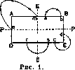
non-polarizable electrodes to a galvanometer. Then, along the wire connecting these points, a current flows, the direction of which is determined by the following rule. Let EE be the cross-section dividing the muscle into two symmetrical parts; EE is called its equator. The current flows in the external circuit from point a (or respectively c), lying closer to the equator, to point b (or respectively d), lying closer to pole P located on the line PP passing through the centers of the muscle bases. Points r and s, symmetrical with respect to the equator EE, produce no current. The strongest current is obtained if one of the equatorial points m is connected to point n (center) of the cross-section. An analogous resting current is obtained in a nerve. The maximum electromotive
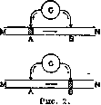
force of a muscle obtained during the resting current is equal to 80 millivolts. During tetanic stimulation of a muscle, the current obtained from connecting point m of the longitudinal surface and point n of the transverse surface weakens, sometimes to such an extent that the galvanometer shows no current. This phenomenon, discovered by Du Bois-Reymond, is called the negative variation of the muscle current and is explained by the fact that the excited spot becomes electronegative in relation to the unexcited one; consequently, the excitation, passing through the equator, makes point m electronegative in relation to point n. When the current spreads to point n, the latter does not change its negativity in relation to m, and thus in the arc connecting m and n, currents of the reverse direction arise, which, summing up, reveal a weakening of the current on the galvanometer. On an undamaged muscle, if the current is deflected from two of its points A and B (Fig. 2), as the excitation runs from M to N, point A first becomes negative (the current flows in the external circuit from B to A), then point B (the current flows in the external circuit in the opposite direction from A to B). This explains the appearance of the diphasic action current. Recorded with the aid of a string galvanometer, the diphasic current has the appearance shown in Fig. 3. Later experiments showed that not only muscle and nerve, but also other excitable tissues behave in an analogous manner; if the retina is stimulated by light, the excited part of the retina becomes electronegative in relation to its unexcited parts, and the current in the external circuit flows from parts located close to the unexcited tissues to the excited tissues. Investigating the mechanical effect of a contracting muscle and simultaneously the electrical phenomena in the latter, one can conclude that the electrical phenomena are stronger the stronger the muscle contracts. It is also proven that the negative variation of the current is observed even before the appearance of contraction, in the latent period. Sensitivity—the ability of a tissue to respond to certain external stimuli, e.g., to the action of poisons (alcohol, nicotine vapors)—is connected with the strength of electrical phenomena in the tissue. The development of research methods and the introduction of devices capable of recording rapidly occurring phenomena (Lippmann capillary electrometer, Einthoven string galvanometer) made it possible to study the time course of electrical phenomena in living tissue. The passage of a direct current through a nerve causes the appearance of currents in the extrapolar spaces that are associated with the special physiological-anatomical structure of the nerve (Fig. 4). If an electric current from element E is passed through the nerve NN by placing electrodes on section c, then in the extrapolar sections a and b one obtains
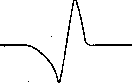
Figure 3.
currents easily detected by galvanometers G1 and G2; the direction of the currents is shown in Fig. 4 by arrows. These currents in the nerve coincide with the direction of the polarizing current in region c. The explanation for this phenomenon lies in the fact that the central axis cylinder of the nerve conducts current better than the neuroplasm; on nerve models consisting of a well-conducting wire surrounded by a poorly conducting electrolyte, one can observe a similar phenomenon.-Vvedensky's research revealed interesting relationships between the period of the stimulating current in the nerve and the period of electrical phenomena observed at its other end. If a nerve consisting of cell Z (Fig. 5), axial process N, and dendrites D is stimulated at B, an apparatus (telephone or string galvanometer) placed at T reveals action currents of the same frequency as the period of the stimulating current; only in the event that the frequency of the stimulating current exceeds a certain limit (thousands of oscillations per second) does the current in T not correspond to the period of the current in R. If, however, the stimulating section P is separated from the place where currents are picked up T by nerve cell Z, then, whatever the period of the stimulating currents in R, T always produces the same period of electric current corresponding to the period of activity of the nerve cell. If one studies spontaneous processes in the central nervous system by the telephone method, they appear as periodic discharges that produce in the telephone, connected to the nervous system by needles, a series of successive noises. Processes of an electrical character accompany not only phenomena occurring in the animal organism; plants also display a series of electrical phenomena analogous to those in animals. Finally, a special section is formed by the doctrine of electrical phenomena in certain fish (electric ray, electric catfish). The electric organs of these animals, studied histologically by Babukhin, are
analogs of muscles, and the electrical phenomena in them reach such proportions that their discharges in water can not only stun but even kill an animal. The study of these phenomena, begun by Matteucci, was continued by Du Bois-Reymond, and after him by Bernstein. The explanation of the phenomena occurring upon excitation must be sought, as the ionic theory of excitation shows, in the appearance of ions in the excited region, which create a definite potential difference between the excited and unexcited tissue. Chagovets was the first to quantitatively prove the possibility of explaining the electromotive forces of a working muscle by the emergence of ionized products.-From what has been said, it is evident that although the study of electrical phenomena does not lead to an understanding of the difference between living and non-living nature, the existence of bioelectric currents turns out to be important for studying phenomena occurring in the living organism. Recently, the study of electrical phenomena in the heart has developed into an extensive chapter of electrophysiology; the results obtained serve for the diagnosis of heart diseases. Finally, in conclusion, one must mention the static charges of the human body, which arise upon changes in the position of muscles of an isolated human body. Plants also demonstrate the appearance of charges on them.
P. Lazarev
Related articles
Mentioned in
Cite this page
“Animal Electricity.” Soviet Medical Encyclopedia. English translation of Bolshaya Meditsinskaya Entsiklopediya, 1st ed. (Moscow, 1928–1936), ed. N. A. Semashko. https://sovietmedicalencyclopedia.pages.dev/article/animal-electricity/