Infusion
Historical document, translated for reference. It reflects medical knowledge of the 1920s–30s and is not medical advice.
Summary
This article from the 1928-1936 Soviet Medical Encyclopedia describes the medical procedure of infusion, including its various types (subcutaneous, intravenous, intracavity), techniques, equipment, indications, and complications. It provides detailed instructions for performing infusions and discusses specific solutions used for different conditions.
Encyclopedia article (1928–1936)
INFUSION (ini'usio), introduction of large quantities of solutions into tissues, cavities, and organs (subcutaneous I. of physiological saline solution, intravenous I. of glucose solution, I. of ether or rivanol into the abdominal cavity, etc.); under I. is also often meant the intravenous introduction of small quantities of solutions (5-10 cubic cm) with therapeutic (e.g., neosalvarsan and urotropin) or diagnostic purpose (e.g., indigo carmine). Introduction of small quantities of solutions is also called injection (see below). Types of I.: subcutaneous, intravenous, and intracavity (into the free abdominal cavity, into the bladder, into the renal pelvis and ureter, etc.). Subcutaneous I. are performed (in most cases) on the anterolateral surface of the thigh, into the subcutaneous tissue of the abdominal wall or into the subaxillary region. Intravenous I. are performed most often in the veins of the elbow bend and in rare cases - in the veins of the dorsal surface of the hand and lower extremities. Intracavity I. are performed under special conditions: into the abdominal cavity during an operation for peritonitis, into the bladder through a catheter during cystoscopy, into the renal pelvis through a ureteral catheter during pyelography, etc. Technique and practical instructions.-Subcutaneous I.: the skin at the place where the needle prick should be made is disinfected in the usual way. A sterile solution (physiol. NaCl or Ringer's solution) is poured into a boiled, hermetically sealed, graduated and thermometer-equipped apparatus (for example, Bobrov's, see figure 2) or, if not available, into a large funnel with a capacity of 250-300 cubic cm (see figure 1). In the stopper (rubber) of the apparatus there are openings for tubes and a thermometer. The outer end of the short tube, containing a loose plug of cotton wool, is connected to a balloon (double rubber), by means of which air is compressed in the vessel. Due to this compression, the solution is displaced from the vessel through another tube reaching the bottom of the vessel. A rubber tube with a needle is connected to the second long glass tube. If there is a third opening in the stopper, a thermometer is inserted into it. The lumen of the tubes is regulated by taps or clamps (see figure 2). In simpler instruments or when working with a funnel, pressure is achieved by raising the apparatus to a height of approximately 1 m above the level at which the needle is injected (see figure 3). The solution should be heated to 38-39°, for which it is better to place the vessel in a container filled with water of the corresponding temperature to avoid cooling. Before injecting the needle, it is necessary to carefully check that there are no air bubbles in the system of tubes (so that the latter does not get into the subcutaneous tissue). For this, several drops of liquid must be released, and then proceed to inject the needle. The injection should not be too superficial (to a depth of 3-4 cm) to avoid resistance to the flow of liquid, as well as superficial necrosis of the skin, but also not too deep, to avoid the needle getting under the fascia (wide fascia of the thigh), as this can cause severe pain and as a result of this, rapid absorption of the liquid does not occur, and when introducing a large amount of liquid, one can even get necrosis of a section of the fascia. I. should be performed slowly, with light massage of the resulting swelling. At one time, 300-400 cubic cm can be injected into one place. For intravenous I., the skin preparation is the same. I. is performed in the area of the elbow bend; it is recommended to wash the iodine off with alcohol after smearing it, in order to better see the vein showing through under the skin. The arm above the elbow (on the shoulder) is tied with an elastic bandage or tourniquet or towel so as to obtain swelling of the veins (but not so much as to compress the artery!). If the vein is clearly visible and lies superficially with thin fatty tissue, then using the same apparatus as for subcutaneous I., but with a less thick and, if possible, sharp needle, one can make an injection into the vein directly through the skin (see figure 4). As soon as the needle is inserted into the vein, the tourniquet from the shoulder must
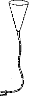
Figure 1.
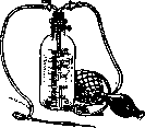
Figure 2. Bobrov's apparatus.
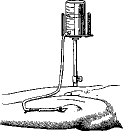
Figure 3. Infusion into the subcutaneous tissue of the thigh.
not get into the subcutaneous tissue). For this, it is necessary to release a few drops of liquid, and then proceed to inject the needle. The injection should not be too superficial (to a depth of 3-4 cm) to avoid resistance to the flow of liquid, as well as superficial necrosis of the skin, but also not too deep, to avoid the needle getting under the fascia (wide fascia of the thigh), as this can cause severe pain and as a result of this, rapid absorption of the liquid does not occur, and when introducing a large amount of liquid, one can even get necrosis of a section of the fascia. I. should be performed slowly, with light massage of the resulting swelling. At one time, 300-400 cubic cm can be injected into one place. For intravenous I., the skin preparation is the same. I. is performed in the area of the elbow bend; it is recommended to wash the iodine off with alcohol after smearing it, in order to better see the vein showing through under the skin. The arm above the elbow (on the shoulder) is tied with an elastic bandage or tourniquet or towel so as to obtain swelling of the veins (but not so much as to compress the artery!). If the vein is clearly visible and lies superficially with thin fatty tissue, then using the same apparatus as for subcutaneous I., but with a less thick and, if possible, sharp needle, one can make an injection into the vein directly through the skin (see figure 4). As soon as the needle is inserted into the vein, the tourniquet from the shoulder must
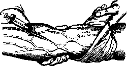
Figure 4. Infusion into the subcutaneous vein in the elbow bend (according to Kirschner-Schubert).
be removed. For intravenous I., it is especially necessary to ensure that there is no air in the tube. For this, you can first insert the needle into the vein, having removed it from the tube, and, having made sure that the needle is in the vein (blood flowing from the needle), connect the needle to the tube, while releasing liquid from the latter drop by drop. If at the very beginning of the infusion a swelling appears at the injection site and in its vicinity, this means that the needle is not in the lumen of the vein, but the liquid is going into the tissue. The error must be corrected immediately, i.e., remove the needle and perform the infusion into another vein, since the entry of some solutions under the skin causes a violent inflammatory reaction and necrosis of the tissue and skin. If the vein is not visible due to well-developed subcutaneous tissue, then with a certain skill, the needle can be inserted by touch, since the swollen vein is palpated in the form of an elastic cord. But in this case, it is better to isolate the vein (see Phlebotomy). Infusion should be performed slowly (10-15 minutes - one liter). The average dose is 1 liter. With large fluid loss (algid stage of cholera), several liters are infused, on average - 3.5. In cholera, when veins collapse, preliminary warming of the arm in hot water greatly helps in getting the needle into the vein. At the end of I., the cannula is removed; if the vein has been isolated, it is ligated and sutures are placed on the skin. The latter method is applicable only for single I., but in general, and especially with repeated I., it is better to use the method of simple needle prick through the skin. For intravenous I., special measures must be taken to prevent air from entering the vein. If in a healthy person the entry of small amounts of air into the vein, even one close to the chest, does not pose a danger to life, then in a weak patient even small portions of air can cause embolism of the branches of the pulmonary artery.-A frequent complication when introducing a saline solution into a vein (and sometimes under the skin) is the so-called "saline fever". In children it is common. Its appearance is explained in part by bacterial contamination of stale, distilled water or physiological solution. During sterilization, bacteria die, but the protein of their bodies causes general phenomena (the so-called "water defect" - Wasserfehler of German authors). With saline fever, the temperature with chills rises soon after infusion. Hot water bottles, hot drinks, caffeine under the skin help. Subcutaneous and intravenous single I. are used in shock, after bleeding, in acute anemia, when it is not required that the solution contains various blood salts, and it is quite sufficient with ordinary physiological solution (0.9% NaCl). Single and repeated intravenous I. of 0.9% NaCl solution are used, mainly, in the treatment of the algid period of cholera and dysentery. In septic and toxemic diseases, intravenous I. of 1% calcium chloride solution, 300 cubic cm, is used every 2-3 days. Intravenous I. of 10% glucose solution are used in the treatment of diabetic acidosis, but more often in this case intravenous I. of 3% soda solution is used. This same solution is sometimes also infused intravenously in azotemic uremia. I. of 50% glucose solution in the amount of 100 cubic cm is used during brain operations to reduce the intracranial pressure of cerebrospinal fluid (slow infusion at 3 cubic cm per minute). For the same purpose, infusion of 35% Ringer's solution and 15% NaCl solution is used. 0.75% hedonal solution is introduced into the vein in the amount of 100 cubic cm for anesthesia; rivanol solution 1:2,000 in the amount of 100-200 cubic cm - in sepsis; solution of sodium salt of tetrabromophenolphthalein in 40 cubic cm of water (calculated at 5.0 per 60 kg weight) - for cholecystography. Solution of 25% sodium bromide - for pyelography. In addition, urotropin (40% - 5 cubic cm) is commonly used for intravenous administration in pyelitis and epidemic encephalitis, indigo carmine (0.4% - 5 cubic cm) - for chromocystoscopy, strophanthin and digalen - for heart diseases, electragol - for sepsis, neosalvarsan - for syphilis and malaria, hypertonic solutions of glucose (33%) and sodium chloride (10%) - for hemoptysis and bleeding, as well as some vaccines and therapeutic sera. When infusing intravenously and subcutaneously large quantities of solutions, if these solutions are prepared on distilled water, it is desirable that they be prepared on freshly distilled water.
Related articles
Mentioned in
Cite this page
“Infusion.” Soviet Medical Encyclopedia. English translation of Bolshaya Meditsinskaya Entsiklopediya, 1st ed. (Moscow, 1928–1936), ed. N. A. Semashko. https://sovietmedicalencyclopedia.pages.dev/article/infusion/