Condurango and Limbs
Historical document, translated for reference. It reflects medical knowledge of the 1920s–30s and is not medical advice.
Summary
This article provides a historical overview of Condurango bark, once used as a stomachic, and a detailed anatomical description of the limbs of vertebrates, tracing their evolution from fish fins to the pentadactyl limbs of land animals. It covers the pharmacological properties of Condurango and the skeletal structure of vertebrate appendages.
Encyclopedia article (1928–1936)
CONDURANGO, Cortex Condurango, the bark of the creeping shrub Marsdenia Condurango, family Asclepiadaceae, which grows in Ecuador and Peru. Under the name Condurango, the bark of other species (Gonolobus Condurango, etc.) also enters the market, but only the bark of Marsdenia C. is used in pharmacy (Pharmacopoeia of the USSR). The dried bark appears as tubes or grooves 5–10 cm long, 1–3 cm wide, and 1–6 mm thick. Its surface is grayish-brown and bumpy; the fracture of the outer part is fibrous, while the inner part is smooth. The odor is faint but intensifies when moistened with hot water. The taste is bitter and scratchy. Condurango contains tannins, resin, and the glycoside condurangin (C40H60O16). This is a yellowish powder with an aromatic, bitter taste. It is more soluble in cold water than in hot, which is why a cold infusion of Condurango (1:5) becomes cloudy upon heating and turns transparent again upon cooling. The bitter taste is still discernible at a dilution of condurangin of 1:20,000. Experimental study of condurangin on animals has revealed that it acts on the central nervous system, causing ataxia, increased reflexes, and convulsions. Death occurs with symptoms of paralysis of the respiratory center without symptoms of direct action on the cardiovascular system. Condurango was proposed in the 1870s as a remedy used in South America against stomach cancer. After an initial fascination with this remedy, a skeptical attitude toward it emerged. Later clinical observations established that Condurango, by no means being a specific remedy against cancer (in the period when Condurango began to be used, cancer diagnosis was often erroneous), can be considered a good stomachic of the amara mucilaginosa group (see Bitters). Condurango is still often prescribed for loss of appetite and other dyspeptic disorders in the form of a fluid extract (Extr. C. fluidum), 20 drops several times a day, in the form of wine (Vinum Condurango consists of Extr. Condurango 10 parts, Vinum xerens 80 parts, Tinct. aromat. 1 part, Sacch. 9 parts) by the teaspoon or tablespoon, and in the form of a maceration decoction, which is strained after cooling. The resorptive effect of condurangin when Condurango is administered in these doses is highly questionable. V. Karasiv.
LIMBS of vertebrates represent either the sole or auxiliary organs of movement in them. In fish, unpaired and paired limbs (fins) are distinguished. Both are organs here that serve mainly as rudders, regulating the direction of movement. Unpaired fins arise in the form of a continuous skin fold encircling the entire body, with the exception of the head and belly. This fold breaks up into a certain number of individual fins, into which musculature grows (from myotomes) and in which a special internal skeleton then develops from a series of cartilaginous or bony rays, supplemented by horny or bony rays of cutaneous origin. Paired fins develop in a similar way and differ from unpaired fins, besides their position, especially in the existence of a more solid support inside the body in the form of limb girdles. Normally, there are 2 pairs of fins: the anterior, or pectoral, are located behind the gill apparatus; the posterior, or pelvic, lie initially directly in front of the anal opening. In higher fish, the pelvic fins often move far forward, under the head. In lungfish and lobe-finned fish, the paired fins also serve as support organs when crawling along the bottom of water bodies. In land vertebrates, the paired fins are transformed into pentadactyl limbs, i.e., into complex levers that move the body on land. The skeleton of the paired limbs consists of the skeleton of the free limbs and the girdles: the pectoral and pelvic. In fish, the pectoral girdle has the appearance of an arch encompassing the body from the sides and below. The primary cartilaginous or bony girdle consists of a dorsal-scapular and a ventral-coracoid section and is supplemented in fish with a bony skeleton by a series of dermal bones, of which the most important are the clavicula (collarbone) on the ventral section and the cleithrum on the dorsal (Fig. 1). The right and left halves of the girdle are connected by their ventral ends to each other along the midline. The pelvic girdle of fish consists of a paired, usually triangular plate lying entirely in the ventral body wall (Fig. 2). The skeleton of the free limb in fish has a different
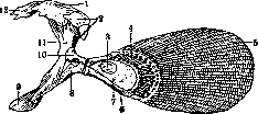
Figure 1. Pectoral girdle and pectoral fin of a ganoid fish (Polypterus) according to Gegenbaur: 1—supracleithrale; 2—postclaviculare; 3—mesopterygium; 4—radii (bony rays); 5—lepidotrichia (dermal rays); 6—metapterygium; 7—basale; 8—coracoid; 9—clavicula; 10—scapula; 11—cleithrum; 12—posttemporale.
form, but at its base, it reduces to a series of rays of the internal skeleton lying at the base and usually supplemented by longer dermal rays supporting the fin lobe itself (Figs. 1 and 2). In land vertebrates, the internal skeleton develops and differentiates much higher. The much more significant mechanical requirements placed on the pentadactyl limb are associated with the requirements for more significant support inside the body itself and, consequently, with the progressive development of the girdle skeleton. The pectoral girdle is strengthened on the rib cage through the medium of the sternum. Furthermore, by expanding both the scapular and coracoid sections, an increase in the support area is achieved. The girdle itself ossifies initially from three centers, giving rise to the scapula in the dorsal section and the coracoid (coracoideum) and procoracoid (procoracoideum) in the ventral. The dermal bones of fish, still well developed as dermal bones of the girdle in stegocephalians, have been reduced in modern forms, with the exception of the clavicle, which enters into a closer connection with the internal skeleton. The pelvic girdle has also developed quite significantly. By means of a dorsal (iliac) process, it has attached itself to the ribs of the sacral region of the spine. This connection becomes more solid in higher vertebrates due to the fusion of a number of vertebrae and the merging of ribs with transverse processes. The pelvic girdle also ossifies from three centers, giving rise to the ilium in the dorsal process and the ischium and pubis in the ventral section of the girdle. The skeleton of the free limb is initially very similar in both pairs (Fig. 3). It consists of a rather long proximal section, articulating by its head with the girdle (humerus in the anterior limb and femur in the posterior) and bearing two also rather long elements: the radius and ulna in the anterior limb and both shin bones (tibia and fibula) in the posterior limb, to which the paw (hand, foot) articulates distally. The base of the latter is composed of three transverse rows of small elements, which in lower land vertebrates still retain a fairly regular radial arrangement corresponding to the fingers sitting on this section. There are no more than five of the latter, although rudiments of extra rays (praepollex, praehallux, postminimus) are sometimes encountered along the edges of the paw, indicating the origin of the pentadactyl limb from a more richly articulated fin (Fig. 3). The elements of the anterior and posterior limbs can be compared as follows. Anterior limb
Posterior limb humerus radius, ulna radiale, intermedium, ulnare centralia carpalia distalia: 1, 2, 3, 4, 5 metacarpalia: I, II, III, IV, V phalanges (stylopodium) (zeugopodium) (basipodium) (acropodium) femur tibia, fibula tibiale, intermedium, fibulare centralia tarsalia distalia: 1, 2, 3, 4, 5 metatarsalia: I, II, III, IV, V phalanges The limbs of mammals differ from the limbs of lower vertebrates by a whole series of features and, first of all,
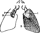
Figure 2. Pelvic girdle and pelvic fins of a fossil ganoid fish: 1—scales; 2—lepidotrichia; 3—bony rays; 4—basal element; 5—pelvis.
Figure 3.
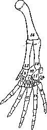
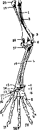
Figure 3.
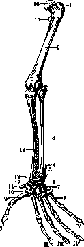
Figure 4. Figure 3. Diagram of the structure of a pentadactyl limb: 1-5-basalia distalia of the digits; 6-post-minimus; 7-centralia; 8-centralia distalia; 9-radiale; 10-intermedium; 11-ulnare; 12-ulna; 13-radius; 14-humerus; I-V-metabasalia of the digits. Figure 4. Skeleton of the forelimb of a marsupial mammal: 1-crista deltoidea; 2-humerus; 3-olecranon; 4-condyl. lat.; 5 and 17-ulna; 6-pisiforme; 7-ulnare; 8-carpale IV-V; 9-metacarpale V; 10-metacarpale I; 11-carpale I; 12-praepollex; 13-centrale; 14-radiale; 15-intermedium; 16-radius; 18-for. entepicond.; 19-fossa bicipit.; 20-caput humeri. Regarding position: in lower vertebrates, the limbs are turned away from the body, and only the paw itself turns forward. In mammals, the limbs move downward, and the characteristic bends of the limbs (elbow backward and knee forward) are much more pronounced. Lower vertebrates crawl, only slightly lifting their body, whereas mammals walk, supporting the body high above the ground. A whole series of morphological features is connected with this: the length of the proximal bones, the shape and position of the hemispherical head of the humerus and especially the femur at an angle to their axis, etc. The most ancient mammals apparently lived mainly in trees, and the mobility of their limbs is connected with this. The skeleton of the forelimb of mammals (Fig. 4) generally retains a more primitive composition than in many lower forms; however, in some respects, it is developed much higher. The proximal bones reach a significantly greater length. The change in the position of the limb led to a sharper development of the cross between the radius and ulna. The olecranon process of the latter (proc. olecranon) reaches a significant size, and finally, with the development of greater mobility, the joints also reach a high degree of differentiation (especially the hemispherical caput humeri, standing at an angle to the axis of the humerus, and the trochlea humeri). In the wrist, there are all the main elements of the primitive limb. However, the os centrale exists only in a single number, and even then not in all mammals (in the adult state), and the carpalia distalia 4 and 5 are always represented by one bone. The skeleton of the hind limb of mammals (Fig. 5) is characterized again by the length of the proximal bones, the position of all its elements in a plane parallel to the medial one, the position of the hemispherical head of the femur at an angle to the axis of the latter, as well as the development of both trochanters (from the proximal ends of the muscular ridge of lower forms) and the presence of a tendinous ossification—the kneecap (patella). The fibula is developed more weakly than the tibia and is sometimes reduced and fused to the latter. Of the proximal bones of the tarsus, there are only two large bones: the talus and the calcaneus (astragalus and calcaneus), corresponding apparently to the intermedium
and fibulare; a significant heel process develops on the latter. There is one distal centrale (naviculare). The last two distal small bones have fused into one (tarsalia distalia 4-5, s. cuboideum). We provide a comparison of human anatomy terms with comparative anatomical nomenclature. Carpus Tarsus Naviculare Radiale (Tibiale?) Lunatum Intermedium Intermedium Astragalus Triquetrum Ulnare Fibulare Calcaneus Centrale Centralia Centralia Naviculare Trapezium Carpale dist. 1 Tarsale dist. 1 Entocuneiforme Trapezoideum Carpale dist. 2 Tarsale dist. 2 Mesocuneiforme Capitatum Carpale dist. 3 Tarsale dist. 3 Ectocuneiforme Hamatum Carpale dist. 4 + 5 Tarsale dist. 4 + 5 Cuboideum. The number of phalanges in mammals is reduced and is expressed by the following formula: 2, 3, 3, 3, 3. In well-running mammals, digitigrade locomotion develops, and the number of digits is reduced (in horses to 1 middle one). In climbing mammals, the first digit is distinguished by great mobility and can be opposed to the others. In others, it is often reduced. In humans, with the transition from climbing to bipedalism, the first digit of the foot lost the ability to be opposed and develops more strongly than the others. The musculature of the paired limbs develops at the expense of the material of the muscle segments of the trunk and forms a dorsal and ventral layer on each fin in fish. In terrestrial vertebrates, the musculature develops in a similar way but reaches a much higher differentiation, with the dorsal layer giving rise to the dorsal muscles of the girdle and the system of extensors of the limb (mm. extensores), and the ventral layer to the ventral muscles of the girdle and the system of flexors (mm. flexores). In addition to this musculature of the limbs, there is also a special secondary musculature, serving for the movement of the shoulder girdle itself and developing later at the expense of the trunk musculature. These are the muscles of the chest (mm. thoracales), which include the muscles that lift the scapula (m. levator scapulae), the anterior serratus muscles (mm. serrati antici), and the subclavius muscle (m. subclavius) of mammals. Finally, the trapezius muscle (m. trapezius and the m. sterno-cleido-mastoideus of mammals derived from it), which has a visceral origin and is innervated by the vagus and accessory nerves, belongs to a special group of secondary muscles. In mammals, the muscles of the limbs are differentiated particularly highly; in particular, at the expense of the extensors of the forelimb, muscles that turn the limb with the palm upward (mm. supinatores) develop, and at the expense of the flexors—muscles that produce the reverse movement (mm. pronatores). The limbs are innervated by spinal nerves that are part of the brachial and lumbosacral plexuses (only the secondary musculature of the shoulder girdle is innervated through the thoracic nerves). The number of nerves included in their composition in fish can be quite significant (especially in cartilaginous fish), but in terrestrial vertebrates, it is reduced, reaching 3-7 pairs in each plexus. The limbs are supplied with blood—the forelimb through the subclavian artery, and the hind limb through the iliac (femoral) and sciatic arteries, which can depart as a common trunk (a. iliaca communis) from the dorsal aorta. Venous blood is collected into veins of the same name.
CONIMETER (dust meter), an instrument for studying air dustiness by the counting method, i.e., for determining the number of dust particles suspended in a unit volume (1 cm3) of air. Two modifications of these instruments are most common, especially in the USA and England: the Kotze conimeter and the Owens dust counter. The action of the Kotze conimeter is based on the principle of the impact of an air jet drawn at high speed against the sticky surface of a screen placed in its path: striking with force against the surface of the screen, which is smeared with vaseline or some other viscous substance, the air leaves the dust contained in it on it. The newest model of the
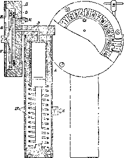
Figure 1.
Kotze instrument—the circular conimeter (Fig. 1)—consists mainly of a valveless pump A with a capacity of 10 cm3 and a chamber equipped with a dial, in which a round glass plate B is placed, pressed tightly against a rubber ring C by means of a spring D. The plate is attached to a toothed bronze ring E, on which 29 divisions are marked, and rotates together with them by means of a screw F. The chamber communicates 1) with the cavity of the pump, the piston of which is set in motion by a spring G, through a narrow channel H and 2) with the outside air—by means of a narrow (0.5 mm in diameter) impact tube I, the inner end of which is at a distance of 0.5 mm from the glass plate. To take air samples, the instrument is preliminarily subjected to thorough cleaning (the opening of the impact tube I is cleaned with a horsehair), the glass plate is covered with a thin layer of purified vaseline and set so that division No. 1 of the dial is opposite the opening of the impact tube, and the piston is pushed into the cavity of the pump until it stops. Then, by pressing the release catch J, the spring G is released, the piston jumps back, and the air, passing through the impact tube with great speed, strikes the glass, on which a spot consisting of adhering dust is formed. By turning the screw F, the next division of the dial is set opposite the opening of the impact tube, and a second sample is taken, and so on. Usually, to obtain the average dustiness of the air in a given place, several samples are taken.
The counting of dust particles is performed under a microscope equipped with a specially adapted rotating stage, using an ocular micrometer with two diameters intersecting at a certain angle (9° or 18°). The number of dust particles located in one sector of the micrometer is counted, then the same sector of the micrometer is set on another part of the preparation and counted again, etc., up to about 5 times; the average number of dust particles in one sector is determined, then—in the entire dust spot (by multiplying by 40 for a 9° sector or by 20 for an 18° sector). The quotient of dividing this value by the number of cm3 of drawn air expresses the number of dust particles in 1 cm3 of it.
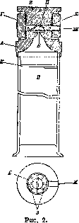
The Owens apparatus is based on the principle of the impact of an air jet against a screen and the condensation of water vapor in the air around dust particles (Fig. 2). Its central part is a cylindrical chamber with a base diameter of 1 cm and a height of 1 mm. The bottom of the chamber is formed by two metallic semi-lunar plates, between which there is a slit B with a length equal to the diameter of the chamber base, i.e., 1 cm, and a width of 0.1 mm (A is the part containing slit B). The side wall of the chamber is formed by a metal ring fastened to the lower base with two groove-like recesses 3 located opposite each other. A round cover glass D (square glasses can also be used, but this is less convenient) is placed on the metal ring, which is pressed tightly against the side wall of the chamber by means of an elastic spring E on the inner side of the apparatus's screw-on lid C, thus forming its upper base (G is a connecting coupling with an internal thread). The chamber communicates a) from below through the slit of the base with a screw-on hollow cylindrical tube I, lined on the inner surface of the wall with filter paper that is moistened before taking the sample of air being studied (K is a spring fixing the filter paper), b) from the side through the groove-like channels 3 with a valve pump of a specific capacity (50 cm3), which screws into the opening Zh in the side wall of the apparatus at the level of the chamber. With a rapid, single pull (to the stop) of the pump piston, a jet of air of a specific volume, equal to the capacity of the pump, i.e., 50 cm3, is sucked into the cavity of the apparatus through tube I at high speed. Meeting a moist environment in the cylindrical tube, the drawn-in air is humidified, penetrates from there under high pressure through the narrow slit into the chamber, where it suddenly expands, the pressure drops, the temperature decreases, the water vapors transition into a liquid state and condense around the dust particles, which strike the glass with great force and adhere to it. The moisture around the dust particles soon evaporates, and as a result, a narrow strip of dry dust corresponding to the slit is obtained on the cover glass, which is examined under a microscope. For this purpose, a cardboard or tin ring is glued onto a clean microscope slide, greased on both surfaces with glue that hardens easily in air and softens easily again upon slight heating. The inner diameter of the ring is slightly larger than 1 cm. By slightly heating the prepared microscope slide by passing it over a weak flame to soften the glue hardened on the ring, it is applied to the cover glass removed from the apparatus with the strip of dust so that it adheres to the ring along its circumference. Counting the dust particles under a microscope is best done with oil immersion (objective 1/12), using a grid eyepiece micrometer with square divisions of 1 or 0.5 mm (Fig. 3 shows a micro-drawing of the preparation: a—at low magnification, b—at high magnification). First, the average number of dust particles located in one strip of micrometer squares across the entire width of the preparation, i.e., between two lines of the micrometer across the entire dust track, is determined. To do this, the dust particles are counted in approximately 5–10 such transverse strips in several fields of view and the average (N) is calculated. If the number of such transverse strips that fit along the entire length of the track under given optical conditions (S) is determined, then the number of dust particles in the entire preparation will be equal to NS. Dividing this by the number of cm3 of drawn air (c), the average number of dust particles (a) in one cm3 of air is obtained: a = NS/c. Practically, the value S under the same optical conditions can be considered constant for any preparation, since it is a function of the track length, which always approximately corresponds to the length of the chamber slit. With a 1/12 objective with immersion, eyepiece 4 [magnification 1,000 (Zeiss)] and a micrometer square side of 1 mm, S is equal to 667; the value c, provided one pump volume is drawn, is also constant (50 cm3); consequently, for given optical conditions, there is a constant coefficient.
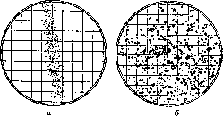
Figure 3. Bronze dust.
With a 1/12 objective with immersion, eyepiece 4 [magnification 1,000 (Zeiss)], S/c = 13.3, whence x = 13.3 N. Under other optical conditions (but with the same eyepiece), e.g., with objective D, eyepiece 4 [magnification 400 (Zeiss)], S = 667 * 0.4 = 267; S/c = 5.3; x = 5.3 Nx. If 2c, 3c, 4c, etc., of air are drawn, then 13.3 N (resp. 5.3 Nx) must be reduced accordingly by 2, 3, 4, etc., times. Besides the described instruments, the Aitken konimeter (see) and so-called coniscopes are often used; the latter instruments are usually used for rough, preliminary determinations of dust, primarily right there at the place of dust collection. Such are the Aitken coniscope, Kotze (one of the old modifications of his konimeter), Hill, and others. In 1926, Burshtein (Odessa Medical Institute) proposed, under the name K., an instrument for determining the "dustiness coefficient" (a term proposed by the author and denoting the average quantity of dust that is inhaled by a worker in a given production facility within 1 hour). The instrument has not gained widespread use to this day.
Related articles
Cite this page
“Condurango and Limbs.” Soviet Medical Encyclopedia. English translation of Bolshaya Meditsinskaya Entsiklopediya, 1st ed. (Moscow, 1928–1936), ed. N. A. Semashko. https://sovietmedicalencyclopedia.pages.dev/article/condurango-and-limbs/