Cholelithiasis
Historical document, translated for reference. It reflects medical knowledge of the 1920s–30s and is not medical advice.
Summary
Cholelithiasis is a disease characterized by the presence of gallstones in the gallbladder and bile ducts. This article explores the historical background, composition and structure of gallstones, etiology and pathogenesis, clinical manifestations, complications, and treatment approaches from a 1930s Soviet medical perspective.
Encyclopedia article (1928–1936)
Cholelithiasis. Contents: Historical data.............171 Composition and structure of gallstones......172 Etiology and pathogenesis of Ch. b. . .........175 Symptomatology and course...........181 Biliary colic.................183 Consequences and complications of Ch. b........188 Obstruction of bile ducts by stones .... 188 Cholecystitis..................193 Cholangitis...................194 Rupture of bile ducts............196 Biliary fistulas................197 Prognosis......................197 Prevention..................198 Treatment......................199 Surgical treatment.............200 Cholelithiasis, a disease having as its basis the presence, mainly in the gallbladder and bile ducts, of special formations called gallstones. For understanding this disease, it is of interest to study the structure of stones, the ways of their formation, but special importance is given to consideration of the pathological phenomena caused by these stones. It must be kept in mind that Ch. b. in the vast majority of cases proceeds latently, gallstones can be in the bile ducts without giving any symptoms. According to Riedel, in 95% of cases gallstones do not manifest clinically, and according to German statistics, in 6.94% of all autopsies gallstones are found without any clinical indications. For the appearance of a clinical picture, special circumstances are needed, which at the present time are sufficiently studied and come down to three moments: 1) active state of stones, their migration, or movement, 2) infection and 3) stagnation of bile. Thus, one must speak both about Ch. b., manifesting clinically, and about the same disease in the broad sense, i.e., take into account latent forms, and in studying the pathogenesis of stone formation, indicate those moments that determine the visible clinical onset of this complex and diverse suffering. Historical data. First observed in mammals, specifically in horned cattle, gallstones were observed in humans (on corpses) as early as by Vesalius. In 1554, Fernelius gave a good description of gallstones and the symptoms observed with them. The earliest information regarding the structure of these stones belongs to Waeter (1796), who described them and gave drawings kept in the rich collection of the Berlin Museum. In the XVIII century, Morgagni, Hoffmann, Boerhaave carefully studied the question of gallstones and paid attention to the disorders they cause; in this same century Poulthier de la Salle and Durand undertook the chemical analysis of these stones, however, the decisive step in this matter was made only in the XIX century by Chevreul, who discovered cholesterol in gallstones. Cruveilhier, Frerichs, Charcot left excellent descriptions of the clinical picture with anatomical drawings of gallstones. The study of the pathogenesis of this disease began in 1882 with the works of Bouchard, who first pointed to the significance of slowed metabolism and the general constitutional factor; in 1892 Naunyn put forward the infectious theory of the origin of stones; this theory found support in the works of Gilbert, Dominici and others. In recent times the works of Aschoff, Bacmeister, Chauffard and his students, the research of Grigaut, Schade and Neubauer have shed new light on the complex pathogenesis of Ch. b. and indicated the significance of diathesis, cholesterolemia, stagnation of bile and infection; finally these works drew attention to the biochemical, colloidal relations of the medium in which stones are formed. Since in the last 25 years Ch. b. has become widely accessible to the surgeon's knife and has entered the border area between internal medicine and surgery, a whole series of brilliant works and research in this field have been obtained from surgeons [the Mayo brothers and Deaver in America, Kehr and Körte in Germany, Hartmann and Gosset in France, Rovsing in Denmark and finally in the USSR Volyn'tsev, Dokhman, S. P. Fedorov and A. V. Martynov]. Finally in recent years the progress of knowledge in the field of Ch. b. has been facilitated by two methods: duodenal probing and cholecystography (radiography of the bile ducts). [See separate table (pp. 223-224), fig. 6]. Composition and structure of gallstones. The most valuable material for research is provided by stones obtained during operation, because in addition to their greater softness and freshness they always correspond to a certain clinical picture, whereas stones obtained from a corpse, as is known, may not have given any clinical phenomena during life. Attempts have been made to obtain stones experimentally in vitro and in vivo. A necessary condition for obtaining stones in vivo is stagnation of bile, which can also be sterile. With stagnation of sterile bile, small stones are obtained, but they fully resemble cholesterol stones with radial structure, described by Aschoff and Bacmeister; these are stones formed without the participation of infection - stones from stasis. Stones obtained experimentally on animals (Mignot - on guinea pigs, Klinkert - on rabbits and Flandin - on dogs) with the participation of virulent microbial cultures contained little cholesterol and consisted mainly of pigment. Ikonnikov (from Fedorov's clinic) by ligating the cystic duct in dogs, cats and rabbits obtained in a period from 3 weeks to 3½ months without the participation of infection not only radially-cholesterol stones, but also pigment-lime stones.- Gallstones differ from each other in size, color, shape and internal structure (see separate table, pp. 175-176). The size of stones varies from a sand grain to a chicken egg; large stones weigh from 25 to 30 g. Blackburn described a stone weighing 48 g. The number of gallstones varies from 1 to many thousands. The largest number of gallstones found during an operation amounts to 14,000. The most voluminous are usually single, solitary stones (monoliths), and they usually reflect the shape of the organ in which they sit; thus, bladder stones are usually round, ovoid in shape, stones of the ductus choledochi resemble the end of a cigar, and stones of the intrahepatic ducts can be branched. Aschoff calls intrahepatic stones obstructive and considers that infection does not participate in their formation. Very often in the gallbladder numerous, faceted stones of various shapes are encountered. It was believed that the formation of facets depends on the pressure of stones on each other when they were young and still soft (Naunyn, Boysen); however, in recent times the main importance is attached to the circumstance that the deposition of new salts occurs more on the free parts of the stones than on the touching parts. In the origin of these stones the influence of infection is considered unquestionable. Furthermore, in a certain percentage of operated cases (5.6%) black pigment stones are encountered; they are more often found inside the liver in the bile ducts, sometimes also in the gallbladder; they are hard, prickly and of branched shape. Rovsing believes that due to constant irritation they cause a peculiar aseptic catarrh in the bile ducts and bladder, contributing to the formation of new concretions. Finally, along with hard stones in the bladder and ducts, yellowish-white sand and even a marked pasty mass are found; the latter is detected especially frequently in the presence of particularly virulent infection. One can observe stones developing in the mucous and even submucous layer of the gallbladder. Then the bladder has a peculiar appearance with granulations on the mucous and accumulation of lumps of cholesterol on its inner surface.- The color of stones is extraordinarily diverse - white, gray, yellow, brown and almost black. The consistency of stones can be hard and brittle or, more rarely, wax-like. Despite the external diversity of stones, common features can be noticed in their structure: when a stone is cut, we observe a core of one structure or another and a shell or body - homogeneous or with striations, either radial or concentric; sometimes a cortical layer can also be distinguished, which in turn is either homogeneous or striated (see separate table, fig. 8). Thus, in most gallstones we observe a certain organization, and on their cut surface, as Chauffard says, their history is written. In this respect gallstones are very similar to urinary stones and sharply differ from concretions found in the pancreas and salivary glands and representing a simple, undifferentiated mixture of materials (mucus, lime salts, etc.). The material for the formation of gallstones consists of organic and inorganic substances. The main components are cholesterol, pigment (bilirubin and its higher stages of oxidation) and lime salts. All these substances can combine in various proportions, but besides them, from organic substances, stones contain a special colloidal substance of protein nature forming the framework of the stone, and from inorganic substances, besides lime salts (carbonate and phosphate), especially their compounds with bilirubin, in gallstones (pigment) iron, copper, magnesium, aluminum and sulfur have been found.
For practical purposes, it is sufficient to distinguish three types of stones based on their chemical composition: cholesterol stones, pigment stones, and mixed stones. 1. Cholesterol, radial stones consist almost exclusively (up to 98%) of cholesterol; they are white, sometimes yellowish in color, round or oval in shape, and range in size from a pea to a large cherry (see separate table, figs. 2,3, 12) (they correspond to the obstructive stones of Ashoff and Baumeister); however, sometimes accumulation of cholesterol masses is observed on the bladder mucosa, having the appearance of yellowish granulations. Cholesterol stones are distinguished by their lightness (they float in water) and burn with a bright flame, which distinguishes them from other concretions. Cholesterol stones just removed during surgery have a smooth surface, but when they dry, they quickly become rough and even granular. On the cut, the periphery of the stone has a coarse radial striation, cholesterol here is crystallized in the form of crossbars arranged radially. In the center of the stone, the crossbars are arranged less regularly, crossing around a microscopic clot consisting of lime salts and pigment. Cholesterol radial stones are usually solitary and occur only in the gallbladder. 2. Pure pigment stones are of two types: a) those occurring in Ch. b. and b) in hemolytic jaundice. Stones of the first type are especially common in herbivorous animals (cows), perhaps in connection with their nutrition. The peculiarities of the latter should explain the fact that pigment stones are more often observed in Japanese people, who, as is known, consume little fat and meat, but more plant food. These pure pigment stones are usually small, from a grain of rice to a pea, black when removed during surgery, turning green in the air, their fracture resembles graphite. They are heavier than water. The structure of these stones is completely homogeneous, without striation or layering. Chemically, they consist mainly of bile pigments with some amount of bile salts, lime, and cholesterol. These stones are usually multiple, occurring in the bile ducts and in the gallbladder (see separate table, figs. 4 and 11). Pigment stones characteristic of hemolytic jaundice are small formations, black in color with a metallic luster; they sink in water, contain 60% bilirubin lime and a small amount of cholesterol and bile salts. 3. Mixed stones, cholesterol-pigment-lime, multiple, faceted (see separate table, fig. 6) occur in tens, hundreds, even thousands. These are the most common, most widespread stones. From the uniform size and color of a series of such stones, it is evident that they form entire generations at once, which serves as proof of the common conditions of origin for stones of each generation. Their color is the most diverse: whitish, grayish, brownish, green. The size of these stones varies from a pea to a forest nut. The shape of faceted stones is polygonal or multifaceted with flat or slightly concave surfaces. Sometimes stones resembling raspberries or rather mulberries are encountered. On the cut, the layered structure can be clearly seen with a central core representing a soft black substance consisting of cholesterol. In older stones, there is a central wide cavity of star-shaped shape. These stones are found in the bladder, less often in the ducts, when they are carried there from the bladder. In the center of mixed stones, fragments of epithelium, foreign bodies (blood clot, dried roundworm, etc.) are sometimes found, around which precipitates precipitating from the bile are deposited. Stones of carbonate lime are extremely rare and are considered by the Ashoff school as a product of the inflammatory process. They are very small, their shape is bizarre, mostly they are provided with spine-like processes (Fedorov). Various combined stones do not require special description, representing a combination of the described groups. Etiology and pathogenesis of Ch. b. Etiology. Ch. b., as daily clinical observations and facts established at autopsies show, is a very common disease. The combined German statistics, summarizing observations of 12 different authors, indicates that in 6.94% of all corpses, gallstones are found. The Danish statistics of the Institute of Pathological Anatomy in Copenhagen gives a higher percentage (up to 8), and if from these figures subjects under 20 years of age (on which only two cases of Ch. b. fall) are excluded, the percentage rises to 11.2. Authors who pay special attention to finding stones in cases of cholecystitis (Scheel, Hansen) consider the percentage of stones even higher - up to 15-25. According to combined data from Moscow morgues for 1923-27, Ch. b. as the main disease (and cause of death) was noted 76 times in 21,383 corpses (aged 16 years and older), i.e. in 0.35% of autopsies of adults. Ch. b. occurs in all latitudes, however, the cold and humid climate predisposes to this disease, and it is more often observed in temperate regions than in hot countries. Races are affected differently, white and black apparently uniformly, but the yellow race, especially Japanese, get sick less often than Europeans. (Japanese statistics by Miyake indicate 3.5% findings of gallstones at autopsies, i.e. approximately 2 times less often than in Europe). Among predisposing causes, age and sex are very important. Information about the influence of age is only approximate, since the onset of Ch. b. often does not coincide with its first manifestation (first attack of colic). 'Clinical age is not the pathogenetic age,' says Shoffar, and often the first attack occurs in subjects who have carried stones in their bile ducts for many years. In childhood, gallstones are rare, although there are cases of congenital cholelithiasis. Such a case was described by Wendel in a newborn who died on the 11th day of life; 90 small cholesterol stones were found in the gallbladder at autopsy. The disease most often manifests itself between the ages of 30 and 55. With each decade of life, the frequency of Ch. b. increases, and in both women and men of 70 years of age, stones are found in every third person (Ker). The first attack of the disease in women occurs on average somewhat earlier than in men. Shoffar indicates an average figure - in women 37 years, and in men 421/2 years. - As for sex, statistics of many countries indicate a predominance of the disease in women (4-5 times more often than in men). In the clinical material of Fedorov, the vast majority of operated patients are women (out of seven patients - six women). This predominance of women is explained by the influence of pregnancies, since, according to Ker, cholelithiasis in girls is a rare phenomenon. 83% of women operated on by Ker in the past had had pregnancy. According to Shoffar's statistics, out of 114 women who developed Ch. b., in 51.7% hepatic colic appeared during pregnancy, in 25% - after childbirth. Pregnancy undoubtedly in various ways increases the predisposition to Ch. b.: mechanically it hinders the emptying of the bile ducts and promotes stasis of bile, under the influence of pregnancy the functional capacity of the liver cell is weakened, finally Grigo, Shoffar and others indicate on hypercholesterolemia, established by them during pregnancy and affecting the development of Ch. disease. Among other predisposing causes, heredity must be mentioned, and one can distinguish the inheritance of a predisposition specifically to Ch. b. and the inheritance of a certain diathesis predisposing to a certain group of diseases, to which Ch. b. also belongs. Shoffar in his material (161 cases) found indications of Ch. b. in the family history of his patients in one third of cases, and it was more often transmitted through the female line. As for the transmission of diathesis, Ch. b. is often observed in the same family together with obesity, gout, urinary sand, kidney stones, arteriosclerosis with hypertension, diabetes, with the pathogenetic factor
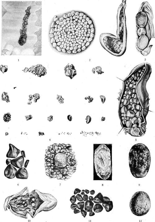
Figure 1. X-ray of gallstones (according to Lommelio). Figure 2. Multiple (white) stones of the gallbladder (on the right - gallbladder). Figure 3. Three cholesterol stones in the gallbladder. Figure 4. Bilirubin stones of various shapes and sizes. Figure 5. Multiple stones of the gallbladder (with obstruction of d. cystici). Figure 6. Faceted stones of the gallbladder (with obstruction of d. cystici and empyema of the bladder). Figure 7. Stones of the gallbladder in the form of mulberry (a large stone obstructed d. cysticus). Figure 8. Sawed stone of d. choledochi. Figure 9. Stone obstructing d. choledochus. Figure 10. Faceted stones in the stage of formation, surrounded by mucous masses. Figure 11. Multiple bilirubin stones of the gallbladder. Figure 12. Cholesterol stone obstructing ductus cysticus. (Figures 4, 8 and 10 from Chauffard; figs. 2, 3, 5, 6, 7, 9, 11 and 12 from the Faculty Surgical Clinic of 2nd Moscow University.)
To the article Cholelithiasis. Special importance is attached to hypercholesterolemia in cholelithiasis. In connection with this, modern authors attribute great significance to the origin of cholelithiasis to a particular disturbance of the chemical processes of tissues (humoral personality of Chauffard). Among the causes causing cholelithiasis, many, since the time of Nawin's work, attribute primary importance to infection. Among the diseases undergone, special attention is paid to typhoid fever, since it is known that Eberth's bacillus can directly act on the bile ducts, being excreted with bile. However, some authors consider the significance of typhoid fever in the etiology of cholelithiasis to be exaggerated and attribute greater importance to local inflammatory processes. If the first etiological factor is considered to be a known diathesis with hypercholesterolemia, the second is infection, then the third important factor is bile stasis. The latter is promoted by a sedentary lifestyle, inappropriate clothing that compresses the liver or restricts diaphragmatic movement, pregnancy, prolapse of abdominal organs, especially the right kidney and liver; in this case, bending of the bile ducts, especially the cystic duct located in the ligamentum hepatoduodenale, may occur. Attention must also be paid to diseases of the gastrointestinal tract. The anatomical proximity of the duodenum gives rise to combined diseases. Swelling of the duodenal mucosa and scarring of its ulcerative processes can compress the opening of the common bile duct and cause bile stasis, not to mention that duodenal catarrhs resulting from gross dietary violations can contribute to both bile stasis and infection of the bile ducts. Habitual constipation affects the bile ducts unfavorably in terms of bile stasis and infection, both mechanically and due to increased intestinal putrefaction. Diseases of neighboring organs, especially tumors and cysts of the liver and pancreas, can also impede bile evacuation and contribute to the development of cholelithiasis; it particularly frequently coincides with gallstones in cancer of the head of the pancreas. More rare causes of bile stasis are intestinal parasites, ascarids, Distoma hepaticum, crawling into the bile ducts from the intestine through the papilla Vateri (particularly frequently in Japan). - The mechanism of bile stasis is particularly thoroughly elucidated in the latest works of Berg, who sees the main cause of stasis in special, mostly congenital, anatomical and functional anomalies of the bile ducts. He draws attention to anomalies of secretion and to the retention of mucus (mucostasis) and to the existence in some patients of an underdeveloped gallbladder. His works also illuminate the great influence of the autonomic nervous system on the emptying of the bile ducts. Thus, one cannot disregard the possibility of bile stasis caused not by gross mechanical, but by functional nervous influences. Köerte in one of his latest works (1924) definitely recognizes the significance of spasm of the sphincter of Oddi in the origin of cholelithiasis. These influences of the autonomic nervous system on the emptying of the bile ducts, thanks to new techniques (duodenal probe-Meltzer-Lyon, cholecystography-Graham), have recently been registered in the clinic more definitely and have even outlined a new area of disorders in the form of so-called dyskinesias, or disorders of movement of the extrahepatic bile ducts. This bile-excreting system begins from the extrahepatic ductus hepaticus, includes all up to the sphincter of Oddi, and also includes the gallbladder. In the neck of the latter, Lütkeins found a special sphincter regulating the entry of bile into the bladder and its excretion back into the extrahepatic bile system. Thanks to both sphincters (of Oddi and Lütkeins), certain relationships between the concentration and excretion of bile—gallbladder and duct systems—can be observed in the clinic. It is possible that dysfunctions of these systems may have significance in the pathogenesis of cholelithiasis. One bile stasis usually does not give stone formation; it is sufficient to point out that prolonged retention jaundice (e.g., Köerte's case—26-year jaundice in a woman due to compression of the ductus choledochus by an aneurysm, etc.) usually does not cause stones; it is necessary to take into account, along with bile stasis, the influence of infection or diathesis or both factors simultaneously. The infectious theory. As early as the mid-19th century, Meckel von Helmsbach, Hein, and Lobstein pointed out that catarrhal changes of the gallbladder play the main role in the origin of stones and that inflammatory products in the form of mucus with the lime contained in it and desquamated epithelial cells provide the basis for stone formation. This theory of stone-forming catarrh of Meckel formed the basis of the infectious theory of Nawin, which gained great popularity by the end of the 19th century. It is necessary to recall that Botkin foresaw the role and significance of microorganisms in the formation of gallstones, 'the life of which gives as a result such chemical compounds that convert substances present in bile in a soluble form into insoluble ones, which then precipitate and deposit with a certain regularity giving the stone its pattern.' The ideas of Nawin can be summarized as follows: the basic condition for stone formation is infection penetrating into the bile ducts due to bile stasis and causing an infectious desquamative stone-forming process (angiocholitis, cholecystitis), which is the direct cause of stone formation. The main source of infection is the lower (duodenal) segment of the ductus choledochus, where the presence of Bacterium coli can almost always be found even in healthy individuals. The obstacle to the penetration of Bact. coli deep into the bile ducts is the normal continuous outflow of bile. As soon as this outflow is disturbed, i.e., stasis develops, favorable conditions immediately arise for the spread of infection deep into. Another path is descending infection circulating in the blood and penetrating into the bladder from the hepatic ducts. The material for stone formation is obtained from the disintegrating epithelium (cholesterol) and the inflammatory exudate of the mucous membrane (lime), with the primary nuclei of sedimentary formations consisting of a special stone-forming magma containing droplets of fat and cholesterol in the form of crystalline drusy spherulites. The infectious theory found support in many clinical observations and in some experiments. Virulent microbes began to be found in stones obtained at operation. Of particular interest is the work of Romantsev from the clinic of Fedorov. This author, using a new method for studying spherulite formations in some rocks, gave a new interpretation to the process of stone formation. Cholesterol more often precipitates in the form of spherulites under the influence of infection. Each new appearance of spherulite deposits in the stone coincides with an exacerbation of the inflammatory process. - Despite the interest and significance of the infectious theory, stone formation cannot be fully explained by infection alone. The same clinical observations indicate the possibility of stone formation without infection (Fedorov), which in many cases is a secondary complicating factor. The chemical (humoral) theory. Indications of changes in the chemical composition of bile in cholelithiasis appeared as early as the beginning of the 19th century, but the connection between lithiasis and slowed metabolism (bradytrophy) and increased precipitation of cholesterol from bile was first clearly formulated by Bouchard in 1882. Aschoff and Baeckmeister recognized the existence of a special cholesterol diathesis, and according to their opinion, every lithiasis begins with the formation of a cholesterol stone in aseptic bile under the condition of its stasis, while the further growth of the concrement is associated with outbreaks of infection. But of particular significance in indicating the role of hypercholesterolemia and insufficiency of the liver cell itself in the pathogenesis of cholelithiasis are the works of Choffard and his school. Choffard's students—Grigo, Flanden—showed an increase in cholesterol in patients with cholelithiasis, as well as during pregnancy, during menstrual periods, during recovery from infections (from typhoid fever in particular), and thus established a connection between hypercholesterolemia and those conditions which are etiologically connected with gallstones. The harmful effect of food rich in cholesterol (fats, brains, eggs) on liver patients has long been known. Choffard cites one fact bordering on an experiment. A young girl, anemic and emaciated, without indications of liver symptoms in the past, due to some signs from the right pulmonary apex for fear of developing pulmonary tuberculosis, was prescribed enhanced nutrition. For three months, she daily ate 11 eggs, which amounts to approximately 2.75 g of cholesterol. In total, she consumed 1,034 eggs. At this time, she had a severe attack of hepatic colic, which was repeated many times later. Choffard considers hypercholesterolemia such a constant companion of cholelithiasis that he finds it possible and even advises using it as a diagnostic sign in the differential diagnosis between calculous cholecystitis and duodenal ulcer. Miasnikov, Vikhrt and Yakovlev confirm the presence of hypercholesterolemia in cholelithiasis. Hypercholesterolemia is accompanied by an increase in cholesterol in bile—hypercholesterolemia. It should however be pointed out that some authors (K.
Zaitseva, Chiray) found upon examination with a duodenal probe, on the contrary, a decrease in cholesterol content in the duodenal contents in the presence of hypercholesterolemia. Grigo explains this fact by the fact that cholesterol circulating in the blood, when secreted by liver cells, undergoes some transformation, turning into a similar product—cholic acid, which is a chemical radical of bile acids. In his most recent work, Chiray and his colleagues state that hypocholesterinolia is an extremely frequent change in the bile of patients with cholelithiasis. It apparently depends on the precipitation of part of the cholesterol from the bile, in which it is in the state of an unstable colloidal solution. This instability of the solution and hypocholesterinolia are absent in some periods of established cholelithiasis. This circumstance forces one to think of two different phases in the course of the disease: a lithogenic stage, corresponding to the formation of stones, and a lithiasic stage, accompanied by signs which the already formed stone gives under the influence of either mechanical conditions or infection. In any case, the pathogenesis of aseptic cholelithiasis comes down to a special form of liver insufficiency. Primum movens of this disorder, which causes cholelithiasis, lies in the liver cell itself, producing cholesterol under conditions that do not allow this lipid to remain in a colloidal solution, so that it easily precipitates. It is possible, although not yet proven, that this colloidal instability depends on insufficient secretion of bile acids, since bile acid salts, as protective colloids, keep cholesterol in solution. The known diathesis, hypercholesterolemia, damage to the liver cell itself, bile stasis and infection—these are the components that give the sum of factors leading to the formation of gallstones. Considering cholelithiasis in the broad sense of the word, with its latent stage, one must recognize the possibility of stone formation in stone carriers without the participation of infection—aseptic lithiasis—in the presence of a known diathesis (hypercholesterolemia), bile stasis and perhaps a special functional insufficiency of the liver cell, but most of the diverse clinical manifestations of cholelithiasis without the participation of infection are impossible. The clinical picture of this disease gives us bright examples of the almost inevitable influence of infection at various stages of this long and diverse suffering. Each individual case requires a special analysis of the above etiological factors both individually and in combinations. Symptomatology and course. Symptoms and clinical manifestations of cholelithiasis are extremely diverse. First of all, one must remember that one can be a stone carrier and not show any signs of the disease. In 1/4 of cases according to Charcot, 1/5 according to Naunin and 1/3 according to other authors, stones give no sign of themselves. However, in recent times, some prodromal signs have been mentioned that make one suspect stones. Moynihan in 1908 pointed out that every time he accidentally found stones in the gallbladder during laparotomies performed for other diseases, he questioned the patients and they indicated a number of digestive disorders in the past, the cause of which was not clear. On the basis of these indications, they began to speak of small signs of cholelithiasis in an inactive state. A more detailed study of this semiotics in connection with data obtained by the duodenal probe method and cholecystography can at present contribute to early diagnosis. On the other hand, one must keep in mind that the disease often manifests itself suddenly without any preliminary symptoms in the form of a severe attack of pain depending on the movement of a stone—an attack called biliary or hepatic colic. Cholelithiasis in an inactive state. Moynihan and Mayo insist that there are no stones without clinical manifestations, and describe in such patients a number of periodically occurring digestive phenomena: a feeling of fullness and heaviness in the epigastric region 1/2 or 3/4 of an hour after eating, with bloating and belching, at times vomiting, which alleviates these phenomena. Patients often cannot tolerate eggs, cheese and fatty dishes. Appetite is preserved and may even be increased. Sometimes there appear dull or cramping pains under the xiphoid process and in the right hypochondrium, usually 3-4 hours after eating. Sometimes—pains in the back and in the right shoulder blade, also after eating. Examination of gastric juice shows either an increase or decrease in acidity. Patients feel slight chills and difficulty, and sometimes pain on the right side of the chest with deep breathing. In some cases, diarrhea is observed; the patient after eating feels pain under the xiphoid process and in the right hypochondrium with a persistent urge to defecate, with severe weakness. The stools are liquid with a large amount of bile. Aerophagia is often observed. Among the objective signs, various painful points are indicated: under the xiphoid process, the area of the gallbladder, the pancreatocholedochal point, the phrenicus point, the point on the shoulder and behind the points of the VIII, IX, X and XI vertebrae, the right scapular point and at the XI rib (Figure 1). Americans pay special attention to Murphy's sign: when pressing deeply with the fingers in the right hypochondrium below the liver, the patient cannot take a deep breath, as this is hindered by the sensitive gallbladder, which descends during inspiration. In recent times, the following signs have also been mentioned: weakening of the respiratory murmur under the lower parts of the lung on the right side compared to the left and tenderness of the intercostal nerves on the right. The diagnosis of the inactive form of cholelithiasis is difficult, but with careful attention to the above small signs, suspicion can sometimes be confirmed by determining hypercholesterolemia and cholecystography. Most often the disease is confused with a stomach ulcer and especially
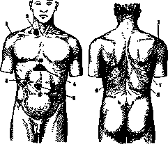
Figure 1. Anterior and posterior pain points in hepatic colic: 1-shoulder points; 2- phrenicus point; 3-gallbladder point; 4-epigastric point; 5-pancreatico-biliary zone; 6-points of VIII-XI thoracic vertebrae; 7-scapular point; 8-point of XI rib. duodeni. For duodenal ulcer speaks the greater definiteness of the pain syndrome (periodicity, late hunger pains), presence of blood in the feces and finally the corresponding data of X-ray examination of duodeni. Confusion with appendicitis is possible inasmuch as cholelithiasis sometimes gives reflex-spastic intestinal symptoms. In appendicitis the pain is localized lower, often there is an increase in temperature, leukocytosis and so on. It is necessary to keep in mind the not infrequent coincidence of both diseases. Duodenal probing is of great help for the diagnosis. With the help of a thin probe one can obtain indications both on the mechanical manifestations of cholelithiasis and on the presence and degree of infection, resp. inflammation of the biliary passages in general and of the gallbladder in particular (increase in protein, polymorphonuclear leukocytes, red corpuscles, mucus in the bile). Absence of bile 'A' and 'B' or only 'B' indicates obstruction of one or the other duct. It is not always easy to diagnose the location of stones. More often they are located in the gallbladder, and the symptoms described above refer to their localization in the bladder. Intrahepatic localization is much rarer, for it dull pains in the hepatic region are more characteristic. Localization of stones in the ducts (d. cysticus and choledochus) is usually secondary, is the result of migration of the stone and usually gives obstructive symptoms, of which more will be said below.-Path.-anatomical changes in the gallbladder in the case of inactive stones removed at operation are negligible: its wall is slightly thickened, the Luschka's canals are somewhat deeper, the folds are smoothed out, a small infiltration under the mucosa and around the diverticula (Luschka's canals) is noted. Biliary colic is a characteristic syndrome of paroxysmal pain, due to the efforts which the musculature of the gallbladder and biliary passages makes in order to push out and into the intestine any foreign body, most often the gallstones located in them. Biliary colic is a great and main symptom of Ch. disease, however in very rare cases it can be observed during the passage through the biliary passages of a blood clot, a worm or a hydatid cyst. In Ch. disease colic usually appears between 30 and 40 years of age, very rarely is it observed in children (in particular it also occurs in them in hemolytic jaundice). The pain can appear suddenly and quite unexpectedly, but sometimes it is preceded by prodromal symptoms in the form of loss of appetite, nausea, heaviness and pain under the xiphoid process, yellowness of the sclera and a feeling of tension in the right hypochondrium (perhaps due to overfilling of the gallbladder). As causes producing biliary colic are indicated: fatigue, emotions, a bumpy ride, cooling of the body, overfilling and irritation of the stomach, especially by some piquant dishes, and much else. In women biliary colic often coincides with menstruation or occurs after childbirth and removal of fibroma. Colic usually begins in the first half of the night, 3-4 hours after eating, at the moment of greatest irritation of the duodenal mucosa by food pulp and greatest outflow of bile. The pain is so severe that it makes even patient patients scream and often requires immediate injection of morphine. The character of the pain is varied: sometimes stabbing, sometimes cutting, sometimes as if tearing apart the right side of the abdomen. The pain is localized in the right hypochondrium and gives a characteristic radiation or irradiation upward and backward to the right shoulder, under the right scapula, into the neck, occiput, head and right side of the chest; rarely is radiation of pain to the left side observed, still more rarely does the pain radiate downward, into the lumbar or iliac regions. The pain is usually diffuse at the beginning of the attack, but soon three maximum points can be noted: the gallbladder point (at the crossing of the outer edge of the right rectus muscle and X rib), the epigastric point and the point at the lower angle of the scapula. The intensity of the pain is explained by the abundance of nerve fibers surrounding the biliary passages, and of course varies depending on the sensitivity of the given subject. The acuteness of the pain is greater in neurasthenics and less in old people. In perforating cholecystitis the pain soon becomes diffuse over the whole abdomen, as is observed in perforations and other abdominal cavity organs. The pain is always accompanied by dyspeptic phenomena: nausea and vomiting. Vomiting is at first of food, then of mucus and bile; the latter is sometimes excreted in large quantities, and in rare cases small gallstones are vomited up. This reaction of the stomach makes it impossible to administer any medicine internally. Nausea and vomiting are caused partly by reflex from the peritoneum, since during colic other signs of irritation of the peritoneum are often observed, such as: rigidity of the right rectus muscle of the abdomen, meteorism, constipation; partly nausea and vomiting depend on irritation of the mucosa of the bladder, ducts and duodeni, as these symptoms occur without any signs of peritonitis. As a rule constipation is observed. Fedorov points out that in gallstones the pain does not always have a colicky character; sometimes patients complain of constant dull pains in the epigastric region or girdle-like pains, increasing after food. The origin of these pains is explained by increased pressure in the bladder or by adhesions of the gallbladder with the omentum, the pylorus or duodenum. It also happens that an enlarged gallbladder sometimes compresses the pylorus and, hindering the passage of food into the intestine, thereby causes pain (Fedorov). Sometimes during colic the pains are so severe that patients faint, the activity of the heart may be weakened; even Botkin noted this reflex influence of biliary colic on the heart and considered that under its influence cardiac asthma may develop. The pulse is usually somewhat slowed, but often it becomes rapid, corresponding to the occurring fever, sometimes arrhythmia is noted. In persons with a weak heart an attack of biliary colic with severe pains may lead to death. Among the general symptoms (besides nervous excitement and at times convulsions) it is necessary to point out the febrile condition, which is short-lived, although t° reaches after chill 39° and even 40° and then subsides. The fever disappears with the cessation of pain, if the process is not complicated by infection; in the latter case ta often acquires an intermittent character. Leukocytosis from 12,000 to 15,000 with polymorphonuclear shift is often observed, indicating infection of the gallbladder.-Objective examination of the patient during an acute attack is almost impossible, and usually it has to be postponed until the pains subside somewhat. However, distention of the abdomen, especially of the epigastric region, and marked defensive tension of the right rectus muscle and tenderness in the above-mentioned specific points can be determined. At the same time the sensitivity of the vermiform appendix and the right kidney can be tested in order to avoid a gross error in diagnosis. As soon as palpation of the abdomen becomes possible, enlargement and tenderness of the liver, especially of the gallbladder, can be easily determined; the latter in some cases can be felt enlarged as a round, firm body. As for the crunching of stones, this sign is almost never observed. It was sometimes possible to feel one large stone, the presence of which was confirmed by operation, and in one case the exit of a large stone the size of a mandarin through an internal fistula from the anus. The spleen is usually not enlarged.-With the end of the attack of pains the patient usually excretes a large quantity of light urine, sometimes containing traces of sugar.-The attack of pain on average lasts from 6 to 12 hours, and sometimes one or two days and even longer. Its duration depends on the one hand on whether morphine was used, and on the other hand the pain can drag on and pass into a new phase if the stone is pushed from d. cysticus into d. choledochus and gives a new attack of pain. Sometimes the pain drags on during the passage of the stone through internal fistulas into the intestine, duodenum or stomach, and finally in connection with an attack of biliary colics perforations are possible, which give pains characteristic of perforating peritonitis. Much more often the attack ends suddenly, and the patient after two or three days returns to his former state. At the end of the attack two signs often appear which quite determine the diagnosis. These-jaundice and the exit of stones with the feces. Jaundice is not always observed, approximately in half of the cases, since the presence of stones in the gallbladder does not hinder the free outflow of bile from the liver through the hepatic and common bile ducts. It is necessary to keep in mind that jaundice does not always depend on obstruction of the bile ducts by a stone, it is often observed with localization of the stone in d. cysticus and depends on infectious hepatitis or on inflammatory swelling of the mucosa of the biliary passages or, as Ker points out, on swelling of the head of pancreas hindering the free outflow of bile.
It is self-evident that the intensity and duration of jaundice depend on the particular mechanism of its origin. In obstruction of the common bile duct (d. choledochi) or its opening at the papilla Vateri, jaundice is intense, with complete discoloration of the stool and other accompanying symptoms (skin itching, bleeding, bradycardia, and other cholemic phenomena); in these cases, it is often eliminated only by surgery. In the absence of a mechanical factor, jaundice may pass quickly, and its course varies individually depending on the conditions of its origin in each case. Sometimes the end of colic, if it is caused only by the movement of stones, is completed by the passage of stones with the stool. Finding stones gives certainty of at least temporary recovery. They can be found in the stool 1-2 days after an attack, and sometimes even after 5-6 days. Therefore, it is recommended to carefully monitor the stool and search for stones by washing the feces through a sieve. The size of the found stone does not always correspond to the suffering it caused during its movement. A stone up to 14 mm wide can pass through the common bile duct and duodenum; stones of larger sizes usually pass through internal fistulas. In cases where no stone is found in the stool, it must be assumed that it has either returned from the cystic duct back to the gallbladder or has become stuck in the bile ducts. Then an obstructive picture develops, which will be discussed below. The question of the possibility of dissolving gallstones by intestinal enzymes and juices on the basis of Naunin's and others' experiments is answered negatively. Based on the clinical picture, it is not always easy to localize the process, i.e., to speak of biliary colic or of proper hepatic colic. Indeed, in some cases, less intense pain, absence of jaundice, and the palpation of a distended gallbladder as an elastic tumor speak for a gallbladder process; on the other hand, more intense pain, persistent obstructive jaundice lead one to think of a stone in the common bile duct. Choffard speaks against such differentiation, thinking that in every colic the gallbladder participates as a reacting organ. Biliary colic itself can give a number of complications in relation to the most important organs. Not to mention that a stone can rupture into the abdominal cavity and give a severe picture of perforating peritonitis, in people with weak hearts, severe colic can give, by reflex, severe phenomena from the heart and blood vessels: pulse fall, fainting, attacks of cardiac asthma or angina pectoris and even sudden death (Choffard). From the nervous system, convulsions, epileptic seizures, and paralyses have been described. In the urine, transient glycosuria is often observed, and sometimes also albuminuria. Pathogenesis of biliary colic. Most clinicians (Choffard, Fedorov) adhere to the classic viewpoint that colic is of mechanical origin; it depends on the migration of the stone and is considered as a reaction to an obstacle to the emptying of one or another organ with muscular walls. To defend this opinion, such indisputable arguments are brought as the finding of stones in the feces as a result of a painful attack and the pictures that surgeons observed during operations and pathologists at autopsies when they saw stones on the path to exit from the bile ducts. Two mechanisms can contribute to the movement of the stone: on the one hand, the natural flow of bile (according to Heidenhain's indications, it goes under weak pressure of 110-220 mm of water column in a dog), and on the other hand, the contraction of the muscular wall of the ducts and gallbladder. The ability of the gallbladder to contract has been especially studied by recent authors (Chiray and Pavel); painful attacks similar to biliary colic were easily caused by duodenal probing by irritating the mucous membrane of the duodenum at the duct opening with a drop of vinegar. At the same time, this irritation caused abundant bile secretion. It is necessary to pay attention to the instability and special sensitivity of the autonomic nervous system in patients with cholelithiasis. This can explain the fact that any cause that can excite autonomic reflexes, such as fear, grief, sadness, can play a role in the origin of an attack. Other authors (Riedel, Heineck, Paviot), not agreeing with the mechanical theory, explain the painful syndrome by local inflammation of the peritoneum, similar to what is recognized in relation to appendicitis. In defense of the inflammatory theory, such facts are cited as the finding by surgeons, in the presence of typical colicky pains, of inflammatory phenomena in the absence of stones. It is necessary to recognize the possibility of participation in the pathogenesis of biliary colic of both factors - mechanical and inflammatory; sometimes they combine, and sometimes each of them separately can give a pain attack. In some cases, based on pronounced muscular defense on the right side, persistent vomiting, and absence of jaundice, one can speak of infection and local peritonitis; in others - the appearance of jaundice with discoloration of the stool and especially the finding of stones in it allow one to think only of the mechanical factor. Differential diagnosis. Although in typical cases biliary colic is not difficult to recognize (localization of pain, its typical irradiation, onset of pain after eating, persistent vomiting, chills, high temperature, negative results of urine analysis, jaundice, etc.), many abdominal colics can cause confusion. 1. In renal colic, the pain is localized lower in the lumbar region and radiates to the groin, genital organs, and leg; dysuria, oliguria, anuria, blood in the urine, and sometimes the passage of sand are often observed; vomiting is not so persistent, temperature is lower; one must not forget that both colics can exist simultaneously. 2. In lead colic, which is more often observed in men, the pain is localized in the middle of the abdomen, is diffuse, is relieved by deep pressure, the abdomen is usually drawn in and tense, blood pressure is elevated, and there is a typical line on the gums. 3. Intestinal colic is characterized by periodic pains with rumbling and sometimes accompanied by diarrhea. 4. Tabetic crises give less intense pains, vomiting during them is more abundant, there is no increase in temperature, and tabetic symptoms are present (Argyll-Robertson, Romberg, Westphal symptoms, etc.). 5. Sometimes, if biliary colic occurs suddenly at night with abundant food vomiting, one can think of acute poisoning. The diagnosis is helped by questioning the patient or those around him and examination and study of the vomit. 6. In acute appendicitis, attention is drawn to the localization of pain (McBurney's point), tension of the abdominal wall below the navel (defense musculaire), more frequent pulse, etc. In less pronounced forms of biliary colic, there can be talk of confusion with gastralgia, stomach ulcer, and duodenal ulcer. Due to the anatomical proximity of the duodenum, duodenal ulcers and periduodenitis are often confused with biliary colic. The diagnosis is helped by detailed analysis of the pain syndrome, pain points, and X-ray examination. As was indicated above, the cause of biliary colic is almost always stones, however, in rare cases, the same painful attack can originate from an ascaris stuck in the ducts or an echinococcus cyst. The diagnosis is helped by analysis of the stool for worms and the presence of other symptoms of helminthic or echinococcal disease. Prevention. In preventing an attack, the rest of the nervous system, the elimination of jolts (bumpy riding), a suitable diet with restriction of spicy and fatty dishes, fractional eating with sufficient fluid intake, and elimination of constipation are important. - Treatment of biliary colic consists in the energetic application of heat to the liver area in the form of dry hot-water bottles or compresses; if the patient cannot tolerate heat, ice application can be tried. Pain-relieving ointments are useless. Belladonna and morphine are prescribed orally. But usually vomiting does not allow giving medicine orally, and generally, with pronounced colic, it is more correct to make injections of morphine under the skin (0.01) which act more correctly and quickly; the addition of 1/2 mg or 1 mg of atropine is advisable from the point of view of its relaxing effect on the spastic contractions of the duct musculature and from the point of view of neutralizing some undesirable side effects of morphine. - Prognosis. An attack of biliary colic usually ends favorably in the sense of cessation of all painful phenomena for a longer or shorter period. Relatively rarely, attacks bring the patient recovery from the disease - this is possible in the case if a solitary gallstone is thrown into the intestine and exits successfully with the stool and the stone-forming process in the bile ducts ceases. Much more often the stone either returns back or gets stuck in the bile ducts. Then in the picture of the disease new symptoms appear in the form of so-called obstructive symptoms or a whole series of processes caused by an infection complicating the disease. Consequences and complications of cholelithiasis. Obstruction of the bile ducts by stones. Stones can get stuck in the bile ducts during their passage, or they, which happens much less often, are formed in place at various points of the bile ducts, rarely in the hepatic ducts, often in the neck of the gallbladder, in the cystic duct, and in the common bile duct (at its beginning, middle, in its seg- Figure 2.
Various locations and positions of gallstones (schema): significantly more often in the gallbladder, in the papilla Vateri; fig. d. hepat.; in d. cyst.; fig. 2). The clinical symptoms caused by this circumstance are characteristic for each of these points. In clinical practice, obstruction of the d. cystici and d. choledochi is most often observed. Obstruction of the d. cystici by a stuck stone can give two variants: either the stone is positioned in such a way that, acting as a valve, it can allow bile to enter but the latter cannot exit the bladder, or the stone completely and permanently blocks the canal, so that the gallbladder is completely excluded from the circulation of bile. In the first case, bile gradually accumulates in the bladder, it stretches and sometimes reaches enormous sizes, containing up to 4 liters of bile (Fedorov), in the second case the gallbladder fills with fluid that gradually loses the color of bile, and hydrops of the gallbladder (hydrops vesicae felleae, s. muco-cele) forms. It must be noted that not only the mechanical moment, namely the obstruction by a stone, plays a role in the formation of this hydrops of the gallbladder; here there is a specific inflammatory reaction of the gallbladder, in which infection participates. Ashoff distinguishes two forms of inflammatory hydrops of the bladder—primary and secondary. He calls primary the one that arises after the very first attack of colic when the cystic duct is closed by a stone, and secondary the hydrops that forms in the later course of Ch. b. after repeated attacks and also as a result of obstruction by a stone of the same duct. Surgeons point out that as early as 7 days after obliteration of the duct, they have seen on operation the contents of the bladder completely colorless. The question of the so-called white bile and how the bile contained in the gallbladder, when its duct is obstructed, loses its color and so quickly transforms into a fluid whose osmotic concentration approaches that of blood plasma has long interested clinicians. It is known that the normal mucous membrane of the bladder does not absorb or weakly absorbs bile pigments, which is why bladder bile is thicker than liver bile. Apparently, with complete obstruction, a specific inflammatory change of the bladder's epithelium occurs, thanks to which the mucous membrane quickly absorbs the remaining bile pigments, produces mucus, and soon transforms into such a membrane that quickly (by dialysis) equalizes the osmotic concentration of the bladder's contents with the osmotic concentration of blood plasma. (Description of inflammatory changes of the bladder wall—see Cholecystitis). The clinical picture of obstruction of the d. cystici is often very distinct. After one or a whole series of attacks of biliary colic, when the pain is no longer so severe but more constant, when there is no jaundice, no itching, no slowing of the pulse, no passage of stones in the feces, one can notice in the patient the appearance in the right hypochondrium, in the area of the gallbladder, of a firm tumor, sensitive to pressure, extending downward along the outer edge of the m. recti in the form of an oval cylindrical body resembling a large cucumber or eggplant. This tumor moves with the liver, i.e., follows the movements of the diaphragm. Regarding jaundice, it should be kept in mind that slight jaundice may occur, but it depends not on difficulty of bile outflow from the liver, but on inflammatory swelling of the mucous membrane of the bile ducts or on infectious hepatitis. The onset of hydrops of the bladder can be acute, and it can quickly disappear as soon as the stone returns back into the bladder or passes into the common duct. Sometimes it disappears slowly, over 2-3 weeks after an attack, and often remains for many months, during which fluctuations in the size of the tumor can be observed; the bladder then empties and then refills again; during this time, repeated attacks of pain are usually observed. One often has to observe a prolonged course of this hydrops of the gallbladder—it does not disappear for several years; however, with the passage of time, the tenderness becomes less and less due to atrophy of the nerve fibers enveloping the bladder; in these cases, the 'hydrops' of the bladder does not resolve without surgical intervention. However, disappearance of chronic hydrops of the gallbladder under the influence of duodenal probing with irritation of the duodenum by a solution of magnesium sulfate and introduction into a vein or per os of a large amount of physiological solution of NaCl (drainage of the bile ducts) has been observed. As for diagnosis, sometimes an enlarged gallbladder can be confused with hydronephrosis, with a cyst of the right kidney or pancreas, but one must remember that these tumors, unlike tumors of the gallbladder, do not follow the movement of the diaphragm. The thought of echinococcosis might more readily come to mind, but the latter usually does not give pain attacks, sometimes eosinophilia is found in the blood and a positive Weinberg reaction, Casoni reaction, etc., are obtained. Hydrops of the gallbladder is often simulated by the liver portion hanging over it, enlarged due to limited hepatitis caused by the irritating influence of Ch. disease, so-called Riedel's lobe, described earlier by Trousseau. Riedel's lobe sometimes exists simultaneously with hydrops of the gallbladder. One must keep in mind the circumstance that if at the time of obstruction of the cystic duct there is a more virulent infection in the gallbladder or the latter is subsequently added by the hematogenous route to the aseptic hydrops, then the enlargement of the bladder occurs faster and the hydrops collection (hydrops, mucocele) turns into a purulent one (pyocele). In the purulent process, a more acute and a more chronic course must be distinguished, accordingly there is one or the other clinical picture. In the more acute course, higher temperature with chills, more severe pain, leukocytosis are observed, and sometimes symptoms of general sepsis are added. In the chronic course, all phenomena proceed more calmly, and often an empyema of the gallbladder is found on operation in the chronic course of Ch. b.—When establishing indications for operation in hydrops of the gallbladder, one must not forget about this possibility of suppuration, and in case of suspicion of its presence—operation should not be delayed. Therapeutic treatment consists of diet, laxatives, application of heat and the above-mentioned duodenal probing. Stones in the d. choledochus most often penetrate from the gallbladder, more rarely—from the hepatic ducts, and sometimes:also very rarely—form in place; most often they are single, round in shape or elongated—like the end of a cigar; if there are many stones, they are faceted. If a stone stuck in the d. cysticus manages to pass into the d. choledochus, the colic usually temporarily ceases, because the d. choledochus is wider than the cystic duct. However, this lull usually does not last long, because the stone approaches a narrower part of the duct and then becomes a serious obstacle to the movement of bile. It blocks the duct; spasm of the wall, and even more so inflammatory swelling of the mucous membrane, intensifies the obstruction. If the latter becomes absolute, then the overlying bile ducts significantly dilate, the d. choledochus reaches the width of a loop of small intestine, the d. hepaticus stretches so much that it reaches the size of a second gallbladder. Due to thinning of the walls of the bile ducts, their rupture may occur. The gallbladder rarely dilates, more often it is reduced, atrophied due to pericholecystitis and sclerosis of the wall caused by long-standing cholecystitis; it contains little bile, watery fluid or pus. The liver is enlarged, in it a picture of biliary cirrhosis develops with sclerosis around the bile ducts, which appear dilated. On section, the liver parenchyma is greenish in color, on it yellow spots are noticeable in places—foci of 'bile apoplexy'. At the very stone in the d. choledochus, changes of the mucous membrane and wall of the duct of a sclerotic nature are found, the epithelium from cylindrical turns into cubical, and soon disappears completely. Often suppuration is added, which gives rise to perforation, which can cause symptoms of local and general peritonitis, and often fistulas form between the d. choledochus and neighboring organs. In the presence of infection, inflammation of the bile ducts throughout the liver (angiocholitis) can arise, which either ends in proliferation of connective tissue (hypertrophic biliary cirrhosis), or with the action of more virulent microbes, purulent miliary scattered foci form in the liver (purulent periangiocholitis). Infection can also spread to the pancreas in the form of chronic or acute purulent or even hemorrhagic pancreatitis. The main symptom of obstruction of the d. choledochi is jaundice, which can be of varying degrees, but when the stone penetrates into the papilla Vateri it is usually complete with all its features (see Jaundice): with intense coloring of the skin and mucous membranes, with golden serum, brown urine (without urobilin—law of Fr. Muller) and with complete discoloration of the feces, with absence in them even traces of stercobilin.
Indeed, the color of the skin of these patients does not have a green or bronze tint, as in mechanical jaundice caused by cancer of the head of the pancreas, however, in them too one can observe cholemic phenomena with skin itching, slowing of the pulse, and bleeding. In most cases, it is a matter of patients who have been suffering from biliary colic for many years or have chronic dyspepsia with dull pains characteristic of inactive cholelithiasis; more rarely, acute obstruction occurs as a result of the first severe attack with rapid onset of jaundice. Even more rarely, obstructive jaundice develops without preceding painful attacks. In case of obstruction of the common bile duct by several stones, allowing bile to pass between them, jaundice may not be sharply expressed. Usually in all cases, high fever is observed, in the blood leukocytosis-- up to 30,000 and even up to 50,000. The liver appears enlarged, but the gallbladder is small and not palpable, as it is in a state of chronic inflammation and its walls are sclerosed. On the contrary, in obstruction of the bile duct due to cancer of the head of the pancreas or papilla Vateri in 92% the gallbladder is enlarged (Courvoisier's law). Duodenal probing confirms that bile does not enter the intestine. Obstruction can occur acutely or develop gradually. The course of the process varies depending on the site of obstruction and the degree and severity of secondary infection. In this stage of cholelithiasis, the prognosis is always serious, and without timely operation recovery is doubtful. Delayed jaundice gives progressive cholemic phenomena with bleeding, with changes in the liver and signs of liver failure (icterus gravis). Secondary infections often cause cholangitis with miliary abscesses and a picture of general sepsis and pancreatitis. Spontaneous passage of a stone most often occurs through a choledochoduodenal fistula. The diagnosis in acute obstruction is not difficult, in chronic--it has to be differentiated from obstruction due to cancer of the head of the pancreas. The latter is more common in men, without preceding pains, jaundice in it has a dirty green bronze tint; there are no characteristic painful points, as in cholelithiasis; there is an enlarged gallbladder (see art. 21, fig. 1). Obstruction of the hepatic duct is observed comparatively rarely. Stones in it most often come from the gallbladder and only after the common bile duct is full of stones. The clinical picture is the same as in obstruction of the common bile duct. In other cases, stones can penetrate into the hepatic duct from intrahepatic ducts, while one of its primary branches may be obstructed; in this case, a rare clinical picture can be observed: enlargement of the corresponding lobe of the liver, jaundice and bile pigments in the urine and at the same time normally colored stool (Fedorov). The role of infection in cholelithiasis. If infection is one of the important conditions for the formation of most stones, then subsequently, when stones have already formed, infection in its various manifestations determines and colors with bright colors the clinical picture of the subsequent stages of cholelithiasis. The transition from a hidden stage to an obvious one, in other words--the beginning of the actual clinical picture of cholelithiasis, is usually associated with a push, which infection gives. When describing the symptoms associated with stone migration, the influence of infection was indicated both in the described characteristic temperature jumps, and in the general phenomena, leukocytosis, and in that complex range of reactive phenomena that each attack of biliary colic gives. It is known that the inflammatory swelling of the duct above the t»s trapped stone, complementing the phenomena of obstruction, and subsequently the phenomena of local perivesicular peritonitis are caused by infection. In the further course of the disease, all complications depend on the nature and virulence of the infection. Various forms of cholecystitis (catarrhal, sclerosing, purulent, gangrenous), cholangitis, biliary hepatitis, liver abscesses, formation of perforations, fistulas and finally the development of general septic processes with metastases--all these complications owe their origin to infection. Cholecystitis. It is known that stones most often form in the gallbladder and with a long course of cholelithiasis, its various clinical forms depend on the reactive-inflammatory processes that take place in the gallbladder itself or around it. To clarify and differentiate these diverse clinical pictures and their corresponding anatomical changes, it is useful to draw an analogy between them and the anatomical-clinical processes that the clinic of appendicitis gives. Just as in appendicitis, so in cholecystitis there are three similar moments: a closed cavity, infection of varying virulence, and this or that reaction from the adjacent peritoneum. The course of cholecystitis can be acute, subacute and chronic; chronic forms with periods of exacerbation are more often observed. Depending on the three mentioned moments, one observes: simple catarrhal cholecystitis without a special reaction from the peritoneum, in which, in addition to the inflammatory swelling of the wall with all the signs of catarrh of the mucous membrane, very often there is dropsy of the gallbladder. Chronic sclerosing forms are characterized by the development of connective tissue in the wall of the bladder itself and at the same time the involvement of the peritoneum with the formation of adhesions and adhesions with neighboring organs (pericholecystitis); most often the gallbladder is small, atrophic, reduced, hidden among numerous adhesions and adhesions with neighboring organs: the liver, the exit part of the stomach and duodenum; its mucous membrane is atrophic, the wall is thickened, the cavity is reduced and contains a small amount of dark bile or mucopurulent fluid. More rarely another form is encountered--hypertrophic form of sclerosing chronic cholecystitis, in which the gallbladder is of normal size or even larger, its wall is thickened and thickened, around it a plastic peritoneal process develops with tissue proliferation, sometimes with the formation of infiltrates and exudate. This process proceeds in spurts: then exacerbating then subsiding. Often in these forms, with the spread of infection to the pancreas, as a complication, chronic pancreatitis develops. One should not forget that these chronic cholecystitis with deep anatomical changes of the mucous membrane can give rise to the development of cancer (Kehr, Czerny, Kimmell indicate that in 10% of cases of chronic cholecystitis cancer develops; other authors--Siegert and Courvoisier--consider this percentage to be much higher--from 80 to 90). Purulent cholecystitis is most often a subsequent complication of the same chronic cholecystitis or develops as a result of the transition of 'dropsy' of the gallbladder into suppuration (pyocele); of course, suppuration occurs as a result of the appearance of a new or an increase in the virulence of the old infection. The gallbladder can enlarge or remain small if the surrounding adhesions prevent it from stretching, the wall thickens, is edematous, on the section sometimes small abscesses are noticeable, the mucous membrane is often covered with ulcers; with more active infection and weak reactive protection, perforation is possible with all its serious consequences, with the formation of limited peritoneal processes or even diffuse perforative peritonitis. Purulent cholecystitis corresponds to a more acute clinical picture: high temperature, sometimes of hectic type, increased pulse, dryness of the tongue, more severe pains, leukocytosis, etc. (in detail--see Cholecystitis).- A more virulent infection can cause an even more severe gangrenous form, which almost always ends in perforation with the phenomena of general or localized peritonitis. The gangrenous form is more often in patients with chronic cholelithiasis, who have signs of sclerosing atrophic cholecystitis. This severe complication is detected by even more severe general symptoms--increase in temperature, change in color and features of the face, dryness of the tongue, fall of the pulse, etc., sometimes with signs of peritonitis; in rare cases, bulging of the right hypochondrium with the appearance of tympanic sound can be established, i.e., the formation of a gas abscess (pneumocholecystitis Pendi). Gangrenous forms as a rule inevitably lead to a fatal outcome.- A histological feature of cholecystitis in cholelithiasis, according to Ashoff, is the development in the wall of the bladder of the so-called Luschko's canals, the formation of bile granulation foci and the new formation of mucous glands. Cholangitis. Infection in cholelithiasis sometimes spreads in another direction, namely--along the hepatic duct and along the intrahepatic bile pathways, and gives severe complications from the liver itself: subacute, acute and purulent cholangitis. The entry of infection into the bile ducts in these cholangitis can also be by the hematogenous route, however, more often these processes occur by the ascending route along the branches of the bile ducts. The subacute form usually proceeds favorably, and this cholangitis resembles the form which is called catarrhal jaundice. Jaundice often appears after an attack of biliary colic, accompanied by an increase in temperature, swelling and sensitivity of the liver on palpation: it depends partly on the inflammatory swelling of the mucous membrane of the bile ducts (mechanical jaundice), and partly on the infection of the liver cell itself (parenchymatous jaundice).
The course of these subacute cholangitis is usually favorable. Acute cholangitis is distinguished by a more violent course and more sharply expressed manifestations of inflammation of the bile ducts and is accompanied by the formation of ulcers, scars, and strictures. It should be remembered, however, that the latter are not always of purely inflammatory scar origin, but may also result from primary cancerous neoplasms of the ducts, which are very difficult to diagnose clinically. The most severe and usually hopeless complication in terms of fatal outcome is purulent cholangitis, arising as a result of a more virulent infection. The lumens of the ducts become clogged with desquamated epithelium, detritus, and thick purulent bile; in severe cases, the intrahepatic ducts are also affected and give rise to multiple abscesses of the liver. Histological research allows for the differentiation of two kinds of abscesses: angiocholitic, or biliary, abscesses, which have developed in the cavity of an expanded bile canaliculus, and periangiocholitic abscesses, which have developed in the liver tissue itself and are surrounded on all sides by a pyogenic membrane. The liver in purulent cholangitis is always enlarged; on cross-section, its parenchyma is dark greenish in color, with multiple cavities from which pus mixed with bile and sometimes biliary sand is expressed. Abscesses may rupture into the abdominal cavity and cause purulent peritonitis. The purulent process may spread to the serous covering of the liver, thence under the diaphragm and spread above the diaphragm to the right pleura and cause the development of empyema. Purulent cholangitis gives a clinical picture of a severe disease: high intermittent fever (fievre intermittente hepatique Charcot). Chauffard named this fever fievre bilioseptique in order to note its infectious origin and to indicate an analogy with other intermittent fevers associated with a septic process, for example with uroseptic fever. In some cases, the fever may be remittent or constant. The fever is usually accompanied by a polynuclear leukocytosis (from 18,000 to 20,000). A constant symptom of cholangitis is jaundice, which may vary in intensity, while the stools are often colored and may contain even an increased amount of urobilinogen in the feces. The liver is enlarged and painful on pressure. The general condition of these patients is very severe and bears the characteristics of a septic process with all possible local and general complications. More favorable forms of cholangitis, if the infection subsides, may end in the formation of biliary hepatic cirrhosis with enlargement of the liver and spleen and chronic jaundice. Thus, we see how significantly and diversely infection participates in the prolonged course of cholelithiasis; it participates in all three acts of the disease: in the first act—in stone formation as a predisposing factor, and in some cases (pigment-calcareous) as the main factor; in the second act, infection gives the impetus for the disease to pass from a latent stage to an overt one; and finally in the third act, infection gives rise to various complications. The paths by which infection spreads are different. Most often it is the ascending path, from the intestine along the bile ducts to the gallbladder. Another path is hematogenous. The liver, as an excretory organ, excretes many microbes with the bile, which the blood delivers to the liver via the portal vein, the hepatic artery, and possibly by a retrograde path (in case of stasis) through the hepatic veins. Just as we speak of bacteriuria, we can also speak of bacteriocholia, and just as in bacteriuria urine excretes bacteria, while the urinary tract and kidneys are often not affected, so in bacteriocholia the liver may remain undamaged. Finally, the last path of infection spread is the lymphatic system, which more often gives local complications (pericholecystitis, subdiaphragmatic abscesses, etc.). The microbes playing a role in cholelithiasis are diverse. Often it is Bacterium coli commune (in 2/3 of all cases), staphylococci, streptococci (some races of these species, which, according to Rosenow, have a special elective affinity for the gallbladder), sometimes pneumococcus, pneumobacillus, Bacillus tetragenes, Pfeiffer's coccobacillus, especially enterococcus, which, according to Meyer (K. Meyer), together with Bact. coli is the main cause of infection of the bile ducts. In putrefactive and gangrenous processes, strictly anaerobic bacteria such as Bacillus perfringens (Halle), Bacillus ramosus (Zuber and Lereboullet) or facultative ones such as Proteus vulgaris (Bezancon) are found. In other cases, these are the specific causative agents of various diseases: typhoid, paratyphoid A and B, tuberculosis, etc. It should be kept in mind that in patients who have had typhoid, cholera, dysentery and develop cholelithiasis, the infection of the bile ducts is often caused not by the specific causative agents of these diseases, but by the same pyogenic microbes, for example Bact. coli, streptococcus, and staphylococcus. Ebert's bacillus was found more rarely in patients who had had typhoid many years before (46 years in the case of Buschke). It is necessary to mention those rare clinical syndromes that arise as a complication of cholelithiasis with the participation of the same infection, namely—ruptures of the bile ducts with subsequent local or general peritonitis, biliary fistulas, when as a result of suppuration and adhesions a communication is formed between the gallbladder or more rarely between the d. choledochus and neighboring organs; in these cases, the stone begins to move outside the bile ducts. Another complication of cholelithiasis deserves special mention—biliary ileus, or intestinal obstruction, which is sometimes caused by gallstones that have penetrated naturally or through fistulas into the intestine. Rupture of the bile ducts is observed very rarely during cholelithiasis; it can occur during a severe attack of biliary colic; the gallbladder is more often subject to rupture than the d. choledochus or d. cysticus. This complication is facilitated by strain during severe coughing, childbirth, or trauma, as well as the inflammatory condition of the bladder (suppuration). The clinical picture depends on whether the bile is infected or not and whether there are sufficient adhesions around the gallbladder and ducts. In the latter case, an encapsulated abscess usually forms. With the free outflow of aseptic bile, a picture of sudden shock with abdominal pain, its enlargement due to bile filling, and later with the appearance of jaundice due to the absorption of bile from the abdominal cavity results. With the outflow of infected bile, signs of perforative acute peritonitis quickly appear. Biliary fistulas can be external and internal. External biliary fistulas are formed almost always from empyema of the gallbladder where it comes in contact with the anterior abdominal wall. They run slowly (over several months) and usually open at the edge of the false ribs on the right or near the navel or in the navel itself. The skin swells, stretches, turns red; a true abscess forms, which ruptures, from which bile mixed with pus and stones or sand is discharged. Sometimes after the stones are discharged, the fistula heals quickly, but more often the suppuration becomes prolonged, healing does not occur, and the patient dies from exhaustion or from the accompanying sepsis or its complications. When the gallbladder or ducts rupture into the abdominal cavity, stones can become lodged in the abdominal cavity in an encapsulated abscess, where they are found during surgery, or in case of adhesions with neighboring organs, communications, anastomoses, or internal fistulas can form between the gallbladder, d. choledochus, more rarely d. cysticus, and the gastrointestinal tract. The communication can be direct or through an encapsulated abscess that has formed in the peritoneum between the mentioned organs. Fistulas are most often observed between the gallbladder and duodenum, more rarely the connection occurs with the colon or the pyloric part of the stomach. When an anastomosis is formed, the stone gets the opportunity to move further, and under favorable conditions it is expelled with the feces or, if the fistula opens into the stomach—with vomiting, and recovery occurs. This spontaneous formation of anastomoses between the gallbladder, stomach, and intestine was viewed as a manifestation of the healing power of nature. Indeed, it is better for the patient if the stone moves through the anastomosis than if it gets stuck in the abdominal cavity, however, recovery does not always occur even with internal fistulas. Sometimes the stone, due to its size, cannot pass through the Bauhinian valve or stops higher up in one of the loops of the small intestine, and then a severe picture of intestinal obstruction (biliary ileus) develops with abdominal distension, characteristic colicky pains, tension of the intestinal loops, vomiting, etc. In these cases, emergency surgery is required. Much more rarely, fistulas form between the bile ducts and the thoracic cavity (in the case of formation of a subdiaphragmatic abscess, rupture of the diaphragm and establishment of communication with the right bronchus).
The prognosis in cholelithiasis must be strictly individual in each observed case and varies depending on the stage of the disease the patient is in, the strength and influence of infection, whether there is obstruction of the bile ducts and whether it can be removed, what complications exist, the body's ability to resist, whether the moment for operation has been missed, etc. It is known that cholelithiasis lasts for years, that in the vast majority of cases it remains latent (in 95%), its manifestations are extremely diverse, and in rare cases limited to attacks of colic that expel stones through normal pathways into the intestinal canal; much more often the accompanying infection, on the one hand, and obstruction of the bile ducts due to blockage of the ducts by stones on the other, give such varied pictures that the prognosis changes sharply; in this case, infection especially worsens the prognosis, since the inflammatory processes complicating the course of this disease are too varied and their final outcomes are too different. It is difficult to give a percentage of mortality in cholelithiasis; Ker in general for clinically evident disease expresses it as 6%, but it must be remembered that different clinical forms give different percentages of mortality, mainly depending on the virulence of the infection and on cholemia (e.g., gangrenous cholecystitis gives up to 100% mortality, suppurative less, stones in the d. choledochi greater than stones in the d. cystici, etc.). Prevention comes down to those measures that promote thinning of the bile, dissolution of its components, i.e., reduction of cholesterolemia, elimination of bile stasis, and destruction of infection. It is far from always easy to carry out these preventive measures, since it is almost impossible to change the conditions of hereditary constitutional diathesis. However, a rational diet, general hygiene, certain medications, and the use of mineral waters bring certain benefits. The diet should be such as to reduce cholesterolemia, not cause digestive disorders, prevent intestinal fermentation, and eliminate the possibility of ascending infection of the bile ducts from the intestine. Therefore, dishes rich in cholesterol are prohibited, such as eggs, especially yolks, fatty mutton, kidneys, brains, liver, fatty fish, ham, sausages, etc. It is better to drink skim milk. Various vegetarian dishes and vegetables are useful except those containing a lot of cholesterol (like peas, carrots, cauliflower); river fish can be given boiled, poultry and veal, fruits (grapes). To avoid intestinal fermentation, alcoholic beverages, cheeses, game, etc., are prohibited. It is necessary to recommend fractional eating, more often but in small portions, as it is the best cholagogue. The daily amount of fluid should be abundant to increase secretion and thin the bile. It is important to eliminate all causes contributing to bile stasis (e.g., tightening with a belt); with severe ptosis, wearing a corset is necessary; one should fight constipation by prescribing an appropriate diet, enemas, and mild laxatives. With a sedentary lifestyle, with nervous system overexertion, it is necessary to prescribe exercise in the fresh air, light sports, and periodic rest. The treatment of developed cholelithiasis with internal remedies is mainly symptomatic and consists of calming pains and reducing inflammatory phenomena (application of heat, sodium salicylate and sodium benzoate, urotropine, morphine and other narcotic agents). (On cholagogues - see Cholagogues.) We have no means for dissolving stones. Durand's drops (from 2 to 4 g pro die) well dissolve stones in a test tube, and in the body, if they act at all, it is rather not by dissolving stones but by promoting emptying of the gallbladder. Provence oil in doses of 100-150 g per day on an empty stomach has the same emptying effect. In recent years, when using a duodenal probe, the beneficial effect of certain agents that excite the bladder reflex and thus facilitate emptying of the gallbladder has been confirmed. In some forms of cholelithiasis, it is useful to apply a duodenal probe (see) with the introduction of magnesium sulfate or Witte peptone or pituitrin. Mineral waters occupy a very important place in the treatment of cholelithiasis. One cannot think that these waters dissolve stones or expel them, however, treatment with mineral waters with rational prescription has a very favorable effect on the course of cholelithiasis. Especially useful is treatment at resorts (Essentuki, Borjomi, Zheleznovodsk, Karlsbad, Vichy, etc.), where the climate, appropriate setting, dietary and general regime improve general metabolism, reduce the cholesterol diathesis, eliminate bile stasis, regulate intestinal functions, and by all this translate the disease into an inactive state. The use of baths and mud is of great importance. The latter act favorably on the accompanying inflammatory phenomena in and around the gallbladder. As for drinking mineral waters, the consumption of large amounts of warm water promotes bile secretion, and the bile becomes less thick and viscous. Especially useful are carbonic-alkaline waters (Borjomi, Vichy), salt-alkaline (Essentuki Nos. 17 and 4, Kissingen), sulfur-alkaline (Karlsbad), and alkaline-earth (Izhevsk, Zheleznovodsk, especially Burovaya No. 16) and others. Thus, waters of diverse mineral composition are offered; it is known that salts are contained in them in negligible quantities, and greater importance than the chemical composition is the amount of drinking, the high temperature of the waters, and their radioactivity. It is recommended to send the following patients to resorts: 1) with initial stages of cholelithiasis, with periodic colic, but without fever, 2) obese patients leading a sedentary lifestyle and eating a lot and incorrectly, 3) overstrained patients, 4) with a greatly congested liver, 5) operated patients. All of them receive significant relief with repeated resort treatment. Resort treatment is relatively contraindicated for patients 1) with recent and very intense colic (less than one month before sending), 2) with frequently recurring colic, 3) emaciating patients, 4) with cholangitis accompanied by fever, 5) with suppurative cholecystitis. It is absolutely contraindicated to send the following patients to the appropriate resorts: 1) with obstruction of the d. choledochi, 2) with chronic jaundice, 3) with cachexia, 4) with secondary cancer, 5) with severe secondary infection of the bile ducts (cholangitis), 6) very elderly patients.
M. Koichalovsky. Surgical treatment of cholelithiasis. From a surgical point of view, it is important to distinguish between stones of the main duct (ductus hepatico-choledochus) and stones of the accessory apparatus-gallbladder and cystic duct. While stones of the bladder in the vast majority of cases are not dangerous to life, stones of the main duct often pose an immediate threat to life. Indications for surgical treatment in uncomplicated gallbladder lithiasis are very relative. An operation can be offered mainly in cases of frequently recurring attacks that interfere with work, when internal therapy remains ineffective. If the patient has to frequently resort to morphine, the indications become more urgent. Complications with cholecystitis, pericholecystitis, and cholangitis strengthen the indications. Repeated jaundice with an increase in temperature always makes one think of the possibility of a stone in the main duct even in cases when the stools are colored, i.e., the obstruction is incomplete. Operations for bladder stones include: removal of the bladder (cholecystectomy), removal of stones with subsequent suturing (cholecystotomia idealis, s. cholecystendysis), incision of the bladder and removal of stones with the creation of a fistula (cholecystostomy). At present, the most common, ordinary operation for bladder stones is its complete removal. For the vast majority of cases, this is a radical operation in the sense of a final cure from stone formation, since stones in humans are formed precisely in the bladder and only in exceptional cases in the ducts inside the liver. Together with the bladder, many surgeons also remove the cystic duct, because 1) the cystic duct, being a continuation of the bladder, can be a site of stone formation, 2) in the absence of the bladder, the duct stretches, as if taking on the function of the bladder, which in itself can lead to a resumption of pain, as well as infection with all its consequences. Others leave part (Maio, Fedorov) or even the entire cystic duct, fearing another complication-a lateral fistula of the bile duct and considering that the above indications for removing the duct are unfounded (see Gallbladder-surgery). Cholecystostomy is performed rarely. It may be indicated in cases when it is necessary to quickly complete the operation, while removal of the bladder due to extensive adhesions would require a significant amount of time. It is especially important to shorten the operation time in the complication of cholangitis with jaundice, as well as pancreatitis, where the bladder sometimes needs to be preserved for possible anastomosis with the stomach. "Ideal cholecystotomy" is now almost never performed. It could be considered indicated in cases where the stones are in an uninfected, unchanged bladder. However, such stones rarely give rise to an operation, and even in such cases, the surgeon usually does not dare to leave the bladder due to the risk of recurrence. Single large aseptic stones, usually cholesterol stones, are sometimes an accidental finding during laparotomy for another reason. In such cases, the stone is sometimes removed and the bladder sutured. However, cholesterol stones are formed predominantly in the bladder compared to others. Cholecystendysis cannot be erased from the list of operations on the bile ducts, if only because an incision of the bladder sometimes has to be made for research purposes. Stones d. cysticus get there from the bladder and contribute to inflammatory changes in the bladder (if they did not exist there before) or cause retention of the gallbladder contents-hydrops s. empyema. Isolated operations on the cystic duct are not performed. Either an incision is made during removal of the bladder or part of the duct containing the concrement is removed along with the bladder. Treatment for stones of the bile duct can only be surgical. If there are no serious contraindications, the operation should be offered immediately after the diagnosis is clarified. Delaying the operation can lead to serious and even ch fatal complications: head pancreatitis, decreased blood clotting due to changes in the liver, multiple abscesses of the liver in acute cholangitis, in chronic course-to biliary cirrhosis, etc. A properly performed operation in itself is not very dangerous. The normal type of operation is opening of the bile duct (choledochotomy), removal of stones, and drainage of the hepatic duct. For stones d. hepatici, an attempt is made to bring the stones down to the incision d. choledochi or remove them through this incision with a spoon advanced into the hepatic duct. Sometimes this is not possible with impacted stones, and it is necessary to make an incision d. hepatici (hepaticotomy). The d. hepaticus must be opened with the greatest caution due to the possibility of injuring the hepatic artery or its right branch (see Gallbladder-figure 3). A stone stuck at Vater's papilla can sometimes be pushed up and removed through an incision in the bile duct above the duodenum. If this is not possible, either the duct is opened behind the duodenum after preliminary mobilization of this intestine, or the stone is removed through an incision in the duodenum. In strictures of the lower end of d. choledochi, caused by scars or pancreatitis, the operation of anastomosis d. choled. with the duodenum-choledocho-duodenostomia externa-deserves great attention. "Internal anastomosis" (choledocho-duodenostomia interna) is performed in strictures of the very mouth of the duct at its entrance into the intestine. (For details of operations-see Gallbladder-surgery.)
Related articles
Mentioned in
Cite this page
“Cholelithiasis.” Soviet Medical Encyclopedia. English translation of Bolshaya Meditsinskaya Entsiklopediya, 1st ed. (Moscow, 1928–1936), ed. N. A. Semashko. https://sovietmedicalencyclopedia.pages.dev/article/cholelithiasis/