Epiconus
Historical document, translated for reference. It reflects medical knowledge of the 1920s–30s and is not medical advice.
Summary
An anatomical description of the epiconus region of the spinal cord from the 1920s-1930s Soviet medical encyclopedia, detailing its structure, relation to nerve roots, and clinical syndromes associated with its trauma or disease.
Encyclopedia article (1928–1936)
EPICONUS, the part of the spinal cord located between the fourth lumbar root and the third sacral root. The epiconus corresponds to the lower sections of the lumbar enlargement (latum.escentia lumbalis) of the spinal cord (see figure). In the vertebral canal, the epiconus lies at the level of the lower half of the twelfth thoracic vertebra and the upper part of the first lumbar vertebra. In its development, chemical, and morphological structure, it does not differ in any way from other sections of the spinal cord. On a cross-section of the spinal cord at this level, one is struck by the very rich development of gray matter in both the anterior and posterior horns and the relative poverty of white
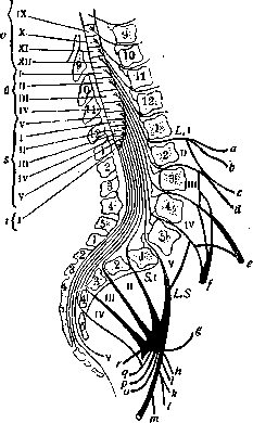
Anatomical relationship between the segments of the spinal cord and the vertebral bodies, as well as between the spinous processes and the exit of the roots. Root structure of the plexuses: v, u, s, t--thoracic, lumbar, sacral, coccygeal segments; r--nerve for m. levator ani; q--for m. obturat. int.; p--for m. sphincter ani ext.; o--n. pudendus; n--n. ischiadicus; m--n. cutan. femor. post.; l--nerve for m. quadrat. femor.; h--for m. gemellus inf.; i--for m. gemell. sup.; k--for m. piriformis; g--for m. gluteus sup.; f--n. obturator.; e--n. femoris; d--n. genito-femoralis; c--n. cutan. femor.; b--ram. cutan. lat. n. ilio-hypogastr.; a--n. ilio-hypogastr. (According to Dejerine.)
matter due to the fact that some descending tracts have already ended in higher sections (such as, for example, the direct pyramidal tract, fasc. vestibulo-spinalis), while many of the ascending tracts have not yet formed (Gowers' tract, Flechsig's tract); the posterior roots have not yet entered in sufficient quantity into the posterior columns of the spinal cord. The roots originating in this section of the spinal cord take part in the formation of the cauda equina; upon exiting the vertebral canal, they form, together with the fourth sacral root, the sacral plexus; from this plexus emerge the nerves innervating some muscles of the pelvic girdle and the sciatic nerve; thus, the epiconus controls the movements of the foot and partly of the lower leg, as well as the sensitivity of a significant part of the lower extremity (for the scheme of the segmental distribution of sensitivity, see Dermatomes). Disease of the epiconus gives a characteristic symptom complex, first isolated by Minor in 1900. The main and most frequent etiological factor is trauma—a fall from a height, extreme tension of the lumbar musculature when trying to lift a load or maintain balance during a forward fall; compression by a neoplasm, inflammatory processes, etc. are also possible. Autopsies are very few; in trauma, hemorrhages into the gray matter of the brain were found (see Haematomyelia). The onset of the disease is usually sudden; particularly persistent and severe paralyses are present in the region of the peroneal nerve; they are accompanied by atrophies and changes in electrical excitability; severe pain, sensory disturbances, sphincter disorders, and the absence of the Achilles tendon reflex are noted; the patellar reflex is almost always preserved, sometimes even increased. In the further course of the disease, rapid improvement sets out; first of all, sphincter functions are restored, then movements, but a more or less clearly expressed defect always remains in the region of the peroneal nerve, which indicates that the maximum of the lesion falls on the nuclei and roots of the peroneal nerve. In case the disease spreads to the medullary conus and cauda equina, the clinical picture is obscured by the overlay of new symptoms. Treatment depends on the etiological factor (see Spinal cord).
Related articles
Cite this page
“Epiconus.” Soviet Medical Encyclopedia. English translation of Bolshaya Meditsinskaya Entsiklopediya, 1st ed. (Moscow, 1928–1936), ed. N. A. Semashko. https://sovietmedicalencyclopedia.pages.dev/article/epiconus/