Electrodiagnosis
Historical document, translated for reference. It reflects medical knowledge of the 1920s–30s and is not medical advice.
Summary
Electrodiagnosis is the application of electrical energy for disease diagnosis, particularly of the nervous system and motor apparatus. It uses galvanic and faradic currents to assess nerve and muscle excitability, with quantitative and qualitative changes indicating various pathological conditions.
Encyclopedia article (1928–1936)
Electrodiagnosis, the application of electrical energy for the purpose of recognizing diseases. Electrodiagnosis has the greatest significance in diseases of the nervous system, particularly of the motor apparatus. For electrodiagnosis, galvanic and faradic currents are usually used. High-voltage currents and Franklinian sparks have no diagnostic value. The law of contraction formulated by Flügers, based on experimental bipolar irritation of an exposed nerve, encounters certain difficulties when applied to humans. With the monopolar irritation of the nerve used in the clinic, the current does not spread exclusively along the nerve but partly runs obliquely and across the nerve. The point of current entry is called the polar zone, and the point of exit is called the peripolar zone (Fig. 1). The current partially exits from the nerve not far from the point of application of the active pole. This opposite current is called the virtual current. When any point of the body is irritated by the cathode, the current tends toward the anode along several lines, spreading throughout the body—in current loops (Fig. 2). Nevertheless, the organ near which any pole is placed is under its predominant influence. This forms the basis of the monopolar method of research used in the clinic for electrodiagnostic purposes. Nerves and muscles are irritated by electric current through an active electrode at certain points corresponding to the most superficial location of nerves and the points of entry into muscles (Fig. 3 and 4). The passive indifferent large electrode is placed on a part of the body devoid of large nerves or muscles, for example, on the sternum or sacrum. With weak currents, contraction occurs only when the cathode is closed, since the irritating effect of the cathode on the nerve exceeds that of the anode. With medium-strength currents, contraction appears when the anode is opened and with almost the same current strength when it is closed; in the latter case, when the circuit is closed with the anode located on the active electrode, virtual cathodes come into play, which have a significant effect on the neuromuscular apparatus. With strong currents, irritation by the cathode causes tetanic contraction; the influence of the virtual anode, despite its relative weakness, is still sufficient for contraction to occur when the cathode is opened. In electrodiagnosis, the strength of current required to produce contraction, the pole, and the moments of closing and opening, as well as the nature of the muscular response, are taken into account. For judgment of normal and deviations from it, Stinzing's tables are used, which provide the average values of excitability for each nerve and muscle, the extreme highest and lowest figures that still lie within normal limits, and the difference in excitability between nerves and muscles of both sides. For example, the lower limit of galvanic excitability of the facial nerve is 0.8 µA, the upper limit is 2.8, and the average value is 1.75; the normal figures for galvanic excitability of the common extensor of the fingers of the hand range from 0.6 to 3.0 µA. Changes in the strength of current capable of causing contractions constitute the quantitative aspect of electrodiagnostic research and have particular significance in the investigation of nerves. Changes in the nature of muscular contraction itself are revealed primarily upon irritation of muscles and form the qualitative aspect.
Figure 2. Distribution of current through the human body. (According to Rleger.)
A decrease in nerve excitability is expressed by three phenomena: 1) to cause minimal contraction, currents of significantly greater strength than normal are required, 2) the strongest currents cause only weak contractions, 3) some elements of the Breinor formula (see Brenner-Erba formula) do not occur even with the strongest currents (for example, KZS).
In neurogenic lesions of the motor apparatus, the data from electrodiagnosis constitute distinguishing features of flaccid and spastic paralysis. First, the law of contraction formulated by Flügers is checked for an individual nerve and muscle. For this, the same nerve or muscle point is first acted upon by a faradic current, and then alternately by the cathode and anode of a galvanic current at the same current strength. Thus, the minimal contraction obtained from irritation is established, and the weakest current necessary for such contraction serves as the electrodiagnostic measure. In pathological cases, when irritated by the same pole of the electric current, quantitative changes in excitability are mainly discovered; qualitative changes are revealed when the poles are changed. The combination of qualitative and quantitative changes is called the reaction of degeneration. It occurs when the connection between the neuromuscular apparatus and its trophic center is disrupted, i.e., in diseases of the peripheral neuron. Conversely, as a rule, the reaction of degeneration is not observed in primary diseases of the musculature and lesions of the central neuron. In the reaction of degeneration, the excitability of the nerve to both currents and the faradic excitability of the muscles decrease, while the galvanic excitability of the muscle in the first period of the reaction of degeneration may even be increased and only then falls. The most important sign of the reaction of degeneration is the sluggishness of muscular contraction. In the severe form of the reaction of degeneration, the nerve completely loses its excitability; the faradic excitability of the muscle also disappears, and only its galvanic excitability to strong currents remains. The Brenner formula in the reaction of degeneration of moderate and severe degree often changes; AZS and KRS approach KZS and ARS in strength. The latent period increases with sluggish contraction up to four times. In normal conditions, the greater the muscle contraction at the same strength of irritation, the closer the irritant is applied to the point of nerve entry (Ranvier's motor points);
23 .22
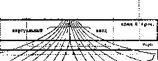
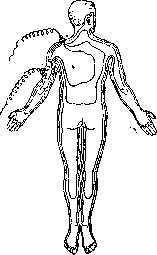
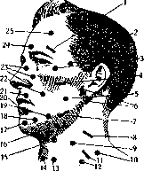
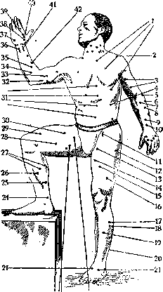
Figure 4. Diagram of motor points on the trunk and limbs: 1- m. pectoralis; 2-m. deltoideus; 3- m. biceps; 4- m. obliquus abdominis externus; 5-m. supinator longus; 6-m. extensor carpi ulnaris; 7-m. flexor carpi radialis; 8-m. extensor digitorum communis; 9-m. extensor indicis; 10-m. extensor pollicis longus; 11-n. femoralis; 12-m. tensor fasciae latae; 13-m. sartorius; 14-a. obturatorius; 15-m. adductor longus; 16-m. vastus lateralis; 17-m. peroneus longus; 18-m. gastrocnemius lateralis; 19-m. tibialis ant. et extensor dig. communis longus; 20-m. extensor hallucis longus; 21-m. extensor digitorum communis brevis; 22-m. interosseus dorsalis I; 23-m. sartorius; 24-n. obturatorius; 25-n. tibialis; 26-m. flexor digitorum communis longus; 27-m. gastrocnemii; 28-m. adductor longus; 29-m. vastus medialis; 30-m. rectus femoris; 31-m. rectus abdominis; 32-n. ulnaris; 33-n. medianus; 34-m. flexor digitorum profundus; 35-m. flexor digitorum sublimis; 36-n. ulnaris; 37-m. flexor digiti minimi; 38-m. abductor pollicis; 39-m. adductor pollicis brevis; 40-m. opponens pollicis; 41-n. medianus; 42-m. flexor carpi radialis. In reactions of degeneration, sometimes there is a displacement of motor points (the Wertheim-Salomonson phenomenon). Besides its diagnostic significance, the reaction of degeneration also plays a major role in prognosis, formulated by Erb in the following propositions. With the same disease form and the cause producing it, the more significant the lesion, the longer the duration of the disease, the less favorable the outcome, the more pronounced the reaction of degeneration. A partial reaction of degeneration gives a better prognosis than a complete one. The reaction of degeneration in the late stage of the disease is prognostically more unfavorable than in the early stage. Increased excitability as an independent phenomenon is observed in tetany, where sometimes contraction occurs with AR and KR with a current below 5 μA (the Mann-Timikh phenomenon). The neurotonic reaction (the reaction of Remak and Marin) consists in the appearance of tetanus upon irritation of the nerve with weak currents not only with CC but also with AC. The neurotonic reaction was observed in muscular dystrophy, epidemic encephalitis (Krol).-Myasthenic reaction (Jolly) consists in the gradual extinction of muscular excitability with successive direct and indirect irritations. After a short pause, the previous excitability is restored. With repeated irritation, the sequential extinction is repeated again. The myasthenic reaction is encountered in myasthenia, obliterating endarteritis, epidemic encephalitis.-The cadaveric reaction consists in the complete disappearance of excitability to both currents, occurring during an attack of periodic paralysis of the limbs. The myatonic reaction consists in a decrease in faradic excitability of the muscles with preservation of galvanic excitability. It is sometimes encountered in congenital amyotonia (myatonia).-In the myotonic reaction, the excitability of nerves for medium currents is normal. The electrical excitability of muscles is sharply increased for both currents. With faradization of the muscle even with a weak current, a prolonged contraction occurs. With a galvanic current, the contraction is very sluggish, persists after breaking the circuit and slowly subsides. With prolonged passage of current through the muscle, rhythmic contractions appear in it, spreading from the cathode to the anode. Sometimes they also appear upon irritation of nerves with a strong current. The myotonic reaction occurs in myotonia, epidemic encephalitis and syringomyelia.-The reaction with a gap (Benedict) consists in the fact that upon irritation with a galvanic current, single contractions occur, which do not appear with subsequent irritants; to obtain a contraction, increasingly stronger currents become necessary. The reaction with a gap was noted in progressive muscular dystrophy. In the longitudinal reaction, the muscle responds with a contraction to a galvanic current only when the current is passed along it. The active electrode is placed on the tendon of the muscle being examined and even away from it. Sometimes the longitudinal method is used in cases of loss of faradic excitability of the muscle in the reaction of degeneration. By passing a galvanic current along the entire limb, it is sometimes possible to simultaneously cause a contraction of the muscle with a strong faradic current. The basis of the longitudinal reaction is the property of a degenerating muscle to lose excitability first at the points where the nerve enters it and last in its terminal parts. The motor effect in the longitudinal reaction is sometimes more extensive than in the reaction of degeneration. Sometimes the longitudinal reaction for a long time replaces the reaction of degeneration. E. in application to changes in sensitivity has much less significance than in changes of the motor apparatus. In the examination of the latter, E. operates on the contraction of the musculature, subject to observation and precise measurement; in E. of sensitivity, the results are controlled only by the subjective sensations of the subject. According to the principle of specific energy, although an adequate stimulus, the electric current causes specific forms of sensation from the sense organs themselves and their nerves. Therefore, in the electrical examination of such nerves as the optic, auditory, gustatory, olfactory, it is impossible to distinguish between irritation of the sense organ itself and its nerves. Somewhat greater significance is attached to E. of cutaneous nerves. Faradic cutaneous sensitivity does not always run parallel with other forms of sensitivity. Its pathways and endings are not exactly established. The electro-myesthetic sensitivity of Duchenne in the form of the painful sensation of muscle contraction is not entirely identical with the sensation of ordinary muscle contraction. Similar to the figures of Stintzing's table for motor nerves, Hoffmann established the minimum current figures necessary to obtain a sensation upon irritation of sensory nerves. A sensitive reaction of degeneration, analogous to the motor one, is sometimes observed in tabes dorsalis and herpes zoster. Faradic cutaneous sensitivity is absent or diminished in it, galvanic sensitivity may be increased, the anode is somewhat more painful than the cathode. Irritation with a faradic current of a nerve exposed during operation or as a result of trauma causes a sensation of itching. A galvanic current passed through an exposed nerve produces a sensation of heat; with simultaneous passage of galvanic and faradic current, a sensation of pressure appears. This sensation is sometimes strictly localized in the area of the irritated nerve, sometimes it extends beyond its limits.-For diagnostic purposes, the degree of resistance of the skin to electric current is used. Increased resistance is characteristic of Basedow's disease, more rarely it occurs in hysteria and traumatic neurosis. Increased resistance is observed in scleroderma, myxedema, elephantiasis. The resistance of the skin to current is determined by its moisture, depending on vascular factors and the degree of perspiration. Electrodiagnosis on the brain exposed during operation is used to determine individual motor and sensory centers.
Related articles
Mentioned in
Cite this page
“Electrodiagnosis.” Soviet Medical Encyclopedia. English translation of Bolshaya Meditsinskaya Entsiklopediya, 1st ed. (Moscow, 1928–1936), ed. N. A. Semashko. https://sovietmedicalencyclopedia.pages.dev/article/electrodiagnosis/