Glaucoma
Historical document, translated for reference. It reflects medical knowledge of the 1920s–30s and is not medical advice.
Summary
An overview of glaucoma from the 1930s Soviet medical encyclopedia, detailing its primary symptom of elevated intraocular pressure, clinical forms such as inflammatory and simple glaucoma, and associated pathological changes.
Encyclopedia article (1928–1936)
GLAUCOMA, glaucoma (from the Greek glaucos—shining, bluish-green color of the sea), is one of the most complex diseases of the eye, not so much in its clinical picture as in its etiology and pathogenesis. The essence of the disease consists in an increase in intraocular pressure (tensio—Tn). Left to itself, glaucoma always leads to total loss of vision. Various factors play a role in the increase of pressure, and therefore glaucoma cannot be considered a disease in the proper sense of the word, but rather a concept uniting a whole group of diseases based on increased intraocular pressure. Large fluctuations in intraocular pressure in terms of the degree of elevation, rapidity of onset, duration, as well as the causes of the elevation, entail a diverse clinical picture of this disease. In a number of cases, there is an elevation of pressure due to some other disease of the eye, i.e., secondary glaucoma. If the cause of the pressure elevation is unknown, it is called primary glaucoma. The elevation of pressure in secondary glaucoma constitutes a complication of a pre-existing disease just as in primary glaucoma, and leads to the consequences characteristic of this disease. Eye diseases that usually lead to an increase in intraocular pressure include: occlusio pupillae, leucoma corneae adhaerens, luxatio lentis, intraocular tumors, etc. As further research into the pathogenesis of glaucoma progresses, the boundaries between these two forms will become blurred. It is even more difficult to draw precise boundaries between the various clinical forms of primary glaucoma, since many transitional forms exist in life. Nevertheless, two clinical forms of glaucoma can be distinguished that differ sharply from each other: glaucoma inflammatorium (inflammatory glaucoma) and glaucoma simplex (simple glaucoma). Inflammatory glaucoma is characterized by attacks of lesser or greater intensity occurring at various intervals. A sudden increase in intraocular pressure entails severe pain in the eye, which radiates along the first and second branches of the trigeminal nerve. Patients complain of headaches, sometimes completely unaware that the pain originates from the eye. Their general condition is poor, appetite is absent, and vomiting sometimes occurs. Vision drops sharply during the attack, the visual field narrows, and eyelid edema, chemosis, and conjunctival hyperemia appear. The cornea becomes edematous, having the appearance of frosted, misted glass; the pupil dilates, its reaction is absent, and the anterior chamber becomes shallow. The attack lasts several hours, days, or a week, after which improvement occurs, but vision does not always return to the state it was in before the attack. Attacks may differ in intensity, duration, and frequency of occurrence. Sometimes patients complain only of periodic blurring of vision, rainbow halos, and mild pain. Such mild attacks may appear several times a day, once a week, or less often. In other cases, attacks acquire an acute form, as described above. In the intervals between them, the eye has its usual appearance with the general signs of glaucoma, which are discussed below. This clinical form of glaucoma is called glaucoma inflammatorium acutum during the attack and chronicum in the intervals between them. Glaucoma with mildly expressed attacks also refers to chronic inflammatory glaucoma. In contrast to the form described above, another is encountered that creeps up unnoticed on the patient, all the more so since it affects one eye first and, with the good vision of the other, remains unnoticed by the patient for a long time. Simple glaucoma is characterized mainly by changes in the posterior segment of the eye, a drop in vision, and a slight increase in intraocular pressure, which previously remained unnoticed, as a result of which simple glaucoma was then regarded by some ophthalmologists as optic nerve atrophy with cupping of the disc. Simple glaucoma is diagnosed rather on the basis of visual impairment and the ophthalmoscopic picture of the fundus. Its course is slow, almost painless, and therefore patients consult a doctor only when the visual impairment becomes more noticeable. According to the new terminology, glaucoma with inflammatory signs in the anterior segment of the eye is called uncompensated glaucoma, and without them—compensated. Clinically, as indicated above, all kinds of transitions from simple glaucoma to inflammatory are observed, and therefore forms are often encountered in practice that are difficult to assign definitively to one or another group of this disease. Having begun as simple glaucoma, the course subsequently acquires an inflammatory, uncompensated character. All this indicates that we are dealing with one and the same cause, i.e., an increase in intraocular pressure. The main symptom of glaucoma is elevated intraocular pressure. This symptom is present in all forms of glaucoma. Normal intraocular pressure, expressed in millimeters of mercury, lies between 18 and 30 mm. However, exclusive significance should not always be attached to absolute pressure figures, and in cases where the intraocular pressure figure does not exceed the norm, it is necessary to pay attention: 1) to the difference in Tn in both eyes: normally the figures are almost identical, the difference should not exceed 2–21/2 mm, and therefore an intraocular pressure of 25 mm in one eye may appear suspicious in the presence of 18 mm in the other; 2) to daily fluctuations, the swings of which should also not exceed 2–21/2 mm normally, and 3) to fluctuations in Tn from various influences (psychic, medicinal), i.e., in other words, monitoring the stability of intraocular pressure is necessary, since a disorder in this regard in itself indicates a pathological state of Tn. Plotting daily curves of intraocular pressure sometimes plays a major role in diagnosing glaucoma. Tn, which is normal during the day, shows a sharp rise during the hours of its highest elevation (6–8 a.m.), and it is quite possible that many glaucomas proceeding as if with normal Tn show an elevation in the period least convenient for examination (night, early morning). The study of subtleties in intraocular pressure fluctuations has dispelled the notion of the existence of glaucoma without elevated Tn. All other symptoms of glaucoma can also be explained by the increase in intraocular pressure. When it exists for a long time, one of the important symptoms is excavation, a deepening of the optic nerve head due to the backward displacement of the lamina cribrosa, i.e., that part of the sclera which is located
at the site of entry of the optic nerve, is pierced by numerous openings, and therefore yields first of all to the increased intraocular pressure. Upon ophthalmoscopy, the optic nerve head appears depressed compared to the neighboring parts, which is recognized by the bending of the vessels at the place where they pass from the disc to the retina [see Figure 1 and the color table (pp. 91–92), Fig. 1]. The bending of the nerve fibers together with high pressure causes their atrophy, which imparts a pale gray-white color to the disc and is the main cause of the decrease in visual

acuity. Impairment of peripheral vision also occurs quite early. Characteristically, there is a constriction of the visual field from the nasal side, i.e., loss of function of the temporal part of the retina, which is explained by its greatest distance from the optic nerve head, i.e., from the entry into the eye of the vessel nourishing the retina. One of the earliest symptoms, pointed out by Bjerrum, must be considered the enlargement of the blind spot, which acquires a peculiar vertical-crescentic shape. This enlargement is often a great aid for
Figure 1. Glaucomatous excavation.
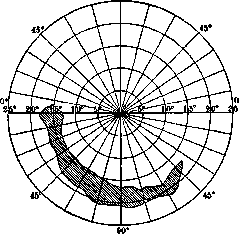
Figure 2. Crescentic shape of the blind spot.
diagnosis of glaucoma in doubtful cases (see figure 2). In addition to the above-described main symptoms of glaucoma, there is also a whole series of secondary ones that may occur or be absent depending on the form and course of the disease. The most frequent include: dilation of the ciliary veins of the eyeball, corneal edema, a shallow anterior chamber, a wide pupil, and the absence of its reaction to light. Among subjective symptoms, one must note halos around lights and pain. Glaucoma in which vision is completely absent is termed glaucoma absolutum. In the subsequent course, degenerative changes set in within the blinded eye, called glaucomatous degeneration. The cornea becomes cloudy, gets covered with vitreous deposits, the corneal epithelium is lifted up, blisters form (keratitis bullosa), sometimes the cornea ulcerates, and the lens becomes cloudy. Severe pain in such an eye is an indication for surgical intervention, namely neurectomia optico-ciliaris, i. e., excision of a piece of the optic nerve with the ciliary nerves, leaving the eye in place. The entire symptomatology of glaucoma is fully explained by the elevation of intraocular pressure, while the cause of the latter remains unclear. No single theory can yet explain all cases of glaucoma, and meanwhile, knowing the cause of the elevation of intraocular pressure means unraveling the essence of glaucoma. Most ophthalmologists divide all theories of the origin of glaucoma into two groups: the first includes theories that see the cause of glaucoma in an increased influx of fluid into the eye (secretory), and the second includes those theories that see the cause in a hindered outflow of fluid (retention). Of course, it is difficult to fit all theories into any strictly limited classification, but some base the division of theories into groups on a different principle: one group includes all theories considering that the cause of glaucoma lies within the eye itself, and the second includes theories that place it outside the eye. Among the hypotheses of the second group deserving the most attention is the theory that links glaucoma with changes in the angle of the anterior chamber, which occupies an important place in the process of fluid outflow from the eye. Knies found in the glaucomatous eyes he examined a closure of the spaces of Fontana due to the adhesion of the root of the iris to the cornea. This obliteration of the spaces of Fontana is, in his opinion, the cause of the origin of glaucoma. Weber holds the same opinion. The difference between him and Knies lies in their understanding of the cause of the obstruction to outflow from the anterior chamber:
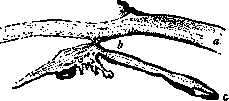
Figure 3. Normal angle of the anterior chamber: a - cornea; b - angle of the anterior chamber; c - iris.
Knies sees it in adhesive inflammation, and Weber in the pressing of the root of the iris against the cornea due to the swelling of the ciliary body (see figures 3 and 4). Confirmation of this theory is experimental glaucoma, which can be caused by the injection of provencal oil or india ink into the anterior chamber. Recently, in connection with the invention of the slit lamp, new data have been obtained in favor of the Knies-Weber theory. Koeppe, engaged in vital microscopy of the eye, noted an interesting phenomenon in glaucomatous eyes, namely, the displacement of retinal pigment and the blockage of the angle of the anterior chamber of the eye by it. The Knies-Weber theory and Koeppe's pigmentary glaucoma are typical of the retention theory. The secretory theory was proposed by Graefe, who saw the cause of glaucoma in hypersecretion of the uveal tract due to irritation of the secretory nerves entering it. Among modern theories, Seidel's secretory theory must be included here, as he assigns the primary role in the secretion of intraocular fluid to the epithelial cells of the ciliary body. But what role the epithelium of the ciliary body plays in the origin of glaucoma is difficult to say, since its secretory function has not yet been fully proven at all. As for the theories placing the cause of glaucoma outside the eye, it must be said that the adherents of these theories by no means neglect the processes taking place within the eye: great importance is attributed to them, but
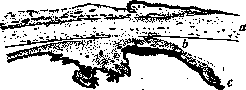
Figure 4. Angle of the anterior chamber in glaucoma: a - cornea; b - angle of the anterior chamber; c - iris.
The primary cause of glaucoma is placed outside the eye, with changes in blood circulation and vascular walls taking first place. The question of the role of circulation in the etiology of glaucoma is not new. Graefe, seeing the cause of glaucoma in serous choroiditis, thereby already emphasized the major role of the vascular system in the pathogenesis of this disease. The most interesting question has been the relationship between intraocular pressure and general blood pressure, and the role of the latter in the origin of glaucoma. A whole series of works has been devoted to this question, but ophthalmologists have not reached a unanimous conclusion. Thus, Wessely saw a direct connection between intraocular pressure and general blood pressure. The same opinion is held by Klezkowsky, who observed an increased blood pressure in all glaucomatous patients. Kümmell and Gilbert noted an increase in only 75%. On the other hand, Römer, Magitot, and Elschnig see no direct connection between these phenomena. All discrepancies in the results of various researchers indicate the absence of a direct link between intraocular and general blood pressure. Having failed to find the cause of glaucoma in elevated blood pressure, some ophthalmologists turned their attention to the composition of the blood. Hertel began to attach special importance to blood concentration after discovering its decrease in all cases of glaucoma. A lowered blood concentration disturbs the isotonicity between it and the tissues. Fluid begins to exude from the vessels, the amount of fluid in the closed cavity—the eye—increases, and intraocular pressure rises. According to the studies of N. A. Pletneva, a decrease in concentration was observed in only 20%, but the intravenous administration of a concentrated salt solution (10% NaCl) after some time (20–30 minutes) lowered the intraocular pressure by a significant amount. The low intraocular pressure did not last long, and the intraocular pressure soon reached its initial height. Subsequently, Hertel somewhat complicated his view on the origin of glaucoma. He based it no longer on low blood concentration, but on changes in the function of the vascular wall. In recent times, the question of the role of circulation in the origin of glaucoma has been shifted to a somewhat different plane: great importance has begun to be attached to the peripheral system, since all vital processes between tissues and blood take place in it, while veins and arteries are merely tubes bringing blood to and carrying it away from the capillaries. The lack of equilibrium (dysergia) of the vascular system occurs in many cases of glaucoma and is often its cause. If the enormous importance of peripheral circulation, both local and general, in the origin of glaucoma is not exaggerated, the question of the cause of this disorder arises first of all. Where does this lability of the vascular wall come from? It is quite possible that the constitutional factor should be placed in first place, as well as disorders of internal secretion, which plays no small role in maintaining the tone of the vascular wall. In the clinic, there are many facts confirming the lack of equilibrium on the part of the vascular system in glaucomatous patients. First of all, one must note the highest incidence of glaucoma during the climacteric period in women, i.e., in the period when the vascular system is in the least equilibrium. The lability of the vascular wall also explains those large fluctuations in intraocular pressure shown by glaucomatous patients, and the attacks of glaucoma arising from the consumption of alcoholic beverages, sexual excesses, sleepless nights, and various psychological influences. The infatuation with chemistry has recently penetrated into ophthalmology as well. Meesmann observed an increased alkalinity of the blood in glaucomatous patients, which gave him the right to attach great importance to hydrogen ion concentration in the etiology of this disease. The low intraocular pressure in pregnant women, in whom acidosis is observed, seems to confirm the stated point of view, but on the other hand, a number of researchers, such as Schmerl, have not obtained similar data, and the question of the role of pH in the etiology of glaucoma remains open for now. There is an endless number of theories striving to explain the origin of glaucoma, but none is capable of explaining all cases of glaucoma. The question of the pathogenesis of glaucoma is far from resolved, and meanwhile its resolution has great, not only theoretical, but also practical significance, since until the pathogenesis of this disease is clear, treatment cannot be directed against the primary cause, but is rather symptomatic. All measures in glaucoma have the purpose of lowering intraocular pressure. Glaucoma was considered an incurable disease until Graefe in 1856 proposed iridectomy. The success of this operation forced many ophthalmologists to hold the view that all cases of glaucoma must be operated on, and the earlier the better. Most oculists adhere to this view, but it must be said that recently the enthusiasm for surgical treatment has greatly weakened due to the fact that the operation in many cases, which relate mainly to simple glaucoma, does not achieve its goal. Every ophthalmologist has in his memory more than one case when a patient, having appeared to him with a visus of a few tenths, leaves him after the operation with hundredths. The best effect from surgical treatment is obtained in cases of acute glaucoma; in chronic or simple glaucoma, according to Römer's statistics, an arrest of the process occurs in 33.7%, and in 66.3% there is a deterioration. Schmidt-Rimpler points out that in acute glaucoma, improvement from iridectomy was observed in 80% of cases, in chronic glaucoma—somewhat more than 50%, and in simple glaucoma—in 33%. These figures clearly show that good results of surgical treatment occur only in acute glaucoma. Dealing with simple glaucoma, before proposing surgery to the patient, many circumstances should be weighed. One can easily decide on an operation on the first eye; in the case of disease of the second, the question of treatment methods becomes significantly more complicated, and before proposing surgical treatment, it is in any case desirable to try the entire stock of conservative means. First place is occupied by miotic agents, such as pilocarpine and eserine. Causing pupillary constriction, they restore the outflow of fluid from the eye, which is hindered by the pressing of the root of the iris against the cornea. Recently, adrenalin therapy (glaucosan) has become widespread. Linking the etiology of glaucoma with a decrease in the tone of the sympathetic nerve (Hamburger), they quite naturally turned to adrenalin as an agent toning up the sympathetic nervous system (see Glaucosan). The desire to find the cause of glaucoma in a disease of the entire organism led to an infatuation with general therapeutic agents, such as pituitary preparations, ovarian preparations, intravenous infusion of CaCl2, and the use of Gynergen. With the development of the idea of the connection of glaucoma with a disease of the entire organism, general therapy is growing. For now, it must be recognized that conservative treatment very often fails to achieve its goal, and in these cases, one has to resort to surgical treatment. Among operations for glaucoma, iridectomy, scleral trephining according to Elliot, cyclodialysis, iridencleisis, and sclerotomy should be noted. Hydrophthalmus represents glaucoma of childhood. Elevated intraocular pressure stretches the elastic young sclera, and the eye greatly increases in size (buphthalmus—ox eye). The cornea becomes larger, very often dull and clouded, the anterior chamber deep, the intraocular pressure elevated, and glaucomatous excavation is sharply expressed on the fundus. The disease is either congenital or develops in the first years of life and almost always in both eyes. In most cases, the cause is developmental anomalies, mainly the absence of Schlemm's canal. Treatment is mostly fruitless, since neither conservative nor surgical methods can restore the pathways of outflow from the eye.
Related articles
Mentioned in
Cite this page
“Glaucoma.” Soviet Medical Encyclopedia. English translation of Bolshaya Meditsinskaya Entsiklopediya, 1st ed. (Moscow, 1928–1936), ed. N. A. Semashko. https://sovietmedicalencyclopedia.pages.dev/article/glaucoma/