Canthoplasty
Historical document, translated for reference. It reflects medical knowledge of the 1920s–30s and is not medical advice.
Summary
Canthoplasty is a surgical procedure to lengthen the palpebral fissure, primarily at the outer angle of the eye. This 1930s article details various techniques for the operation, noting its frequent use in the USSR for treating conditions like trachoma.
Encyclopedia article (1928–1936)
CANTHOPLASTY (from the Greek canthos—angle and plasso—I form), plastic surgery of the angle of the palpebral fissure. In the narrow sense, canthoplasty is understood as an operation to lengthen the palpebral fissure, mainly in the region of its outer angle. Internal canthoplasty is performed extremely rarely in the form of dissecting scars or pathological skin folds at the inner canthus and has nothing typical about it. A distinction is made between canthotomy—a simple horizontal dissection of the outer canthus of the eyelids without suturing—as a provisional procedure, and canthoplasty proper as a completed operative method designed for a lasting effect. Simple canthotomy is used: a) as a preliminary act in operations on the eyeball (cataract, glaucoma) to facilitate access to it and weaken the tone of the orbicularis oculi muscle; b) during enucleation and exenteration of the orbit for freer removal of the bulbus and orbital contents through a narrow palpebral fissure; c) in severe acute conjunctivitis (gonorrhea, diphtheria) to weaken the pressure of swollen, inflamed eyelids on the eyeball, and also as bloodletting for anti-inflammatory purposes; d) sometimes in severe corneal lesions with strong blepharospasm. Nowadays, simple canthotomy is generally performed rarely, and usually, following the dissection of the outer canthus, sutures are carefully applied, i.e., canthoplasty is performed. The indications for canthoplasty are primarily the same as for canthotomy, which it fully replaces as a more perfect procedure. Canthoplasty is recommended for spastic entropion and for all forms of congenital and acquired (burns, wounds) narrowing of the palpebral fissure. But it finds especially wide application in trachoma, either as an independent operation to eliminate blepharophimosis, ankyloblepharon, or as part of a more complex operation against trichiasis, entropion of the eyelids.—Technique of the operation (v. Ammon; 1839). Local anesthesia with 2% novocaine-adrenaline in the region of the outer canthus and the outer third of the upper and lower eyelid (in children—general anesthesia). With the thumb and index finger of the left hand, the canthus of the eyelids is stretched, pulling it at the same time slightly toward the nose. Under the bridge formed in this way, one blade of straight blunt-pointed scissors is inserted into the conjunctival sac, they are positioned in the direction of the palpebral fissure, holding their blade perpendicular to the skin surface of the canthus, and with one stroke, the outer angle is dissected to the bony edge of the orbit (Fig. 1). The incision of 10–15 mm passes through the skin, the orbicularis oculi muscle (or rather, through its tendinous expansion), and the conjunctiva and is accompanied by fairly significant bleeding from the arcus palpebralis, which soon stops. A rhombic-shaped wound is obtained, the inner edges of which are formed by the conjunctiva, the outer ones by the skin. The conjunctiva is dissected in the direction of the eyeball (Fig. 2) and the skin edges are joined.

Figure 1.
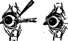
Figure 2.
Figure 3. of the wound with three conjunctival sutures: the middle suture, forming the new angle of the palpebral fissure, grasps the medial and lateral angles of the rhombus (Fig. 3). Two other sutures—above and below—pass in the region of the upper and lower eyelids. The insertion of a blepharostat after canthotomy facilitates the dissection of the conjunctiva and the application of sutures. An aseptic dressing is applied with the insertion of a gauze compress smeared with boric vaseline into the newly formed angle of the palpebral fissure. The sutures are removed on the 3rd–4th day. The operation performed in this way is not always satisfactory. Already during the operation, after the dissection of the canthus, there is not enough relaxation of the muscular, and especially the fascial, ring surrounding the palpebral fissure. Agnew (1875) proposed, after the described dissection of the canthus, an additional section of the tarso-orbital fascia in the vertical direction along the outer edge of the orbit in the upper eyelid. The same incision of the fascia is also carried out downward from the main incision in the lower eyelid. Furthermore, the fragile, scar-shortened conjunctiva easily tears when the main middle suture is applied and tightened, the edges of the incision grow together again,

Figure 4.
And the effect of the operation in terms of its duration is reduced to naught. Therefore, Heuse (1874) and Czermak (1894) facilitate the pulling of the conjunctiva to the angle of the skin wound with a relaxing incision of the conjunctiva bulbi near the cornea (Fig. 4). Pick (1908) achieves the same with two incisions of the conjunctiva, running from the angle of the palpebral fissure to the eyeball and forming a triangular


Figure 5.
flap of conjunctiva, which is then drawn into the angle of the skin incision. But these and similar procedures (de Vincentiis; 1928) do not always help with severe cicatricial shrinkage of the conjunctival sac (trachoma, burns). In these cases, the best results in terms of durability and radicalism of action are provided by the method of Kuhnt (1906), who proposed closing the wide wound after canthotomy with pedicled skin flaps from the lower eyelid (Fig. 5, 6) or from the temple, or with free Thiersch grafts, as well as with mucous membrane from the lip. Blaskovics, on the contrary, attempts to achieve lengthening of the palpebral fissure by pulling the outer canthus toward the temple, for which he adds to the usual incision of the canthus an excision of a triangular section of skin 2 mm from the canthus. The apex of the V is downward, and the base, 7–8 mm long, runs horizontally in the direction of the palpebral fissure. Thus, canthoplasty can be performed by various methods adapted to each particular case. In the USSR, due to the widespread prevalence of trachoma, canthoplasty is one of the frequent eye operations.
Related articles
Cite this page
“Canthoplasty.” Soviet Medical Encyclopedia. English translation of Bolshaya Meditsinskaya Entsiklopediya, 1st ed. (Moscow, 1928–1936), ed. N. A. Semashko. https://sovietmedicalencyclopedia.pages.dev/article/canthoplasty/