Vestigial Organs
Historical document, translated for reference. It reflects medical knowledge of the 1920s–30s and is not medical advice.
Summary
Vestigial organs are underdeveloped structures whose complexity exceeds their apparent function. Their study provides crucial evidence for evolutionary theory, showing how organs lose their primary function over time while sometimes acquiring new roles.
Encyclopedia article (1928–1936)
Vestigial organs, underdeveloped organs. Their structure, despite relative simplicity, is still more complex than would seem to be required by the function they perform. The significance of V.O. is revealed by comparing this organism with other closely related organisms, in which the homologous organ is fully developed and functions normally. Therefore, it is natural to conclude that the structure of V.O. is determined primarily by its history. From this follows the second definition of V.O. as an organ that has lost its main function. The study of V.O. provided the most convincing evidence for the historical validity of organic structures and had extremely great importance for establishing the theory of evolution. The first evolutionists, such as Geoffroy Saint-Hilaire, already paid great attention to them. Subsequently, the study of V.O. became connected with a whole series of particular problems of evolution, such as the question of the inheritance of the results of exercise and non-exercise, and the question of the causes of rudimentation and the complete disappearance of organs in general, which at one time served as material for the historical discussion between H. Spencer and A. Weismann. Vestigial organs may perform some secondary functions, may acquire some new functions that then protect them from disappearance, but in some cases may be devoid of an obvious specific function. In some cases, rudiments of organs perform certain functions in the embryo or larva and are then preserved as 'provisional' organs of temporary significance. Since each organism in its historical development transforms all its parts in accordance with the continuously changing environment, V.O. and parts of organs as survivals of passed stages exist in greater or lesser numbers in any organism. Here we will indicate only some more striking examples. In the skeleton of all vertebrates, a provisional central axis-the notochord or chorda-develops embryologically, which is the permanent axial skeleton of lower chordates. In many adult vertebrates, rudiments of this chorda are preserved in the form of gelatinous masses between the vertebrae (in most fish), inside the vertebrae (in many amphibians and some reptiles), or inside the intervertebral cartilages (nucleus gelatinosus of mammals). Ribs, initially uniformly developed along almost the entire spine, are in terrestrial vertebrates mainly limited to the 'thoracic' region. In the cervical region, they are reduced in size (free cervical ribs are still present, for example, in crocodiles) and their rudiments fuse with the vertebrae at their point of double attachment, which in birds and mammals leads to the formation of a perforated transverse process. In tailless amphibians, ribs are generally reduced and only in some forms are distinct rudiments of ribs preserved. In them as well as in birds, the caudal region of the spine is reduced (forming by fusion of vertebrae the 'urostyle' of frogs and the 'pygostyle' or coccygeal bone in birds). In lizard-like and snakes, limbs are reduced, whose rudiments are preserved in some forms as small appendages (hind limbs of the yellow-bellied snake, boas). More often in this case, remains of girdles are preserved (rudiments of the shoulder girdle and sternum in lizard-like lizards, rudiments of the pelvis in boas), as is also observed in mammals (rudimentary pelvis of whales and sirenians). In non-flying birds, there may be rudimentary wings and girdles (the New Zealand kiwi, the extinct 'moa'). In fast-running vertebrates, the marginal fingers are usually reduced. In mammals, the first finger is very often rudimentary, sometimes also the second and fifth (even-toed ungulates) or even the second and fourth (splint bones of horses). In many mammals, the clavicle is also reduced, and in all mammals, the coracoid bone is preserved as a rudiment attached to the scapula (coracoid process). The fibula is often reduced (birds, ungulates, etc.) and its rudiments are attached to the tibia. Of other organ systems, examples of V.O. can be given as the eyes of many parasitic, deep-sea, and especially underground and cave animals. Among vertebrates, one can name deep-sea hagfish, cave fish of America (Amblyopsidae), underground amphibians-gymnophions, the cave proteus (Proteus anguinus, burrowing snakes (Typhlopidae) and mammals (moles, and especially the Australian marsupial mole), in which rudimentary eyes are preserved at various stages of their reduction.
In many mammals, there are separate rudimentary teeth, sections of the dental system (incisors of sirenians, toothless mammals) or finally the entire dental system has the character of a provisional rudiment (monotremes, anteaters, toothless whales). Various internal organs also undergo rudimentation: the left lung in snakes, the right ovary and oviduct in birds, primary kidneys, etc. Often V.O. acquire a new function and in this case are preserved as already a different, fully functional organ. Such are, for example, in vertebrates the appendages of the testicle-remains of the primary kidney, such are also many endocrine organs; the epiphysis-rudiment of the unpaired eye, the thyroid gland-rudiment of the hypobranchial groove (endostyle) of lower chordates, the thymus gland, developing from the remains of gill slits, etc.-Such is also the significance of the auditory ossicles of mammals, which are fully developed organs that are at the same time rudiments of parts of the visceral skeleton: the stapes-rudiment of the suspensorium of fish, the incus-rudiment of the quadrate bone, and the malleus-rudiment of part of the lower jaw.
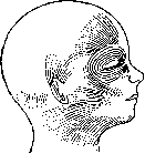
In humans, there are many organs typical of animals that have lost their original significance here and serve as documents testifying to his origin. Such are the remains of the body hair cover, usually inactive (in humans and anthropoid apes) muscles for moving the auricle and facial skin muscles (platysma) (Fig. 1), the caudal region of the spine (as in anthropoid apes), the remnant of the third eyelid, or nictitating membrane of the eye (plica semilunaris) with rudiments of the gland of Harder (caruncula lacrymalis) (Fig. 2), and sometimes also the rudiment of the muscle retracting the eyeball (m. retractor bulbi), the rudiment of Jacobson's organ, the palatal ridges of the newborn, Morgagni's ventricles of the larynx, which in many apes are in the form of vocal sacs, etc. The last molar tooth (wisdom tooth) in humans can also be classified as rudimentary. The vermiform appendix of the cecum, which is also found in apes and some other mammals, is also commonly designated as a rudiment.-Organs are reduced when they lose their biological significance. This may be the result of 1) changes in living conditions (many organs in humans), 2) replacement by other, more perfect organs (primary kidney, unpaired eye, notochord), 3) a sharp change in the function of certain organs (fingers and claws in the bird's wing). V.O. are characterized by great individual variability in all their parts, and their complete disappearance is obviously determined by underdevelopment due to disruption of the coordination of parts already in the embryo.
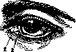
Figure 1. Skin muscles of the human head (platysma).
Figure 2. Human eye: 1-caruncula lacrymalis; 2-plica semilunaris.
Related articles
Mentioned in
Cite this page
“Vestigial Organs.” Soviet Medical Encyclopedia. English translation of Bolshaya Meditsinskaya Entsiklopediya, 1st ed. (Moscow, 1928–1936), ed. N. A. Semashko. https://sovietmedicalencyclopedia.pages.dev/article/vestigial-organs/