Amoebas
Historical document, translated for reference. It reflects medical knowledge of the 1920s–30s and is not medical advice.
Summary
Amoebas are microscopic animals belonging to the class Rhizopoda of the Protozoa type, characterized by their protoplasmic body and one or more nuclei. They move and capture food through pseudopodia and exhibit various forms depending on species and environmental conditions.
Encyclopedia article (1928–1936)
AMOEBAS (from Greek amoibe - change), or amoeboids (Amoebida), an order of microscopic animals belonging to the class Rhizopoda, type Protozoa; consist of a protoplasmic body and one or more nuclei. In the latter case, all nuclei are equivalent. Some amoebas normally have two equivalent nuclei (Pelomyxa binucleata, Sappinia diploidea, etc.), other A., so-called paramoebas, have one nucleus of the ordinary type, and in addition, another formation similar to a nucleus, the so-called "accessory" nucleus (Nebenkern). Of all rhizopods, A. are the simplest in structure. They are completely naked, i.e., they have no external skeleton, shells, etc. Their body is covered with an ectoplasmic layer capable of easily changing its shape due to the formation of pseudopodia (false feet), which can arise on any part of the body surface of A. and serve for their movement and for capturing food. This explains the variability of the body shape of amoebas, which is noted in their very name. However, despite such variability of form, the latter is quite characteristic of each given species. Thus, one distinguishes forms with long finger-like pseudopodia, e.g., A. proteus (see figure 1, a), forms with broad pseudopodia, e.g., A. polypodia, A. limax, Entamoeba histolytica (see figure 1, b, c, d), forms with pointed ray-like pseudopodia, e.g., A. radiosa (see figure 1, e). By artificially changing the degree of acidity or alkalinity of the environment, one can experimentally achieve a change in the external contours of A. The protoplasm of the body of A. represents a fluid mass, which, depending on various conditions, can take on a more or less viscous consistency. In the protoplasm of the body of A., two layers are usually clearly distinguished: an inner, more viscous, opaque, granular layer - the so-called endoplasm, and an outer layer covering it, more liquid, transparent, glassy, without any visible structure - the so-called ectoplasm. The superficial layer of the body of A., directly in contact with the environment, the so-called pellicle, is a denser layer of the same ectoplasm, while retaining its mobility and ability to change shape. These features can be most easily observed in the moment of movement of A. during the formation of pseudopodia, when new transparent ectoplasmic pseudopodia form before the observer's eyes, gradually acquiring the granular character of endoplasm, while at the new place a new ectoplasmic pseudopodium already forms (see figure 1, c, f; see also fig. b, c-k in the article Amoeboid movements). Thus, ecto- and endoplasm are certain colloidal states of the protoplasm of the body of A., which, depending on physiological, resp. physico-chemical, conditions, can pass into one another. The nucleus of A. is a spherical formation surrounded by a membrane. The internal structure of the nucleus varies greatly in different groups of A. The two most characteristic types of structure are: karyosome and reticular. The first type is distinguished by the presence in the center of the nucleus of a large body, the so-called karyosome (see figure 2 a). The second type is characterized by a reticular structure and a small karyosome (see figure 2 b). In many A., division of the nucleus occurs by typical mitosis, in others it proceeds by a different type; so-called promitosis.
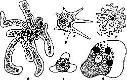
Figure 1. Various types of amoebas.
In addition to the processes of reproduction, the nucleus also plays a major role in the other manifestations of life of A. This is revealed in experiments with cutting A. into parts: parts deprived of nucleus lose the ability to ingest food and soon die. The movement of A. is connected with the formation of pseudopodia. (Details - see Amoeboid movements). Nutrition. A. feed mostly on live food, leading at the same time a predatory or parasitic mode of life. Saprophytic A. ingest organic residues into the body. In this process, pseudopodia and the superficial layer of the protoplasm of A. participate. One of the methods of food ingestion consists in its "flowing around" by protoplasm (see figure 3). In the body of A., food particles are digested inside the digestive vacuole with the help of corresponding enzymes. Respiration. Gas exchange occurs by osmotic means, i.e., over the entire surface of the body. In freshwater A., contractile vacuoles participate in this process - bubbles inside the body of A., rhythmically contracting and expelling water with gases dissolved in it to the outside.


Figure 2. a-nucleus of karyo-reticular type. so-called promitosis. b-nucleus in simplified form,
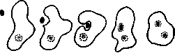
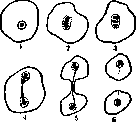
Cysts. Under the influence of sharp changes in the environment, during drying, increase in salt concentration or change in medium reaction - A. encyst, i.e., take on a spherical shape and secrete on their surface a shell impermeable to some substances and possibly semipermeable to others. In the form of such cysts (see figure 4) B.m.e.t. I. amoebas are largely protected from the harmful effects of the environment and even withstand a considerable degree of drying. Inside cysts in some A., asexual reproduction processes occur, and possibly sexual ones as well. Reproduction of A. occurs by division, which in the simplest case consists in division in two of A., in a free, non-encysted state (see figure 5). Simultaneous division into more than two daughter individuals usually occurs in cysts. A sexual process has also been described for some A., but most of these descriptions are unreliable. More precise information exists about the sexual process in Paramoeba chaetognathi, parasitic in the marine worm Sagitta, and in the saprophytic A. Sappinia diploidea. The first reproduces by division, but at the end of the asexual cycle forms flagellate gametes which copulate with each other. The second has two nuclei throughout the vegetative period and reproduces by division; during the sexual process, two 2-nucleate A. are covered by a common shell, after which both nuclei of each A. fuse in pairs, and from the cyst emerges a new 2-nucleate A. (see figure 6). In general, however, very little is known about the sexual reproduction of A.
Figure 3. Successive stages (1-5) of food ingestion by amoeba by flowing around. The food particle (black dot) is enveloped by pseudopodia (1-3) and enclosed in a digestive vacuole (4-5).
Cultures. Many saprophytic amoebas easily multiply on simple agar in mixed culture with bacteria. Cultures of parasitic amoebas and, in particular, Entamoeba histolytica, have been obtained on the medium of Beke and Drbohlav (Boesk and Drbohlav, 1925), consisting of a mixture of egg white or serum and Locke's solution.
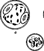
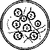
Figure 4. a and b-
cysts of Ent. coli; c-cyst of Ent. histolytica. Distribution. As free-living forms, as commensals and para
sites of various animals, amoebas are widely distributed throughout the world.
Parasitic amoebas are found, mainly, in the digestive tract of various animals, from where they can also enter other organs. Their coexistence can be accidental, when free-living amoebas, entering the host's intestine with water, adapt to living in it, without, however, losing the ability to live freely. Other forms are unconditional parasites and have adapted so deeply to living together with the host that they have completely lost the ability to exist outside its body. Among the latter, a distinction should be made between pathogenic parasites that live at the expense of the host's organism (for example, Entamceba histolytica), which feed on human erythrocytes and destroy its tissues, and harmless commensals that feed on their host's food (e.g., Entamoeba coli in the human intestine or Entamoeba suis in the pig intestine). The spread of parasitic forms of amoebas occurs through cysts excreted from the intestine of one host and swallowed by another host along with food or water. Most A. at all stages of their life move only by means of pseudopodia. Other amoebas have flagellated stages in their life cycle, or possess the ability to form flagella when environmental conditions change, for example the salt composition of the medium. Depending on the formation of flagellated stages in one or another family of amoebas and on the position of these stages in the general life cycle of a given family, the modern taxonomy of amoebas is constructed. Taxonomy of A. Order: amoeboid (Amoebida). Naked rhizopods without a skeleton. Possess amoeboid movements. In some A., flagellated stages occur: 1. Family Amoebidae. Have one nucleus. Never form flagellated stages (examples: freshwater A. proteus, parasitic Ent. histolytica, Ent. coli). 2. Family Paramoebidae. Have an 'accessory' nucleus, form small flagellated stages during reproduction (example: parasitic Par. chaetognathi). 3. Family Bistadiidae. Capable of transforming into flagellated forms, otherwise similar to family 1 (example: free-living Vahlkampfia bistadialis). 4. Family Rhizomastigidae. In the adult state, they constantly have one or more flagella, while also forming Figure 5. Sequential stages (1-6) of division of Amoeba Umax. Nucleus of the karyosome type; chromosomes are formed from the outer part of the nucleus. 6-the nucleus has divided, the protoplasm is constricted in the middle; two newly formed individuals. and pseudopodia. When encysting, they lose their flagella (example: free-living Mastigi-pa vitrea; parasitic M. hylae). Of the four families mentioned above, forms of importance in parasitology and human pathology belong to the family Amoebidae, which consists of 13 genera belonging to four types. Type I-Amoeba, contains one genus: Amoeba Вогу, 1824, em. Ehrenberg, 1830, which includes most large free-living A., for example, A. proteus and one A., parasitic in freshwater polyps, A. hydroxena. The nucleus is karyosome. The type of nuclear division has been insufficiently studied. Type II-Hartmanella, contains three genera: 1) Hartmanella Alex., 1912, 2) Amphizonella Greef, 1866, 3) Dactylo-sphaerium Hertw. u. Lass, 1874, forms are free-living, of medium size. Nucleus


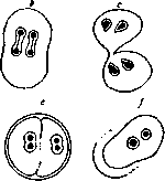
Figure 6. Reproduction of Sappinia diploidea: a-by division; b-by sexual method (c-connection of two individuals; d-their encystment and pairwise fusion of nuclei; e-emergence from the cyst of individuals with two fertilized nuclei).
contains a large karyosome, division is mitotic, during which the karyosome dissolves, and typical chromosomes are formed from the nuclear contents. Type III-Vahlkampfia, includes eight genera: 1) Vahlkampfia Chatt. et Bonnard, 1912,-small free-living and saprophytic amoebas, 2) Pygolimax Kuen. u. Swell., 1917, 3) Endolimax Kuen. & Swell., 1917, 4) Jodamoeba Dob., 1919, 5) Malpighiella Minch., 1910, 6) Pelomyxa Greef, 1874, 7) Sappinia Dang., 1896 and 8) Dientamoeba Jepps, 1918. Of these, forms 2,3, 4, 5, 8 are non-pathogenic parasites, among which in humans are found Endol. nana, Jodam. Butschlii and Dient. fragilis. Forms 1 and 6 are free-living. Form 7 is saprophytic. In most representatives, the nucleus has a large karyosome, divides by promitosis, during which the karyosome is drawn out, and chromosomes are formed from the extra-karyosomal part of the nucleus. Type IV-Entamoeba includes the genera Endamoeba Leidy, 1879, and Entamoeba Casagr. & Barb., 1897, large parasitic, partly pathogenic, amoebas of humans and animals. Representatives: End. blattae Leidy, parasitizes in the intestine of cockroaches; Ent. histolytica Schaudinn, 1903, the causative agent of human amoebic dysentery; Ent. coli Losch, 1875, em. Schaudinn, 1903, and Ent. Hartmanni Prow., 1912,-non-pathogenic parasites of the human intestine, Ent. gingivalis Gros, 1840, parasite of the human oral cavity; nuclear division occurs according to the mitotic type, but has not yet been sufficiently studied. Amebiasis, the aggregate of morbid phenomena caused by the life activity of certain parasitic amoebas. The presence of non-pathogenic parasitic amoebas, as well as the carriage of pathogenic amoebas, if it does not manifest in any clinical forms and is not associated with any anatomical processes, is not included in the concept of amebiasis. As a spontaneous disease, amebiasis occurs in many vertebrates-humans, monkeys, more rarely-dogs, cats, guinea pigs, rats, as well as in frogs.-Human amebiasis is an acute disease with a clearly expressed tendency to chronic recurrent course; it can have the most diverse localization. Clinically, the most significant are intestinal amebiasis (amoebic dysentery) and liver (amoebic hepatitis and amoebic liver abscess). The causative agent of human amebiasis is Entamoeba histolytica Schaudinn, 1903. - Infection occurs by swallowing cysts of this amoeba with water. In the intestine, mainly in the upper part of the small intestine, the cyst wall dissolves; amoebas emerging from the cysts reach the large intestine, enter the Lieberkühn glands, penetrate between epithelial cells into the submucosal tissue, where they multiply. Due to the action of their proteolytic enzyme, the amoebas cause extensive necroses in the surrounding tissue. The latter are the cause of necrosis of the overlying areas of the mucosa and the formation in the intestinal wall of characteristic ulcers with undermined edges, i.e., with a wide base and a narrow opening opening into the intestinal cavity. Through the venous capillaries, amoebas can be carried into the portal vein system; by this path, as well as through the lymphatic vessels, amoebas penetrate into various organs, where they form metastatic foci. The incubation period in amebiasis averages three weeks, but, depending on the body's resistance, it can vary widely-from several days to three months. There are, however, cases when the clinical outbreak of the disease occurs only several years after infection. Intestinal amebiasis most often occurs in the form of alternating acute periods (expressed in diarrhea up to 20 times a day and more) and free intervals, sometimes very prolonged. Such chronic recurrent cases can last for years. Apparently, there is no spontaneous recovery. In some cases, the disease can have an completely unnoticeable onset and atypical course. Often observed are atony, constipation, increased cardiac excitability, asthmatic phenomena, polyneuritis, skin irritations (acne). Intestinal amebiasis most often occurs without fever. It is usually observed only in complications with bacterial infections, as well as in complications from the liver and other internal organs. The most common complication of intestinal amebiasis is liver abscess, which can rupture either outward or into the peritoneum, pleura, etc. Metastatic abscesses of the brain are observed much less frequently. Metastases have also been described in the spleen, diaphragm, lymph glands, gallbladder, kidneys, bladder, testicles, skin, joints, etc. The theory, supported by American authors, of an etiological connection between amebiasis and some forms of arthritis, as well as Hodgkin's disease, still needs confirmation. Amoebic liver abscess can occur not only in connection with typical amoebic dysentery, but also without any known manifestations from the intestine. The mortality rate from amoebas in untreated cases is quite high and ranges from 18 to 40%. The main causes of death are perforations into the peritoneum, hemorrhages, metastases, as well as accompanying infections, the occurrence of which is facilitated by progressive exhaustion and anemia. The introduction of chemotherapeutic agents-emetine and iatren has extremely lowered the mortality rate. Intestinal amebiasis is established, mainly, on the basis of examination of feces, when the question can be resolved by finding in them Ent. histolytica. For this disease, the abundance of eosinophils in the intestinal contents is very characteristic, and, often, Charcot-Leyden crystals. Compared to bacterial dysentery, the excretions in intestinal amebiasis are characterized by a significantly lower content of neutrophilic leukocytes, the presence of large mononuclear cells of the macrophage type, as well as the abundance of mucus. In acute cases, the external appearance of intestinal excretions is very characteristic, often completely colored with blood and resembling raspberry jelly in its mucous consistency and color. In many cases, eosinophilia is also observed in the peripheral blood, reaching 35%. Leukocytosis is usually present only in complications from the liver, etc. Rectoscopy can provide substantial help in the diagnosis of intestinal amebiasis, which reveals typical undermined ulcers, especially in older and more severe cases.-The main role in the treatment of amoebas is played by the following preparations: the alkaloid of ipecacuanha-emetine (see) in the form of subcutaneous injections of its hydrochloride salt (0.05-0.1 pro die) or in the form of a double salt with iodine and bismuth per os; iatren (see)-1.0-3.0 pro die and etorsol (see) per os; neosalvarsan (see) in the vein. After clinical recovery, a person often becomes a carrier of infection for many years, continuing to excrete amoebic cysts with feces.-Epidemiology. The spread of amebiasis occurs through cysts: 1) by contact, through cyst carriers, by contamination of household items and food products, 2) through drinking water, since it provides the best conditions for maintaining the viability of cysts, 3) with the mediation of flies, 4) under certain conditions, infection with cysts through dust is probably also possible.-Geographical distribution. Amebiasis is endemic, mainly, in tropical and subtropical countries, but is also introduced into temperate zones. In Europe, many cases of amebiasis were observed during the war, in connection with the stay of colored troops on the French front, when diseases were also described among the local population. In the USSR, amebiasis is widespread in Central Asia, Transcaucasia, and also occurs in the Volga region.-Statistics. Complete statistics of amebiasis do not exist. Based on local surveys, it is assumed that in the hot zone of the globe, the number of patients with amebiasis in some places reaches 2%, and the number of carriers is not lower than 20% of the total population. In the USSR, there are very incomplete data for 1926: in Armenia, Georgia and Azerbaijan, about 22,000 people were officially registered.-Prevention consists of measures: a) with respect to cyst carriers and b) with respect to cysts excreted from the body. With respect to cyst carriers, it is necessary to accurately determine their contingent and, if possible, exclude them from the preparation and sale of food products, as well as from work on water supply; systematic use of iatren, as well as arsenical preparations per os (606) contributes to the disinfection of the intestine from cysts. Protection against cysts excreted from the body is possible by a) disinfection of feces with a 0.5% solution of cresol, b) protection of drinking water and food products from contamination with cysts, c) destruction of flies, etc., d) avoiding the use of non-boiled water.-The parasitological diagnosis of amebiasis is made on the basis of finding Ent. histolytica in the intestinal contents, in the pus of an abscess, or in patho-histological examination of tissues (see table, fig. 2 a, b, c).
In acute cases of intestinal amebiasis, the diagnosis is based on the presence in freshly passed feces of typical vegetative stages of Ent. histolytica averaging 25-40 μ, rarely 60-80 μ, characterized by 1) energetic mobility associated with the rapid formation of glassy ectoplasmic "hernia-like" pseudopodia, sharply differing in their refractive index from the opaque granular endoplasm, 2) the ability to phagocytize erythrocytes, 3) a transparent nucleus 5-7 μ in diameter with a fine structure of a reticular type, in the center of which is placed a small karyosome, and under its shell lies one row of small grains staining with basic dyes. Compared to other A., the nucleus of Ent. histolytica is poor in staining morphological elements. During periods of clinical quiescence, the mobile phagocytizing forms of Ent. histolytica are replaced by smaller and non-phagocytizing, however still mobile, precyst stages (forma "minuta"), and finally the cysts themselves. At this time the diagnosis is much more difficult, since the mobile precyst stages cannot always be with certainty differentiated from the vegetative stages of non-pathogenic human A. A more characteristic picture is presented by the cysts of Ent. histolytica—round, rarely oval, formations with a transparent shell. In early stages they have 1 nucleus, in the mature form - 4 nuclei. They contain glycogen droplets and characteristic "chromatoid" inclusions in the form of short, thick rods with rounded ends. The size of the cysts varies between 5 and 20 μ, and on the basis of their size, among Ent. histolytica three to five races are distinguished. For microscopic diagnosis, these cysts provide a firm basis. However, it must be taken into account that among non-pathogenic A. of the human intestine some also form 4-nucleate cysts, which in size and structure are sometimes almost indistinguishable from the cysts of Ent. histolytica. Therefore, in doubtful cases morphological investigation may not solve the question. In such cases, resort is had to infecting cats with cysts, in which only a positive result has significance, since many cats are individually non-susceptible. The diagnosis may be complicated by the simultaneous presence of several species of A. in the same host. In differential-diagnostic terms the following non-pathogenic species are important: 1. Ent. coli, Losch, 1875, em. Schaudinn, 1903 (see table, fig. 3 a, b, c). Size 20-40 μ (limits 10-70 μ); differs from Ent. histolytica in slower movements and the absence of sharp differentiation into ecto- and endoplasm. Erythrocytes are usually not phagocytized. If this occurs, it apparently only in very rare cases. Food inclusions—bacteria. The nucleus is similar to that of Ent. histolytica, but has a coarser structure and differs in the abundance of basophilic grains, usually densely lying at the periphery, and a larger (up to 1.5 μ), compact and somewhat eccentric karyosome. In the nucleus there is often a second nucleolar body about 2 μ in size and located even more eccentrically. The vegetative stages of Ent. coli usually present no difficulties for diagnosis. These may, however, appear in the presence of precyst forms (15-22 μ), which are often difficult to distinguish from the corresponding stages of Ent. histolytica. For differential diagnosis the cysts of Ent. coli are very important, in most cases quite characteristic. They differ in larger size from 12 to 22 μ, on average 15-18 μ; in the mature state they have 8 (very rarely 16 or more) nuclei, and like Ent. histolytica may contain "chromatoid" inclusions, however having the appearance of threads, pointed rods, fragments, etc. Also very characteristic are the immature 2-nucleate cysts of Ent. coli, striking in the presence of a large glycogen vacuole that pushes both nuclei against the very shell of the cyst. However, immature 4-nucleate cysts of smaller races of Ent. coli (12-15 μ) may present a certain danger of being confused with cysts of Ent. histolytica. Ent. coli are found in 50-70% of healthy people in all latitudes. American authors have described Councilmania lafleuri, Kofoid and Swezy, 1921, a large A. (20-60 μ) combining some features of Ent. histolytica (movement, structure of protoplasm and nucleus, phagocytosis of erythrocytes) with features of Ent. coli (8-nucleate cysts); from the latter it differs, according to the authors, in that from the contents of the 8-nucleate cyst small single-nucleate A. bud off, emerging through a special pore in the shell of the cyst. It is quite possible that in its description the authors were misled by the presence of a double infection of Ent. coli and Ent. histolytica.-2. Ent. Hartmanni, Prowazek, 1921 (syn. Ent. tenuis Kuenen & Swellengrebel, 1917; see table, fig. 6 a, b). In the vegetative state this is a small A. averaging 6-8 μ in size (limits 4-12 μ). The diameter of the nucleus is 3.3 μ. It resembles Ent. coli in the structure of the nucleus. It feeds on bacteria. It presents particularly great differential-diagnostic interest by the fact that it forms 4-nucleate cysts, 6-12 μ in diameter, almost indistinguishable from cysts of Ent. histolytica. The vegetative stages may have significance in the differential diagnosis of precyst stages of Ent. histolytica. It is found in 15-20% of healthy people in all latitudes.-3. Endolimax nana Wenyon O'Connor, 1917 (syn. Ent. nana W. O'C, 1917; see table, fig. 4 a, b). A small, rather mobile A., averaging about 8 μ in size. The nucleus, about 2 μ in size, has a typical karyosome structure. Food inclusions—bacteria. It forms spherical, rarely oval cysts averaging 5-8 μ. It has chromatoid inclusions in the form of short threads or strands and a small glycogen vacuole. The wide distribution of End. nana and the relatively high percentage of infection (2-39%) give it a certain diagnostic significance due to some similarity of its vegetative stage to small precyst individuals of Ent. histolytica, and especially in determining cyst carriage due to the possibility of confusing its 4-nucleate cysts with the corresponding cysts of Ent. histolytica.-4. Jodamoeba Butschlii, Dobell, 1919 (syn. Ent. Williamsi, Prowazek, 1911; see table, fig. 5 a, b). Small A., 5-24 μ in size, on average 9-13 μ, are rather immobile, although they can form ectoplasmic pseudopodia. The nucleus is 3-6 μ in diameter and contains a large karyosome 1.5-3 μ in size with even edges. Often the karyosome is surrounded by a layer of large basophilic grains. Cysts are mostly oval from 8 to 20 μ, on average 8-12 μ, have a characteristic 2-contour shell, as well as a large glycogen (iodophilic) vacuole, often pushing all the contents of the cyst to one side. Mostly 1-nucleate cysts are found, 2-nucleate cysts are rare. Distribution is universal. Frequency of infection 0.5-10%. The cysts of Jodam. Butschlii may have some differential significance in the diagnosis of cysts of Ent. histolytica. The least differential-diagnostic significance is represented by
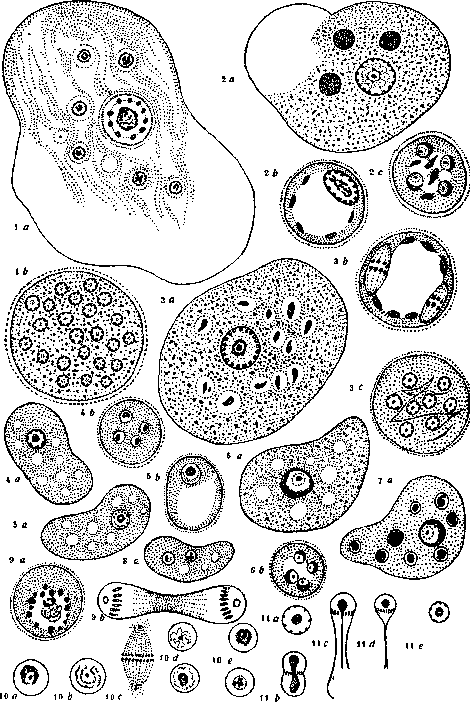
i) A ® 1. Ent. blattae: a-vegetative form; b-cyst.-2. Ent. histolytica: a-vegetative form; b-one-nucleate cyst; c-4-nucleate cyst.-3. Ent. coli: a-vegetative form; b-2-nucleate cyst, nuclei in the state of mitosis; c-8-nucleate cyst.-4. Endolimax nana: a-vegetative form; b-4-nucleate cyst.-5. Jodamoeba Butschlii: a-vegetative form; b-cyst.-6. Ent. Hartmanni: a-vegetative form; b-4-nucleate cyst.-7. Ent. gingivalis.-8. Dientamoeba fragilis.-9. Nucleus of Ent. blattae: a-at rest; b-in mitosis.-10. Nucleus of Hartmanella: a-at rest; b-preparation for mitosis, disappearance of karyosome; c-mitosis; d and e-reconstruction of daughter nuclei, gradual restoration of karyosome.-11. Nucleus of Vahlkampfia: a-at rest; b, c and d-consecutive stages of mitosis; e-reconstruction of daughter nuclei. 5. Dientamoeba fragilis, Jepps & Dobell, 1918 (see table, fig. 8 a), a small, mostly 2-nucleate form averaging 9 μ in size, with a nucleus about 2 μ, constructed on the karyosome type and characterized by the fact that its karyosome consists of several separate grains; cysts have not been described. The parasitological diagnosis of intestinal amebiasis is often complicated by the simultaneous presence of several species of A. The comparative frequency of finding various A. in the human intestine is approximately as follows (the figures below refer mainly to statistics of cyst carriage): Ent. histolytica-7-10%, Ent. coli-36-54%, End. nana-9-13%, Jodam. Butschlii-0.3-15%.
Related articles
Mentioned in
Cite this page
“Amoebas.” Soviet Medical Encyclopedia. English translation of Bolshaya Meditsinskaya Entsiklopediya, 1st ed. (Moscow, 1928–1936), ed. N. A. Semashko. https://sovietmedicalencyclopedia.pages.dev/article/amoebas/