Trichomonas
Historical document, translated for reference. It reflects medical knowledge of the 1920s–30s and is not medical advice.
Summary
Trichomonas are pear-shaped flagellate protozoa of the family Trichomonadidae, measuring 5-30 μm in length. Three species are described in humans: T. hominis from the intestine, T. elongata from the oral cavity, and T. vaginalis from the vagina, with the latter being associated with trichomonas-colpitis.
Encyclopedia article (1928–1936)
TRICHOMONAS, flagellate protozoa of the family Trichomonadidae, pear-shaped in form, 5-30 μm in length. From a group of parabasal grains at the anterior end of the body, a bundle of 3-5 flagella emerges. At this same point begins a flagellum that runs backward along the edge of the undulating membrane, which borders the protozoan's body on one side. This marginal flagellum may extend beyond the posterior pointed end of the body. Along the line of attachment of the undulating membrane at its base runs another flagellum. Along the axis of the body passes an elastic skeletal rod-axostyle, slightly protruding at the posterior pointed end of the body. In the anterior part of the body there is a small cytostome. Near it, behind the group of parabasal bodies, is located the nucleus. Reproduction occurs by longitudinal division, with the flagella being distributed between the daughter specimens, while some are formed anew. In humans, three species of T. are described: T. hominis from the intestine, T. elongata from the oral cavity, and T. vaginalis from the vagina. The independence of these species is disputed. T. hominis (intestinalis), (see illustration), 5-15 μm in length, has 4 (3-5) free flagella, which are in lively motion. The body form is subject to significant changes. Nutrition occurs mainly by the absorption of bacteria and other particles. T. can also absorb red blood cells. T. is well cultivated on serum diluted with a half-percent solution of sodium chloride, or on media used for cultivating the dysentery amoeba. T. hominis is localized mainly in the large and cecum, sometimes extending into the lower parts of the small intestine. Infection occurs through feces, in which, without drying, T. can remain viable for many hours and even several days. Flies, eating feces containing T., can after several minutes excrete motile T. with their own feces and contaminate food with them. T. hominis is found mainly in diarrheal stool. In healthy individuals, it is found in a significant percentage, but it can be found in feces only after the administration of a laxative. The pathogenic role of T. hominis
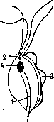
Trichomonas hominis: 1-axostyle; 2-group of parabasal bodies; 3-undulating membrane; 4-nucleus.
is not firmly established, although its penetration into the cavity of the glands of the intestinal wall and the underlying tissues has been described. T. elongata (T. buccalis) is found in the oral cavity both in cases of gum lesions and in individuals with a healthy oral cavity. Morphologically indistinguishable from T. hominis. Found in the purulent discharge of the tonsils, in sputum, in gangrenous areas of the lung, in cancerous tissue in the stomach, in pleural exudates. T. vaginalis is found in the vagina, mainly when the discharge has an acidic reaction. It reaches somewhat larger sizes than the previous two species (up to 30 μm). It is also found in the female and male urethra. It has been observed for a long time in the discharge of an ulcer on the male penis (Marcinowski and Moshkovsky). All described species of Trichomonas are primarily saprophytes, finding particularly favorable conditions for their development in the presence of processes leading to the breakdown of underlying tissue, in which they may play a small secondary role.
Sh. Moshkovsky. Trichomonas vaginalis is quite frequently found in vaginal secretions: according to Hoetme in 28%, according to Haussmann in 40%, according to Schmid and Kamniker in 69.9%. Hoetme was one of the first to indicate that T. vaginalis is the cause of a special type of colpitis, which he named trichomonas-colpitis. The symptom-complex of such colpitis is characterized by abundant purulent discharge, often of a foamy nature; the vaginal mucosa is hyperemic, bleeds easily; the entrance to the vagina is usually irritated, painful, and itching and burning are often noted. Schroder, Loser and others adhere to a different viewpoint, considering T. vaginalis only an occasional companion in the third-degree purity of vaginal secretions. The foamy nature of the vaginal discharge, according to Schroder and Loser, depends on the presence of Micrococcus gasogenes. Hoetme, Schmid and Kamniker refute the viewpoint of Schroder and Loser, since in some cases with the disappearance of T. vaginalis the colpitis is cured, and in other cases T. vaginalis and colpitis are found with the first degree of purity of the vaginal secretion. Some authors seek to reconcile these extreme views to some extent, indicating that T. vaginalis are found on already altered vaginal mucosa and their presence is manifested by certain characteristic features, mentioned above. This viewpoint does not thus refute the pathogenicity of T. vaginalis, and the observations of Hoetme, Schmid, Kamniker and other authors, in which after the removal of T. vaginalis the original flora of the vagina remained, while the discharge from the vagina and the characteristic changes in the vaginal mucosa disappeared, confirm that it is precisely T. vaginalis that can be the cause of colpitis. The possibility is not excluded that the known disagreements are connected with the presence of different types of Trichomonas vaginalis. According to Balassa and Bodocsy, it is necessary to distinguish between T. vaginalis that live in symbiosis with bacteria, and T. vaginalis that are true causative agents of the disease. Schmid, Kamniker and others state that in connection with the presence of T. vaginalis postpartum colpitis develops with an increase in temperature. Therefore, they recommend applying appropriate treatment even before childbirth. The penetration of T. vaginalis into the vagina occurs in various ways-from the rectum, urinary tract, through water (bathing, baths, douching). To detect T. vaginalis, the vaginal secretion is collected with a platinum loop and placed in a drop of physiol. solution heated to a temperature of 35-37°. The drop is covered with a coverslip and then examined under a microscope. Schmid and Kamniker recommend taking the secretion from the vagina with a blunt spoon, passing it lightly along the wall of the vagina. Observations show that T. vaginalis is particularly often found between not yet completely shed superficial cells of the vaginal epithelium. T. vaginalis can also be detected by staining, e.g., by Gram's method, using a carbolic solution of Gentian violet. Trichomonas does not stain red as well as leukocytes and epithelial cells, and a certain skill is necessary to properly examine the preparation. Treatment consists of washing with a 0.1% solution of mercuric chloride; introduction into the vagina of 5-10 cm3 of a 10% solution of boric glycerin or pure glycerin; for this, the folds of the vagina are stretched with a speculum and, gradually extracting it, the vagina is wiped with a cotton swab moistened with glycerin solution. This treatment is continued for two weeks, applying lubrication every 2-4 days. For washing, one can also use lactic acid (1 teaspoon per 4-5 glasses of water), Zinc. sulf., Alumenis aa (1 teaspoon per 5 glasses of water). Recently, treatment has been successfully carried out with the help of biolactin. Certain difficulties in cure and recurrences arise because the spread of T. vaginalis can be very extensive, and according to Rodecurt, T. vaginalis is found not only in the vagina but also in the cervix, in the urethra, in the Skene's and Bartholin's glands.
Related articles
Mentioned in
Cite this page
“Trichomonas.” Soviet Medical Encyclopedia. English translation of Bolshaya Meditsinskaya Entsiklopediya, 1st ed. (Moscow, 1928–1936), ed. N. A. Semashko. https://sovietmedicalencyclopedia.pages.dev/article/trichomonas/