Myiasis (myiasis)
Historical document, translated for reference. It reflects medical knowledge of the 1920s–30s and is not medical advice.
Summary
Myiasis is a disease of humans and animals caused by fly larvae. The article describes various forms of myiasis, including cutaneous, tissue, cavity, intestinal, urinary tract, genital, and ocular myiasis, along with their symptoms, diagnosis, and treatment.
Encyclopedia article (1928–1936)
MYIASIS, myiasis (from Greek myia-fly, other transcriptions: myiosis, myasis), a disease of humans and animals caused by larvae of flies. In some species of flies, the larvae are obligate parasites, in others saprophytes, in still others they live by consuming fresh plant objects. All these biological forms of larvae can be true or false parasites of humans and animals and can cause their host often substantial, and sometimes irreparable, harm. Fly larvae are relatively resistant to various factors, particularly chemical reagents. One can note the ability of the 'rat-tail' - the larva of the fly Eristalis (fig. 1) - to inhabit sewage and other highly contaminated places (accumulation of liquid feces, etc.); larvae of many flies, compared to larvae of other insects, do not die for a long time in solutions of formalin or alcohol. All these characteristics of fly larvae determine their ability to live for a certain period in an environment unusual for them, for example, in the human intestine, where they can enter with food. M. is a general term denoting the presence of living fly larvae in a living host. Forms of M. are classified both according to the location of the larvae in the host and according to the biological properties of the larvae of the corresponding fly species. M. can be, with respect to a given species, obligate or facultative; in the first case it is caused by parasitic flies, in the second by flies whose larvae are not parasites. Among flies of the latter category, the larvae of some live in corpses or in meat, and their parasitism in a living organism is easily accomplished. The larvae of other species are saprophytic or herbivorous; however, they too can be accidental causes of myiasis. According to localization, there are cutaneous, tissue, cavity (ear, nose), intestinal, urinary tract, genital, and ocular myiasis.
Figure 1.
Figure 2.
Figure 3.
Figure 4.
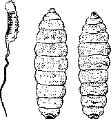
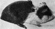
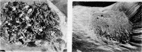
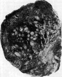
Figure 1. 'Rat-tail'. Figure 2 and 3. Cordylobia anthropophaga: from the back (2) and from the abdomen (3). Figure 4. Musca domestica, larva. I. Cutaneous (dermal) M. in turn has different forms. 1) Epidermal M.; larvae live in the thickness of the epidermis and do not go beyond its limits (e.g., larva of the first phase of the horse botfly Gastrophilus, causing creeping disease). 2) Subcutaneous M. with localization of larvae in the connective tissue layers of the skin, and secondarily the epidermis may also be affected (e.g., the cattle warble fly Hypoderma, whose larvae can also parasitize humans); the corresponding form of M. is called subcutaneous M. with moving swelling of the skin over the migrating larva; larvae of the human botfly Dermatobia and the fly Cordylobia anthropophaga (fig. 2 and 3) cause the development of furuncular M. 3) Subcutaneous M. can pass into tissue M. when larvae of M. enter ulcers or wounds and begin to devour the living tissue of the host's body; ultimately, enormous destruction of soft parts of the body can occur (see separate table, figures 2 and 3) with exposure of bones (figure 5); such forms of M. sometimes take on a generalized character and can cause the death of the host [e.g., the well-known historical cases of 'eating by worms' (i.e., fly larvae) of a person to death]; tissue M., in particular wound M., is caused by both obligate (larvae of the Wohlfahrt's fly) and facultative parasites [larvae of the blowfly (Lucilia), housefly (Musca domestica) (fig. 4), blowfly (Calliphora), Cochliomyia macellaria, Pycnosoma, etc.]
II. Cavity M. Larvae can be located: 1) In the nasal cavity (nasomyiasis), where they sit on the mucous membrane [larvae of the sheep botfly (Oestrus ovis), horse nasal botfly (Rhinoestrus purpureus)] or bore into the soft coverings and destroy them (Wohlfahrt's fly). 2) In the oral cavity in the gums, destroying their soft parts (e.g., Wohlfahrt's fly); this phenomenon is apparently so common that in the people toothache was etiologically associated with the presence in the mouth of 'worms'. 3) In the auditory canal (otomyiasis) - Wohlfahrt's fly and others; M. here usually develops on the basis of any diseases of the ear; larvae can destroy the eardrum, middle ear, penetrate into the mastoid process, venous sinus and reach the meninges (consequences - up to fatal outcome). 4) Of the urinary tract and bladder - larvae of Anthomyia (Fannia) canicularis, Musca domestica. 5) Of the genital organs - larvae of Wohlfahrt's fly (in the vulva of sheep).
III. M. of the eye (ophthalmomyiasis) - larvae in the conjunctival sac of the eye (Oestrus ovis, Rhinoestrus purpureus and Wohlfahrt's fly in humans; the latter is especially dangerous, as its larvae can pierce the wall of the eyeball and completely destroy the eye).
In all these cases, infection of the host occurs passively: some flies deposit their larvae in its cavity, in the eye, in ulcers or wounds; others lay eggs there, from which larvae hatch already in the host's body; larvae of some species (Cordylobia) (fig. 6) actively bore into human skin; there are cases of even the pupal stages being found in the host; therefore, the concept of M. applies to all cases of finding any stages of metamorphosis of flies from egg to puparium in or on the host.
IV. Intestinal myiasis is the term for cases of living fly larvae being present in the stomach or intestine of humans. The symptomatology varies. When larvae are located in the stomach, there may be nausea, vomiting and sharp pain in the epigastric region; when they are in the intestine, abdominal pains, hemorrhages and typhoid state come to the fore. With prolonged presence of fly larvae in the intestine, colitis develops. Various species of flies can live in the human digestive tract: housefly, blowfly, blowfly, cheese fly (Piophila casei), Anthomyia (Fannia) canicularis, Anthomyia scalaris, Eristalis, Philaematomyia, etc. The diagnosis of intestinal M. must be made with caution to distinguish true cases of M. from false ones. The latter include cases when patients intentionally mislead the doctor, bringing 'parasites' that allegedly came out of the intestine; furthermore, when examining feces for examination, foreign objects, including fly larvae, can be accidentally captured; finally, flies can lay eggs in already excreted feces, and larvae may have time to hatch from the eggs before the feces reach the doctor for examination; in the latter case, all larvae will be very small (about 1 mm), while adult larvae reach 1-1½ cm in length, and sometimes more.
Infection of the digestive tract with larvae occurs through swallowing them with food or with some other material. Larvae of the cheese fly (Piophila casei) live in salted fish, cheese and can be easily eaten; other flies can lay eggs in already prepared food (e.g., boiled meat), and on the same substrate the larvae also develop. Cases of getting larvae with vegetables, etc., are not uncommon. Treatment of cutaneous myiasis consists in removing botfly larvae from the tunnels they make, extracting these larvae from wounds and ulcers, applying tampons with chloroform, antiseptic treatment of ulcers and wounds; botfly larvae are removed from the eye with tweezers or by washing, from the stomach - by washing, from the intestine - by various anthelmintic agents.
Prevention of M.: treatment of ulcers on the bodies of domestic animals to destroy fly larvae in them, treatment of ulcers and wounds so that they do not attract flies by their purulent discharge; protection of food products from flies; consumption of well-washed and cleaned vegetables, especially if they are used in fresh form; burning of corpses and carrion.



Related articles
Cite this page
“Myiasis (myiasis).” Soviet Medical Encyclopedia. English translation of Bolshaya Meditsinskaya Entsiklopediya, 1st ed. (Moscow, 1928–1936), ed. N. A. Semashko. https://sovietmedicalencyclopedia.pages.dev/article/myiasis/