Microtome
Historical document, translated for reference. It reflects medical knowledge of the 1920s–30s and is not medical advice.
Summary
A microtome is an apparatus for preparing thin sections of specimens for microscopic examination. The article describes various types of microtomes including manual, student, freezing, sliding, Jung's inclined plane, brain, and Tetradent microtomes, detailing their mechanisms and applications in histological research.
Encyclopedia article (1928–1936)
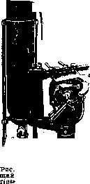
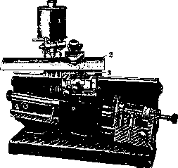
Microtome (from Greek mikros-small and temno-I cut), an apparatus for preparing sections suitable for microscopic examination. The general principle of the microtome's design is that the object, by means of a micrometer screw, is raised vertically in either a direct or inclined plane by a small amount, expressed in microns; in the horizontal plane, a knife moves, held either directly by hand (in the simplest models) or clamped in a special holder. For cutting on a microtome, the object must necessarily be made as dense as possible and embedded in a homogeneous medium, for example in paraffin or celloidin; the object can also be frozen by using atomized ether or liquid CO2. Depending on the embedding and the properties of the object being examined, the microtom makes it possible to obtain sections up to 1 μ in thickness (which is unachievable when cutting directly with a hand razor), as well as to obtain a series of sections of uniform thickness without gaps. Types of microtomes. The manual microtome consists of a thick-walled metal cylinder with a well-polished disk on the upper side, across which an ordinary razor or special knife is drawn by hand. Inside the cylinder there is a fine screw thread; in it moves a micrometer screw with a head at the bottom, on which are divisions indicating how far the specimen has advanced in the cylinder. At the upper end of the screw there is a platform on which the object to be cut is secured. The instrument is inexpensive and suitable only for rough sections. The student microtome is screwed to the table and consists of a sturdy metal frame in which a micrometer screw with an indicator of its movement and a platform for securing the object or a clamp for a wooden or stabilizing cube to which the specimen is glued is fixed. In the upper part of the frame there is a polished arc along which the knife holder, equipped with a clamp, moves. The movement of the specimen occurs either by hand or automatically with each withdrawal of the knife backward, for which purpose the screw has a rack gear connected to the knife holder and provided with a regulator that allows the specimen to be advanced upward by a certain number of microns. Such a microtom makes it possible to obtain fairly thin sections. A similar microtome is used for preparing sections of objects frozen with liquid CO2 ("freezing microtomes" of the firms Jung, Reichert; fig. 1). For this purpose, a sprayer for liquid CO2 is attached to the object platform, which comes here through a flexible metal tube from a steel cylinder, which is usually equipped with a reducing valve. In the sprayer itself there is a tap that allows the required stream of liquid CO2 to be turned on or completely closed and its flow stopped. Sections from frozen objects have recently become widespread, and for some methods of processing histological material they are completely irreplaceable, as they make possible the processing of fresh, unfixed material with various reagents. In addition, the "freezing microtome" eliminates the troublesome and prolonged embedding, which allows for rapid preparation of the necessary specimen for path-histological research (fig. 2). The sliding microtome (Schanze and other firms). On a heavy cast iron base is fixed a thick vertical plate, to the left side of which another similar plate is attached at an acute angle, thus forming a receptacle for a wedge-shaped massive block, on which the microtome knife is secured by means of a screw and special holder. On this wedge-shaped block and on the sides of the angle formed by the mentioned plates, polished strips are ground, coinciding with each other and allowing the knife holder to slide smoothly in these guides. On the left side of the base there is a nut in which the micrometer screw moves. It stands vertically and has a disk on the upper surface of which a series of divisions is marked; the value of the latter is engraved on it or on a special bar along which a spring index moves, counting the number of teeth by which the micrometer screw disk is turned. Each tooth corresponds to a certain number of μ, so that the desired amount of microns for raising the screw head can be set immediately. The head of the micrometer screw presses against the object clamp, which moves up and down in a groove. This clamp, by means of axles secured by screws, can be set in the desired position relative to the horizontal plane. A handle is attached to the holder, allowing it to be raised or lowered by any amount without the aid of the screw during the initial setting of the object and selection of the cutting plane. The microtome is very sturdy, stable, and very convenient for sections up to 5 μ thick. It is already difficult to obtain thinner sections on it. Jung's microtome with an inclined plane (fig. 3). Here on the vertical plate of the microtome on the left is fixed a second plate, also forming an acute angle, but it is positioned so that it slopes downward slightly toward the operator. On it are polished strips along which a wedge-shaped block with an object holder slides. In other models (for example in Reichert's) instead of a plate on the left side of the microtome there is an inclined groove in which the object holder moves. Closer to the operator is the micrometer screw, whose pointed end presses against the object holder or a bar connected to it. The combination of the inclined plane and the micrometer screw makes it possible to raise the object by 1 μ and even fractions thereof. For convenience and continuity of work, the screw can rotate on a horizontal axis and act with one end then the other. In the newest models of this type, the screw nut is made in the form of tongs, which allows the screw to be moved during setting without rotation. Connected to the screw is a disk with divisions in the form of teeth and the height of elevation is engraved for 1 tooth. In large models of this type there is an attachment for automatically turning the screw by a certain number of teeth in connection with the movement of the knife backward. This attachment is most conveniently arranged in Reichert's models, where the knife driver presses against a movable plate fixed to the front end of the guides. Pressure on it is transmitted by a rod to the screw, and the plate is returned to its place by a spring. The microtome with an inclined plane is very convenient and makes it possible to obtain sections up to 1 μ and even thinner (Apati). The object holder is equipped with a clamp for the specimen, which by means of screws, rack gears, or a ball head (inconvenient) can be well oriented when selecting the cutting plane. The brain microtome is designed for obtaining large sections of the brain as well as other organs. Here it is not necessary to obtain a very thin section, but a section of large area is needed. Such a microtome is usually arranged with a vertical screw and a very large platform for securing the object. Since sections are most often made from material embedded in celloidin, a flat reservoir for alcohol (or water) is arranged around the table, and all cutting takes place in liquid, which facilitates the preservation of the integrity of the sections. Corresponding to the large size of the sections, knives for such a microtome are used with a large cutting edge. For cutting large objects, both frozen and embedded in celloidin or paraffin, the firm Jung has recently released a new microtome called the Tetradent. This microtome consists of a heavy four-sided metal frame on legs. Between the upper parallel beams
Fig. 1. Freezing microtome "Sarto-cylinder with carbonic acid; 2-metal tube leading to the sprayer (3) with table for the object to be frozen; 4-knife; 5-tap to the sprayer.
Fig. 2. Sliding microtome: 1-knife; 2-object of study; 3-one of the polished guide strips; 4-cup for spent alcohol with which the knife is moistened; 5-handle for coarse lowering and raising of the object; 6-disk with divisions.
Fig. 3. Jung's microtome with an inclined plane: 1-reservoir with alcohol; 2-knife; 3-object of study; 4-plate with inclination; 5-mechanism transmitting the setting with the object holder.
rial embedded in celloidin, a flat reservoir for alcohol (or water) is arranged around the table, and all cutting takes place in liquid, which facilitates the preservation of the integrity of the sections. Corresponding to the large size of the sections, knives for such a microtome are used with a large cutting edge. For cutting large objects, both frozen and embedded in celloidin or paraffin, the firm Jung has recently released a new microtome called the Tetradent. This microtome consists of a heavy four-sided metal frame on legs. Between the upper parallel beams
Fig. 4. Microtome with a rigidly fixed knife (1) and a moving object (2).
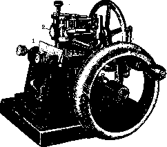
To the frame of the microtome, to which the knife is immovably attached, there is an attachment for securing the object to be cut (a holder or platform); this part is brought into motion toward the knife by means of a special lever, and the object is automatically raised by a micrometer screw to a certain number of microns. When cutting objects embedded in celloidin, the microtome knife must be moistened with 70% alcohol. To avoid excessive alcohol consumption and for even application to the knife, a dropping apparatus is very convenient. It consists of a closed reservoir on a stand. A long tube with a tap delivers alcohol drop by drop from the apparatus, the rate of which can be regulated by the tap. In the models described, the object remains stationary while the knife moves. There is another type where the knife is stationary and the object moves, being mounted on a table that is positioned either horizontally, as in the Leitz model, or vertically, as in the Minot model, which is driven by a wheel similar to that of a sewing machine (Fig. 4). These models offer no particular advantages. On the same principle as the microtome, an apparatus for making thick sections for macroscopic examination (macro-tome) is constructed. For obtaining preliminary sections from fresh organs, a double knife can be used. It consists of small razor blades with handles fastened together by a screw. At the rear end of the handles, another screw moves a thin wedge, allowing the distance between the blades to be set, which are then secured in a fixed position parallel to each other by the aforementioned screw.
Related articles
Mentioned in
Cite this page
“Microtome.” Soviet Medical Encyclopedia. English translation of Bolshaya Meditsinskaya Entsiklopediya, 1st ed. (Moscow, 1928–1936), ed. N. A. Semashko. https://sovietmedicalencyclopedia.pages.dev/article/microtome/