Avitaminoses
Historical document, translated for reference. It reflects medical knowledge of the 1920s–30s and is not medical advice.
Summary
Avitaminoses are diseases resulting from the absence of vitamins in food. This article describes the different types of avitaminoses, their symptoms, and treatment methods as understood in the 1930s.
Encyclopedia article (1928–1936)
AVITAMINOSES, diseases that arise as a result of the absence of vitamins in food. Vitamins (see), which are organic food substances of still unknown chemical nature, despite being contained in food in very small quantities, are absolutely necessary for the normal development, growth, and functioning of the animal organism. The name "vitamins" for these substances and the name A. for the diseases that arise when animals or people consume food without vitamins (avitaminous food) was proposed in 1912 by Funk (C. Funk). However, as early as the end of the last century, it was suggested that certain diseases are caused by insufficient, one-sided nutrition, for example, that the cause of the disease "beri-beri" lies in one-sided rice consumption. The discovery by Eijkman in 1897 of polyneuritis gallinarum (i.e., polyneuritis, or in other words, beri-beri in birds) and further research on beri-beri (Grijns, A. Hoist, Nocht, Fletscher, Fraser, Ellis and others) finally proved that beri-beri is caused by "insufficiency" of food, by the absence in it of special specific substances. Beri-beri and other diseases of similar origin were then called "deficiency diseases." Funk and Schaumann (H. Schaumann), through their research on the nature of these substances and attempts to isolate them, created a solid foundation for the doctrine of vitamins and gave impetus to the further study of avitaminoses, which advanced rapidly. Important stages in the further development of the doctrine of avitaminoses were the discovery of experimental scurvy by A. Hoist, the work of Freise, which clarified that Barlow's disease is infantile scurvy, the establishment of the avitaminous nature of keratomalacia (Goldschmidt and Frank), and research on experimental rickets. People and animals contract these various A. when the corresponding vitamins are absent from their food, and conversely, the aforementioned A. are cured by introducing the corresponding vitamins into the food. At present, five different vitamins are distinguished, each of which, when absent from food, can be the cause of the corresponding A.: 1) vitamin A, or anti-xerophthalmic vitamin; its absence in food is the cause of A.-xerophthalmia; 2) vitamin D, or anti-rachitic vitamin, which is connected with A.-rickets. Both of these vitamins are soluble in fats; 3) vitamin B, or anti-neuritic vitamin, the absence of which causes A.-beri-beri or polyneuritis; 4) vitamin C, or anti-scorbutic vitamin; its absence in food leads to the disease A.-scurvy or scurvy; 5) vitamin E, or reproduction vitamin, without which animals lose the ability to reproduce. Besides pure forms of A., mixed forms also occur, arising from the absence of several vitamins in food; beri-beri, which sometimes affects crews of sailing vessels, belongs to such mixed A. The position of pellagra and various nutritional disorders of infancy in the system of A. has not yet been clarified. - The pathological phenomena that result from avitaminous nutrition. The absence of vitamins in food is accompanied by a series of symptoms indicating various disorders in the life of the organism. Here, first of all, symptoms common to all forms of A. come to the fore, namely: loss of appetite, loss of weight, disorders of the nervous system. In young (growing) animals, a characteristic symptom is the cessation of growth, which is later followed by loss of weight. Disorders of the nervous system manifest themselves either in the form of apathy, general weakness, drowsiness, muscle trembling, or, conversely, in the form of constant restlessness, increased excitability, which may be followed by paralysis or convulsions and other such pronounced symptoms of disorders in the function of the nervous system. These symptoms are not strictly characteristic of vitamin starvation; they are also observed in other forms of "insufficiency" of food, for example, when consuming biologically incomplete protein (i.e., when consuming proteins in which certain important amino acids, such as tyrosine, cystine, tryptophan, are absent or contained in very small quantities), in case of poverty of food in certain mineral substances, etc. As for the curve of weight loss, it should be noted that in quantitative starvation the weight curve immediately begins to fall, while in qualitative (in particular vitamin) starvation this decline does not occur immediately, and at first the curve may even rise somewhat. Often animals subjected to vitamin starvation die in the presence of only these general phenomena, i.e., before specific symptoms characteristic of one form or another of A. appear. Here the matter depends on the species of animal, as different animals do not react in the same way to the absence of one or another vitamin in food, as well as on how complete the A. is, i.e., whether the given vitamin is completely excluded from the food or whether it is still contained in it, even in the form of negligible traces. Of the symptoms characteristic of A. and not manifested in other forms of insufficient nutrition, the symptoms of beri-beri and scurvy have been best studied. In beri-beri (see), according to the research of Mac Carrison, Kellaway, Korenchevsky and others, the disease begins with disorders of the gastrointestinal tract. The symptoms characteristic of scurvy (see) are hemorrhagic diathesis (see) and changes in bone tissue. A characteristic symptom of A-avitaminosis is diseases of the cornea of the eyes, etc. A-avitaminosis. In experimental A., caused by removing vitamin A from the food of animals, first of all, if young animals are involved, cessation of growth occurs, followed by loss of weight, continuing until the animal dies from A. But these are general symptoms of food insufficiency; the characteristic and specific consequence of this A. is xerophthalmia (see) and keratomalacia (see), which was established in 1915 by Freise, Goldschmidt and Frank. According to these data, three weeks after starting to feed with food without vitamin A, rats begin to lose their eyelashes and begin to hide from light; in the 5-6th week the sclera becomes dry and the cornea cloudy, and sores form. Histological examination of the cornea reveals complete identity of this eye disease with keratomalacia, which affects infants. According to Mori (1922), in the development of xerophthalmia, the cessation of tear secretion, which always occurs in A-avitaminosis, plays a major role. In young animals the disease develops faster than in adults. As a result of the absence of vitamin A, other disorders in the normal life of the organism are also observed, such as: disruption of the secretory activity of the salivary and sebaceous glands, anemia; it is characteristic, according to Cramer, Drew and Mottram, that the blood is depleted of blood plates. It is also interesting to note that, according to Macht and Stepp, when young animals that received food without vitamin A are illuminated with polarized light, they have convulsions, from which it can be concluded that the absence of vitamin A causes profound disorders in metabolism. - Treatment of A-avitaminosis. In the initial stages of the disease, all symptoms are quickly cured by delivering vitamin A in the form of butter, fish oil, etc. After one or two days, in any case after 3-4 days, noticeable improvement occurs; the eyes, which had remained closed until now, open, inflammatory phenomena disappear; the secretion of tear and sebaceous glands is restored; simultaneously with this, the loss of weight stops, growth and appetite are restored, etc. Recovery is achieved not only with oral delivery of vitamin A, but also with its parenteral administration. A-avitaminosis in humans generally has the same picture as in animals, experiments with which were of great importance for clinical medicine, especially for the clinic of diseases of infancy. But children usually do not have a complete absence of vitamin A in food, but only relative poverty of it. Therefore, the picture of the disease is not as severe as in experimental A. in animals. In children, this A. can lead to a delay in development and growth and even to a halt in the child's development. It occurs in artificially fed children who receive little whole milk, which was often the case during and after the last imperialist war. A. can also occur during breastfeeding, because women's milk can be very poor in vitamin A if the mother's food is devoid of this vitamin or very poor in it. The ability to return to normal development can be restored to such avitaminotic, strongly lagging in their development infants by delivering vitamin A to them in the form of butter, fish oil, etc. In Fig. 1, the weight curves of a normal and an avitaminotic child are presented; when the latter was 6 months old, he weighed only 3 kg (31/2 kg less than a normal child); from this time, butter (10 g per day) and carrot juice (10 g) were added to his food. Immediately improvement occurred, the curve began to rise sharply upward, and at 9 months of age he weighed only 17a kg less than a normal child.
These symptoms are not limited to the disease, and * These data must be compared with the following observation: if animals are injected with such doses of convulsive poisons as do not yet cause convulsions, and then illuminated with polarized light, they immediately develop convulsions. In children, xerophthalmia and keratomalacia further develop, which are also cured by butter and cod liver oil. These eye diseases, which eventually lead to blindness, were very widespread, for example, in Denmark (where blindness was often found among infants) in 1916-17 and, according to the research of Bloch, depended on the fact that the population widely used skimmed milk instead of whole milk, and margarine instead of butter for feeding children, and butter was exported abroad on a large scale during these years. And when from 1917 this export decreased due to the blockade of Germany and due to corresponding government measures, and the population began to consume butter itself, xerophthalmia and keratomalacia declined and almost disappeared.
Figure 1. 1-curve of weight of a normal child, 2-curve of weight of an artificially fed child, the food of which did not contain vitamin A (at the moments marked by arrows, butter was added to the food).
In 1920, the consumption of butter again decreased, and cases of xerophthalmia again became more frequent. This dependence of the spread of A-avitaminosis (xerophthalmia) on the consumption of butter is clearly visible in the curve of Fig. 2. Thus, the characteristic symptoms of the disease affecting children when fed food not containing vitamin A are the cessation of development and keratomalacia, which are cured by consuming foods rich in this vitamin. D-avitaminosis. Vitamins A and D are both soluble in fats; for a long time these two vitamins were identified and vitamin A was specifically associated with changes in bone tissue occurring in rickets. Only relatively recently was it clarified that the antirachitic vitamin is distinct from the anti-xerophthalmic vitamin A, and then the antirachitic vitamin was named, on McCollum's proposal, vitamin D. The first research on experimental D-avitaminosis, or in other words, on experimental rickets, was done by Mellanby. His experiments, conducted on young dogs, showed that although by removing vitamin D from food it is possible to cause in dogs a disease similar to rickets in children, however, vitamin D is not the only factor determining the development of bone tissue, and that the etiology of rickets is more complex.
Figure 2. 1-consumption of butter per day in grams, 2 - number of patients with xerophthalmia per year.
The development of rickets is accelerated under unfavorable living conditions, with a relative abundance of carbohydrates in food, etc. On the contrary, the development of rickets is delayed by such factors as clean fresh air, the possibility of moving freely, an abundance of meat in food, etc. Mellanby already showed that the calcium in food plays a certain role in the development of rickets; he even found that products rich in vitamin B are also rich in calcium (milk, egg yolk, green vegetables), and, conversely, products poor in calcium are also poor in vitamin B (white bread, margarine, potato, rice). Similar studies on experimental rickets in rats were conducted by McCollum, Simmonds, Shipley, and Park. Their research showed that true rickets in its most typical form develops with food poor in phosphates and antirachitic vitamin. Rickets (however, not such a typical form) also occurs if the food, along with its poverty in vitamin B, is also poor in calcium, i.e., if there is a predominance of phosphorus over calcium in it. For the normal development of bones, it is necessary that the ratio between calcium and phosphorus in food represents a certain value and that the food is not particularly poor in vitamin B. A poverty of food in calcium or a poverty in vitamin B causes not typical rickets, but rather osteoporosis. If the ratio between phosphorus and calcium in food is incorrect, then the development of bone tissue is disrupted, and rickets occurs; such rickets can be cured either by changing the mineral composition of food, i.e., by adding to the food the inorganic substance that was lacking, or by increasing the content of vitamin B. The latter can to a certain extent compensate for the poverty of food in calcium or phosphorus. Accordingly, analyses of the blood of rachitic rats showed that the phosphorus content in it is significantly reduced compared to normal (2.8 mg%, instead of 7-8.5 mg%) and the ratio between calcium and phosphorus is changed in the direction of calcium predominance. The best remedy for rickets is cod liver oil, which is richest in vitamin B of all products. Milk fat, on the contrary, is very poor in vitamin B. Under the influence of cod liver oil, the phosphorus content in the blood of rachitic patients increases (and can even exceed normal), which is apparently the reason for the subsequent deposition of calcium phosphates in the bones. For this, such an amount of cod liver oil is sufficient that it constitutes 2% of all food; the addition of butter remains ineffective even if it constitutes 50% of all food. This means that it indeed contains almost no vitamin B. In contrast, vitamin A is contained in both cod liver oil and milk fat in almost equal amounts. If cod liver oil is passed for 12 hours through a stream of air heated to 100°, then cod liver oil loses its ability to cure keratomalacia or prevent it, but fully retains its antirachitic properties. Vitamin B is therefore a more stable substance than vitamin A. Coconut oil contains a little vitamin B and no vitamin A. Cotton oil, corn oil, and olive oil contain neither of these vitamins.
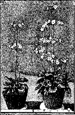
Treatment of B-avitaminosis (rickets). The best therapeutic and prophylactic remedy for rickets is cod liver oil in the amount of 10 to 30 cubic cm per day. The addition of phosphorus to cod liver oil (in very small doses - 0.01 or less per 100 g of cod liver oil) is permissible, but without exerting any significant effect on the bone changes in rickets, it undoubtedly has a favorable effect on the nervous phenomena in this disease. It should be borne in mind that depending on the method of preparation and storage of cod liver oil, its vitamin content can vary greatly. A specific therapeutic agent is also the yolk of a chicken egg, which, according to Gyorgy, can be recommended to children 4-6 months old. The therapeutic effect of ultraviolet rays obtained with a quartz mercury lamp ('mountain sun') is very interesting. For the first time, treatment with a quartz lamp was successfully applied in 1919 by Huldschinsky. His data were confirmed by all subsequent researchers. McCollum, as well as Hess, found that experimental rickets in rats is also cured by illuminating them with natural sunlight or artificial 'mountain sun', and, in Hess's opinion, only a small part of the ultraviolet spectrum with a wavelength from 290 to 300 mμ acts on rickets. Illumination of rachitic patients with a quartz lamp acts on them in the same way as the introduction of cod liver oil: the phosphorus content in the blood increases, the deposition of calcium in bones resumes, and all changes characteristic of rickets disappear from the bone tissue. The reason for the therapeutic effect of the quartz lamp, in the opinion of Hess and others, is that ultraviolet rays cause the formation of vitamin B in the human and animal body (in the skin?). Indeed, the liver of rats illuminated with a quartz lamp is richer in vitamin B than the liver of rats not subjected to illumination. Hess and Weinstock found that the illumination of vegetable oils, as well as other products of plant and animal origin, with ultraviolet rays endows them with antirachitic action or enhances it. The same effect is exerted by illumination with a quartz lamp on the vitamin B content in milk; and dry milk (concentrated), which is particularly important from a practical point of view, becomes strongly antirachitic after illumination. Similar properties are acquired by cholesterol contained in the animal body and phytosterol. The latest research (Windaus) indicates that in all these cases, under the influence of ultraviolet rays, a transformation of a special provitamin into vitamin B occurs. The provitamin is a substance close to cholesterol (and accompanying it) identical with ergosterol (C27H42O). During the transformation of provitamin into vitamin B under the influence of quartz lamp rays, apparently, isomerization or polymerization occurs.
B-avitaminosis. The discovery of experimental polyneuritis was made by Eijkman, but for a long time it did not receive the correct interpretation. Gryns was the first to express the thought that the cause
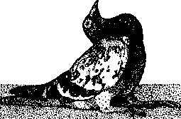
Figure 4. Emaciation in experimental avitaminosis (according to S. Funk).
The similarity between experimental polyneuritis and beriberi in humans, when fed exclusively on white rice, lies in the fact that rice lacks certain substances necessary for the normal metabolism of the peripheral nervous system. This was confirmed by Funk, who in 1911 succeeded in obtaining from rice bran an extract that cured beriberi (polyneuritis) in pigeons (when administered in minimal doses by subcutaneous injection). The active principle of this extract was named vitamin by Funk, and thus the entire doctrine of vitamins was begun. It was later finally established that both human beriberi and experimental beriberi in birds are avitaminoses arising from the absence of vitamin B in the food.
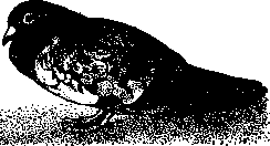
Figure 5. The same pigeon (see Figure 4), cured after 3 hours by administration of 8 mg of yeast vitamins (according to S. Funk). The symptoms of experimental beriberi in birds are as follows: first, the appetite falls, and then the birds (pigeons) completely refuse to eat rice. If feeding is then continued forcibly, after 10-20 days from the beginning of vitamin-free feeding, the characteristic specific symptoms of beriberi appear: pigeons begin to move with difficulty, severe spasmodic phenomena appear, the head is thrown back due to convulsions of the neck muscles, the legs are drawn to the abdominal area, etc.; soon after this the pigeons die. In some animals, spasmodic phenomena do not occur, but general paralyses gradually develop, which lead to complete loss of the ability to move. The weight curve constantly falls. Various metabolic disorders are also observed; for example, oxygen consumption and carbon dioxide output are decreased, tissue respiration is decreased (Abderhalden); creatine content in muscles is increased, oxidative processes are disturbed, carbohydrate metabolism is disturbed, as indicated by hyperglycemia, etc. It is interesting that the need for vitamin B increases with the carbohydrate content of the food. Pathological-anatomical studies of polyneuritic pigeons revealed degenerative changes in the n. vagi and ischiadici and enlargement of the adrenal glands; the secretion of adrenaline is also increased. B-avitaminosis in humans is not common, since vitamin B is widely distributed in plant food products; therefore, food is only in exceptional cases devoid of vitamin B, as for example when fed only on white (polished) rice, which occurred in Japan, Indochina, and other countries. These countries were the places where B-avitaminosis spread. The typical form of B-avitaminosis in humans is the atrophic or dry form of beriberi with typical symptoms from the nervous system, which patho-anatomically manifest as degenerative changes not only in the peripheral nerves but also in the spinal cord. In this form of beriberi, edemas may appear only in very severe and prolonged cases, when due to degenerative processes in the cardiac nerves and heart muscle, cardiac insufficiency occurs. The edematous form of beriberi gives a picture similar to that of edematous disease or hunger edema; sometimes mixed forms of typical beriberi and edematous disease are observed (see). Typical forms (atrophic) of beriberi are easily cured by administering vitamin B, provided the disease has not gone too far. The best effect is obtained from rice bran, yeast, etc. C-avitaminosis.
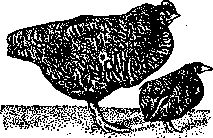
Figure 6. 7-month-old chickens: left - with normal nutrition (weight 2,500g); right - after severe avitaminosis, after being cured with unpolished rice and fish oil - no symptoms of rickets remained, but weight reached only 160g (according to S. Funk).
The consequence of the absence of vitamin C in the food of humans and some animals is their disease with scurvy. This view of the etiology of scurvy has long been held, based on observations of the beneficial effect of various vegetables and generally fresh plant products rich in vitamin C on scurvy. This view was finally confirmed by the research of Holst and Frølich (Axel Holst, Frølich) on experimental scurvy in guinea pigs. They found that when guinea pigs are fed only oats or grains of other cereals, they contract scurvy, the symptoms of which are completely identical to human scurvy. The cause of scurvy lies in the absence of vitamin C in cereal grains; scurvy is cured by cabbage, lemon juice, rich in vitamin C. Heating lemon juice to 100°, by destroying vitamin C, deprives the juice of its healing effect. Monkeys are also sensitive to the absence of vitamin C in their food, and in young monkeys typical infantile scurvy or Barlow's disease can be caused by feeding them condensed milk. Rats do not suffer from the absence of vitamin C in their food, and when fed with food that causes scurvy in guinea pigs, they never contract scurvy. Birds are also insensitive to the absence of vitamin C. The symptoms of experimental scurvy in guinea pigs are as follows: first, as in other forms of avitaminoses, loss of weight and loss of appetite are observed; then the characteristic, specific symptoms of scurvy as C-avitaminosis appear: on the 15th day, pains in the joints appear, making movement difficult for the animals; after three weeks, the animals assume the typical scorbutic position: they lie on their side, stretching out their diseased legs; then the teeth begin to loosen, the hair stands on end; the gums are strongly hyperemic, a scorbutic odor appears from the mouth; often still during life spontaneous fractures of the ribs are observed. Autopsy reveals clearly expressed hemorrhages in the muscles, in the subcutaneous tissue, in the gastric mucosa, phenomena of atrophy of the skin tissue, etc. C-avitaminosis in humans - scurvy - occurs when fresh food products are absent from the diet, namely fresh meat, fresh vegetables, fresh potatoes, milk, eggs, fruits, and when the diet is dominated by canned foods, smoked meat and smoked fish, dried vegetables and bread. Therefore, scurvy usually appeared during long sea voyages, during scientific expeditions to uninhabited places, during wars, etc. The World War also gave quite a few cases of scurvy. Scurvy is cured by lemon juice, fresh vegetables, i.e., products rich in vitamin C. Among vegetables, salad and cabbage are especially rich in vitamin C, but sauerkraut made from it is completely devoid of healing properties. When switching to food devoid of vitamin C, people do not immediately contract scurvy; sometimes months pass before the first signs of the disease appear. This is because some reserves of vitamin C are usually stored in our body. In children, poverty of the diet in vitamin C causes infantile scurvy or Barlow's disease. The work of Frazier showed that the cause of Barlow's disease is always feeding children with vitamin-free food, for example, sterilized or pasteurized milk, which are devoid of vitamin C. That is why Barlow's disease is cured by raw milk or juice from plant products rich in vitamin C. In scurvy, profound disturbances of metabolism are observed, some of which are common to other forms of avitaminoses. Generally, it should be said that the biochemistry of scurvy has been studied better than the biochemistry of other avitaminoses. In experimental scurvy, carbohydrate metabolism is disturbed, which is reflected in the blood sugar curve, which rises at the beginning of scurvy and falls at the end (A. Palladin). Disorders in creatine metabolism manifest as creatinuria and increased creatine content in muscles. Oxidative and synthetic abilities are also disturbed (A. Palladin), etc. E-avitaminosis. Vitamin E was recently discovered by Bishop and Evans (Bishop, Evans), who believe that its absence disturbs the functions of the reproductive organs, which is why they named it the vitamin of reproduction. This vitamin is contained in cereal seeds, especially in their germ part, and in green plants. E-avitaminosis is accompanied by degeneration of the seminal glands in males and cessation of pregnancy in females. The position of pellagra and various nutritional disorders in infancy in the system of avitaminoses has not yet been finally established. For more about vitamins, their distribution, chemical nature and role - see Vitamins; for more about individual avitaminoses - see Rickets, Scurvy, Keratomalacia, Pellagra, Beriberi.
Related articles
Mentioned in
Cite this page
“Avitaminoses.” Soviet Medical Encyclopedia. English translation of Bolshaya Meditsinskaya Entsiklopediya, 1st ed. (Moscow, 1928–1936), ed. N. A. Semashko. https://sovietmedicalencyclopedia.pages.dev/article/avitaminoses/