Pellagra
Historical document, translated for reference. It reflects medical knowledge of the 1920s–30s and is not medical advice.
Summary
A comprehensive historical overview of pellagra, a chronic disease characterized by dermatitis, diarrhea, and dementia, often associated with maize-based diets. The article details the evolution of theories regarding its etiology, ranging from infectious and parasitic causes to the prevailing 1930s understanding of nutritional deficiency and avitaminosis.
Encyclopedia article (1928–1936)
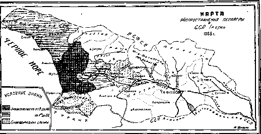
PELLAGRA (from Italian pelle skin and agra rough), a mostly chronically progressing disease with manifestations from the skin, gastrointestinal tract, and psyche (the classic triad of the three "D"s: dermatitis, diarrhea, and dementia); it has an endemic distribution in some countries where maize represents the staple food of the population. The first description of P. under the name Mai de la rosa was given by the Spaniard Casal (Gaspar Casal, 1735). In northern Italy, it was described by Pujati (A. Pujati) under the name "Alpine scurvy." In 1771, Frapolli (Frapolli) described it in Milan and first introduced into medical literature the popular name "pellagra," indicating the skin changes in this disease. In 1776, Odoardi (Odoardi) described this disease under the names: pellarina, scotatura de sole, calore de fegato, etc. Strambio (Strambio) studied P. in detail in a hospital specially founded (in 1786) for its study (in Legnano); he came to the conclusion that P. is a disease sui generis, a disease of the entire organism, and skin phenomena are only symptoms, which may also be absent—pellagra sine pellagra. In 1829, Hameau (Hameau) described P. in France under the name Maladie de la teste. In 1858, Theodori (Theodori) described it in Romania. There are a whole series of other names and synonyms: maidismus, mal del sole, mal' rosso, maladie anserine, der lombardische Aussatz, Erythema endemicum, etc. The etiology of P., despite numerous works, has not yet been clarified. From the very beginning, it was linked to nutrition with maize. Mazari (Mazari) as early as 1810 first definitely pointed to this. Hence the beginning of the zeistic theory of P. (Zea-maize). Guerreschi (Guerreschi) in 1814, considering spoiled maize the cause of P., proposed calling it "raphania maistica." Ballardini (Ballardini) in 1845, finding mold fungi in maize grains, considered them the cause of P.—hence the "Verdeto-theory" (from the green color of the mold). Lombroso (Lombroso) in Italy and Roussel (Roussel) in France developed this theory and considered spoiled, mold-grown maize the cause of P.: the "theory of moldy maize." Since 1906, a wide spread of P. has been observed in the USA; and in America, interest in the study of P. has gradually increased. As a result of this study, two schools have emerged: one considers P. as an infectious disease, the other as a consequence of nutritional deficiency. Since 1912, Funk (Funk), and then others, began to classify P. in the group of avitaminosis diseases. Besides these theories of the origin of P., a number of other theories were proposed by various authors, which now have only historical significance. Alessandrini (Alessandrini) considered contaminated drinking water (parasitic theory) the cause of P. Colloidal silicon found in some drinking waters was also considered the cause of P. (chemical theory, Alessandrini and others). Sambon (Sambon) considered P. a protozoan disease, and its transmitter a blood-sucking insect Simulium (protozoan theory or Simulium theory of Sambon). Since 1912, in the USA, P. was studied by a special commission, which recognized P. as an infectious disease. Many authors supported this theory. McCollum, Simmonds, and Parsons (McCollum, Simmonds, Parsons) considered P. an infectious disease, and attributed to food only a role contributing to the infection. Goldberger and Wheeler (Wheeler) from 1915 worked a lot on clarifying the question regarding the role of animal proteins in the diet of pellagrous patients and until recently considered P. as a result of nutrition with incomplete plant proteins. Only recently has Goldberger also joined the avitaminosis theory. At the present time, only the infectious, avitaminosis, animal protein defect, and general nutritional deficiency theories deserve attention. There are not yet sufficient grounds for classifying P. as an infectious disease; experimental research and epidemiological observations speak against this theory. Nutritional deficiency or defects can take place not only with a one-sided maize diet, but also with any other diet if the food lacks certain components necessary for normal nutrition. The question lies in which specific elements of food play a role in the etiology of P. Funk pointed to the avitaminosis character of P., and in 1917, Chittenden and Underhill (Chittenden, Underhill) induced a pellagra-like disease in dogs, which were kept on a special diet (rusks, peas, and cottonseed oil). Cary (Cary), Goldberger, and others identified a known disease of dogs—"canine typhus" or "canine oral thrush" ("black tongue")—with P. Both diseases arise from a nutritional defect; in both diseases, yeast gives a good therapeutic effect. Based on numerous observations, Goldberger came to the conclusion that the cause of P. is a lack of as yet unknown vitamin-like substances. Goldberger called this substance the P.P. factor (Pellagra preventive). The P.P. factor is found in yeast, milk, meat, tomatoes. This vitamin has not yet been sufficiently studied. The fact that P., in its clinical symptomatological and epidemiological features, resembles other avitaminosis diseases (beriberi, scurvy, xerophthalmia, etc.) also speaks in favor of the avitaminosis theory. According to Funk, in P. there are general symptoms of avitaminosis diseases (degenerative processes of the nervous system, gastrointestinal phenomena, atrophy, edema, indicanuria, hemeralopia, scurvy-like changes). Cherkes also notes that experimental and clinical observations provide grounds to classify P. as an avitaminosis, but considers the question of whether P. represents a mono-avitaminosis state unresolved. All these numerous works on clarifying the etiology of P., although they have clarified that nutritional deficiency and the absence of certain vitamins play an important role in the etiology of pellagra, still leave much that is controversial and unclear in this matter. Epidemiology and geographical distribution of P. People of all ages get sick with pellagra. Cases have also been described in infants (Voegtlin, Kandelaki, and others). Women most often get sick at the age between 20 and 40 years. Similar data were obtained during the study of P. in the SSR of Georgia in 1929 (Figure 1). The first manifestation of the disease, as well as relapses with exacerbations, are usually observed in early spring, sometimes in autumn, rarely in summer. In South Carolina (USA), 80-90% of all cases fall on the period April-June (Goldberger and others), in Italy the first manifestations and relapses are observed in March and April, in the SSR of Georgia—in March-April (Figure 2). The poorest rural population gets sick with P., hence one of the Italian names for the disease—malattici della miseria. Wealthy people get sick with P. as an exception, which once again emphasizes the importance of social conditions in the etiology of P. The geographical distribution of P. usually coincides with the cultivation of maize. Therefore, in Europe, P. is common in Italy (the most affected places are Piedmont, Lombardy), Spain, Portugal, Serbia, Bulgaria, Greece, and Turkey. In Romania, from 50,000 to 75,000 people get sick annually. P. has also been noted in France, Austria, and Hungary, with isolated cases in Germany and England.
seasons of the year. In America, a strong prevalence of pellagra has been noted since 1906 in the USA, Mexico, the West Indies, Brazil, Colombia, and Argentina. For the USA, the total number of pellagra patients is estimated at more than 50,000 (Lavinder) per year. Furthermore, pellagra is prevalent in Turkey, Persia, India, Central Africa, Algeria, Tunisia, and Egypt. On the territory of the former Russian Empire, pellagra was first described in Bessarabia by Martemyanov (1884) and Kholmsky (1887), with a strong prevalence of pellagra being noted, especially in the former Khotin Uyezd (Katsovsky, Prokhorovich). Pellagra in Georgia (Fig. 3) was first noted by Gaudelin in 1888; subsequently, pellagra was discovered in Kherson Governorate (Kazubov). In the USSR, pellagra has recently been noted in the Birzula District (ASSR) (Vaysman, 1929) and the Pervomaysk District (Nekhelis, 1929). In both cases, the spread of pellagra was noted in maize-growing regions. In some surveyed villages of the Libashov District of the Ukrainian SSR, up to 36.6% of all residents were affected by pellagra (Cherkes, Korovitsky, and Kleytman, 1929). Popov (1923) noted 22 cases of pellagra in the Armenian SSR. In the Georgian SSR, sporadic cases of pellagra were noted in the past as well. In the spring of 1929, a significant spread of pellagra was noted in some regions of Georgia where there are maize crops (Chkholaria, Alshibaya, Kandelaki, et al.). Pathological anatomy. The pathological-anatomical picture depends on the stage, form, and intensity of the case. In mild cases, it presents nothing characteristic. In neglected cases, the body tissues bear a senile imprint, the bones are brittle, and the muscles are atrophied (Ruge). The skin is initially inflamed and edematous, and the vessels are injected. Subsequently, hyperkeratosis, desquamation, and later atrophy are noted. In addition to hyperkeratosis, Zhgenti noted in some places a thickening of the papillary layer and atrophy of the remaining layers of the dermis. In many internal organs, atrophic phenomena are noted. In the late stage, the heart muscle is atrophied, the walls are thinned, and the heart is reduced in size. The spleen, liver, and kidneys also undergo atrophy. In the adrenal glands, Zhgenti found a sharp decrease in the amount of lipoids, in particular anisotropic lipoids. In the lungs, there are no characteristic changes. More important changes are found along the gastrointestinal tract. The tongue is inflamed, its surface is hypertrophied, and later atrophied. The mucous membrane of the intestinal canal is initially inflamed and hyperemic, and subsequently atrophied. Ulcerations are noted in the intestines. Sometimes redness, erosions, and subsequently atrophic phenomena are also noted in the stomach. More severe changes are observed in the nervous system. The pia mater is often thickened, with ecchymoses (Funk). Changes in the spinal cord are more constant; in chronic cases, degeneration of the posterior and lateral columns and the pyramidal tract is found; the cells of the anterior horns and Clarke's column may also be involved in the process. Polyneuritis has been described. In the cerebral cortex, various changes are observed in the ganglion cells, up to their complete disintegration, and proliferation of the neuroglia. Phenomena of sclerosis are observed. Thus, pathologically and anatomically, the spread of atrophic changes throughout the entire organism is confirmed, and on the other hand, chronic inflammatory processes, mainly in the gastrointestinal tract.
Course of the disease and symptomatology. Since pellagra for the most part has a long chronic course with exacerbations and remissions, as well as diverse forms in terms of severity, the symptoms and the picture of the disease are different. Some authors divide the course of the disease into different stages, forms, or periods. Funk, just like Roberts, recognizes 4 types of the disease: 1) acute malignant form, 2) mild subchronic form, 3) severe cachectic subchronic form, and 4) chronic form. Babes and Sion distinguish four stages: 1) prodromal or pre-erythematous; 2) erythematous with gastrointestinal and general nervous phenomena, 3) predominance of nervous phenomena, and 4) depressive. Frapolli notes stadium initiatum, confirmatum, and desperatum; Strambio—stadium intermittens, remittens, and continuum; Lavinder—prodromal, first, second, and third stages. All such divisions from a clinical point of view are mostly artificial. In general, the clinical picture of pellagra is characterized by three groups of symptoms: 1) skin phenomena, 2) gastrointestinal disorders, 3) changes in the nervous system and psyche. The prodromal period proceeds with indefinite phenomena and general complaints: headache, dizziness, weakness in the lower extremities, pain in the spine, a feeling of burning in the mouth and stomach, belching, heartburn, salivation, and depressed mood. The first manifestations on the part of the skin (which is considered the beginning of the disease) begin in the spring, sometimes in the autumn, and more rarely in the summer. Skin changes (erythema, dermatitis, pellagraderm, etc.) usually appear on symmetrical, sharply defined parts of the body that are not covered by clothing. Under the action of the first rays of the spring sun, the phenomena of erythema occur immediately: red spots appear on the skin, which disappear upon pressure and then reappear. The redness becomes more intense, sometimes taking on a bluish tint; swelling of the skin is noted; the rash resembles solar erythema. Patients complain of burning and heat in the skin. Blisters or pemphigus-like pustules (pemphigus pellagrosus) may appear; under the influence of secondary infection, ulcerations are sometimes observed. Within 2-3 weeks, the redness gradually disappears, and the swelling decreases. Then the skin thickens (hyperkeratosis) and subsequently peels. On the border of the erythematous and healthy skin, one can notice the so-called "hyperkeratotic zone" (Merk). The skin becomes strongly pigmented and atrophies after repeated relapses. Such parchment-like skin is dry, thin, shiny, and very wrinkled. Skin lesions are usually localized on the back of both extremities, the face, and the neck. Atypical localizations occur in the axillary cavity, knees, elbow bend, on the external genitalia, and on the scrotum. In rare cases, erythema is observed in asymmetrical places and on covered parts of the body. A combination of vitiligo with pellagrous erythema is observed. Regarding the frequency of localization of skin lesions, it was noted (Kandelaki) on the hands in 74.27%, on the feet in 67.7%, in the jugular fossa in 6.7%, on the cheeks in 6.5%; in different places simultaneously: on both extremities in 27%, on both extremities. Figure 3.
and neck in 12.8%, on both limbs, neck and chest in 4.1%. Skin lesions in pellagra in specific places give a characteristic picture: on the forearms—"pellagrous gloves" or "pellagrous cuffs," on the lower leg "pellagrous boots," on the neck—"cervical bow" or "Casal's collar"; from the neck the skin lesion descends to the chest in the form of a triangle with the apex pointing downward; on the face, the nose, cheeks, temples, chin, eyelids, areas behind the ears, above the eyes, and sometimes the lips are affected ("pellagrous mask") (see separate table, figs. 2-7). Gastrointestinal disorders usually appear early. A burning sensation in the mouth, heartburn, and salivation with loss of appetite are noted. Sometimes the appetite is increased. The tongue is coated, inflamed, painful, dry, and cracks; in places, the epithelium peels off, becoming exposed ("bald tongue" - Sandwith's bald tongue). Sometimes very long papillae on the upper surface and a thick coating with cracks give the tongue the appearance of a chessboard (Lavinder). The mucous membrane of the oral cavity is carmine-red and swollen. The gums are red, sometimes loosened and bleeding. Sometimes alveolar pyorrhea is observed. In some cases, changes in the oral cavity resemble scurvy, and changes in the tongue resemble sprue. Patients often complain of pain in the stomach area. Phenomena of chronic gastritis are observed. A decrease or absence of hydrochloric acid, as well as pepsin, is noted. There is an alternation of constipation and diarrhea. In severe cases, there is frequent diarrhea, sometimes mucous-bloody with colic and tenesmus, as in dysentery. Nervous system. Initially, a picture of "pellagrous neurasthenia" is noted. The patient's character changes, the mood often fluctuates, they become irritable, a feeling of anxiety, depression, and malaise is observed, insomnia, confusion, and memory impairment occur. The patient complains of easy fatigability, weakness in the lower limbs; the gait becomes unsteady, ataxic (ataxic paraplegia). Neuralgic pains, paresthesias, and wandering pains are observed. In more severe cases, pellagrous psychoses are described: delirium, hallucinations, inexplicable fear, depression, stupor, stereotypy. Classification of psychic phenomena in pellagra according to Gregor: 1) neurasthenia, 2) stupor, 3) amentia, 4) acute delirium, 5) catatonia, 6) fear psychoses, 7) pictures of manic-depressive states. Kapovsky found an amentive state in 90%, depressive in 8.7%, and catatonic in 1.3%. In a later work, Bonhoeffer considers psychotic states in pellagra as an "exogenous reaction type," common in intoxications. Further, disorders of coordination, ataxia, tremors of the hands, tongue, and lips, convulsions, contractures, and rigidity of the lower limbs are noted. The gait is sometimes spastic or spastic-paralytic; in more severe cases, epileptiform and choreiform convulsions, manic-depressive psychosis. Of the sensory organs, the organ of vision suffers significantly. In many patients, visual impairment and diplopia are noted. Cases of cataract are observed. The sense of taste is altered or perverted; a salty taste in the mouth is often felt (which is why the disease was often called "salso" by the people in Italy). There is a disorder of the sense of smell. Disorders on the part of the genital organs are also noted. These include inflammatory phenomena of the vagina, uterus, and ovary, dysmenorrhea, amenorrhea, and abortions (according to Funk, in 20%). In men, impotence is observed. The blood picture depends on the form and nature of the disease. A decrease in the number of erythrocytes and the percentage of hemoglobin is noted, leukopenia with relative lymphocytosis. There are cases when the number of erythrocytes and the percentage of hemoglobin approach normal (or are normal). Makhviladze notes a decrease in the number of erythrocytes, percentage of hemoglobin, leukocytes, and neutrophils, an increase in the percentage of lymphocytes and mostly normal percentage of eosinophils, basophils, and monocytes. Anding and Sinani note normochromic anemia with leukopenia and relative leukocytosis with protein depletion (hydremia) and acceleration of blood clotting time. The Wassermann reaction and examination of the cerebrospinal fluid give a negative result. In the urine, there is an increase in indican, hippuric acid, and ethereal sulfuric acid (Lavinder). The temperature in pellagra is usually normal; sometimes slight increases are observed in the evenings. The pulse is mostly soft, slightly accelerated, heart sounds are muffled, blood pressure is lowered. The duration of the disease is varied; it can last from one year to several years and even a lifetime. The diagnosis of pellagra is not difficult when the triad of pellagra (erythema, disorder of the digestive tract, and the nervous system) is clearly expressed. For an accurate diagnosis, skin changes are especially important. In the absence of skin phenomena (pellagra sine pellagra), as well as in periods of remission, when skin phenomena and other symptoms have subsided or disappeared, it is necessary to establish observation of the patients and look for characteristic traces of past skin changes. In differential diagnosis, attention is paid to various skin changes: erythema solare, vitiligo, eczema, erythema exudativum multiforme Hebrae (Merk), to intoxication diseases: ergotismus, lathyrismus, acrodynia, and the following diseases: leprosy, scurvy, sprue, beri-beri, syphilis, gastrointestinal and nervous diseases (M. Mayer). With concomitant diseases (ankylostomiasis, malaria, dysentery, typhus, syphilis) that complicate the diagnosis, appropriate laboratory studies are necessary. Prognosis is always serious. Mild cases and initial stages are cured with a timely change of diet. Severe and neglected cases, especially in old people and women, give a high percentage of mortality. Any complication worsens the prognosis. The typhoid form is mostly fatal. The treatment of pellagra must first of all be dietary. A change of diet and improvement of nutrition is an old, proven remedy. Dietary treatment must be early, rational, and prolonged. Only early treatment, before the disease has produced deep changes in the organs, gives good results. A rational diet must contain a sufficient amount of animal proteins, vitamins, and other substances necessary for normal nutrition. Food must be varied. Lavinder places milk
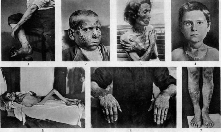
Figure 1. Secondary infection from scratching after mosquito bites. Figure 2. Pellagrous mask. Figure 3. Skin changes on the face, neck, chest, and arms in pellagra. Figure 4. Casal's cervical bow in pellagra. Figure 5. Cachectic form of pellagra; ulcerations on the arms and legs. Figure 6. Lesions of the arms in pellagra. Figure 7. Skin changes on the lower leg and knees in pellagra.
and dairy products; furthermore, veal, liver, kidneys, tender beef, and other meat products are recommended, as well as greens and fruits. According to Funk, liver, kidneys, milk, and eggs contain vitamins against pellagra. Yeast is especially rich in these vitamins and is used in pellagra as a specific remedy. Maize should, if possible, be excluded from the diet and replaced with wheat bread. Gastrointestinal disorders should not interfere with the variety of food. To avoid relapses, dietary treatment must be prolonged. Of medications, preparations of arsenic, iron, as well as strychnine and iodine are recommended. Affected areas are kept clean; one should avoid sunlight if possible, and use gentle ointments if necessary. The oral cavity must be kept clean; in case of a decrease in HCl in the gastric juice, diluted HCl is given. Against nervous phenomena—physiotherapeutic treatment. More or less serious cases require special inpatient treatment (pellagra hospitals, sanatoriums, boarding houses, mountain-climatic stations, etc.). Prevention. Since most authors classify pellagra as an avitaminosis, all preventive measures must be directed toward the rationalization of the population's nutrition. Here, the appropriate organization of public catering and the quality of food must take place. This is especially important during crop failures or other moments entailing a quantitative and, consequently, qualitative change in nutrition. A change in the usual diet plays a large role. Accurate registration of the sick, constant medical observation, and treatment are necessary. The cultivation of maize (corn) should be varied with other crops. And here, state farms and collective farms must and can play a huge role by correctly setting up the sowing of the corresponding cereals and rationalizing public catering.
Related articles
Mentioned in
Cite this page
“Pellagra.” Soviet Medical Encyclopedia. English translation of Bolshaya Meditsinskaya Entsiklopediya, 1st ed. (Moscow, 1928–1936), ed. N. A. Semashko. https://sovietmedicalencyclopedia.pages.dev/article/pellagra/