Yellow Fever
Historical document, translated for reference. It reflects medical knowledge of the 1920s–30s and is not medical advice.
Summary
This article from the 1928–1936 Soviet Great Medical Encyclopedia discusses yellow fever, detailing its history, transmission via the yellow fever mosquito (Aedes aegypti), etiology, geographical distribution, and epidemiology.
Encyclopedia article (1928–1936)
YELLOW FEVER, an acute general infectious disease with primary involvement of the liver and kidneys, rapid development of symptoms of general intoxication, appearance of jaundice, albuminuria, and vomiting of black masses containing blood. Associated with a specific vector and temperature minimum, yellow fever is a disease of hot countries, but under certain conditions, which rely entirely on the well-studied biology of the yellow fever mosquito, it can be carried far to the north. Therefore, yellow fever is of special significance for world communication, and it has long been included in the list of diseases subject to international sanitary conventions. The latest convention of 1926 provides a number of measures for ports and ships regarding their prophylaxis against yellow fever. Vector. The question of yellow fever arose particularly acutely in 1899, during the war between America and Spain. American troops introduced into Cuba suffered heavy losses from yellow fever. This prompted the American government to create a commission to study it. The commission, consisting of Reed, Carroll, Agramonte, and Lazear (Reed, Carroll, Agramonte, Lazear), began by studying the mode of transmission of the disease; for this purpose, the members of the commission experimentally, including on themselves, tested all existing hypotheses: they took the excreta and urine of patients and introduced them to themselves, took mucus from the pharynx of patients and swallowed it, but no one fell ill. Then the commission tested the fact noted as early as 1881 by Finlay (C. Finlay) that wherever there is yellow fever, there are always special mosquitoes, that the epidemic season occurs when there are many of them, and that where the temperature does not allow these mosquitoes to breed, yellow fever does not exist; thus, in hot Rio de Janeiro there is yellow fever, while in Petrópolis, located 45 km from Rio at an altitude of 800 m, with a temperature unfavorable for the development of the aforementioned mosquitoes, it is absent. Indications of the possible role of mosquitoes in the epidemiology of yellow fever were made even earlier—in 1848 and 1853—by the physician Nott and Beauperthuy (Nott, Beauperthuy). With the aim of experimentally verifying Finlay's proposition, after one of the members of this commission (Carroll) fell ill and two others (Lazear and Reed) died of yellow fever under conditions of a volunteer experiment (bitten by an infected mosquito), the commission carried out the following direct experiment. A special, well-disinfected house was divided in half by a metal screen; healthy people were placed in both halves; infected mosquitoes were let into one half; the people placed in the half with mosquitoes became infected, while the people in the second half remained healthy. Subsequently, it was clarified that this insect had been described as early as 1762 by Linnaeus, who named it stegomyia (Stegomyia aegypti L.), which means "living at home." According to modern zoological nomenclature, it should be called Aedes aegypti L.; in domestic literature, it has been proposed to name it in Russian as well—the yellow fever mosquito (Shakhov S., 1926). According to Theobald, it is distributed in both hemispheres between 40° north and south latitude. In this zone, preferring densely populated cities of coastal countries, it sometimes penetrates along major waterways deep into the interior of the country. It is also present in the USSR along the Black Sea coast of the Caucasus (Martsinovsky, 1913; V. Hoffman, 1920). Etiology. Until 1918, more than 20 researchers believed that they had succeeded in discovering the causative agent of yellow fever; however, verification work each time indicated the erroneousness of these conclusions. In 1918, Noguchi, studying 27 yellow fever patients in South America in Guayaquil, discovered in 6 of them a leptospira close in its morphological features to Leptospira icterohaemorrhagiae, the causative agent of Botkin-Weil disease. Recognizing it, on the basis of certain immunity reactions, as the causative agent of yellow fever, Noguchi named it Leptospira icteroides. His view at first found confirmation in the work of his students. However, further study of a number of strains of this leptospira, carried out recently, led to the conclusion that they cannot be reliably differentiated from the causative agent of Botkin-Weil disease either by immunity reactions or by other methods. In the exact same way, the searches for Leptospira icteroides carried out in Brazil (Borges Vieira) back in 1920 proved fruitless. Nor did studies conducted in the same direction in Africa by the British expedition headed by Stokes (Stokes, 1928) yield a single positive finding. These negative results acquired decisive significance in connection with the fact that Stokes, Bauer, and Hudson (Bauer, Hudson) succeeded for the first time in reproducing yellow fever in Macacus rhesus monkeys (see below), while upon infecting them with the blood of yellow fever patients, they never once managed to detect Noguchi's leptospira. Most recently, a report appeared on the discovery by Kuczynski of a special bacillus as the causative agent of yellow fever—Bac. hepatodystrophicans. This report, considering the entire rich history of searches and "discoveries" of the causative agent of yellow fever, requires further confirmation. Nevertheless, the features of the sought-after causative agent of yellow fever are well known thanks to the classical work on yellow fever carried out at the beginning of the 20th century by the American commission on the island of Cuba, the French commission of the Pasteur Institute in Rio de Janeiro, and the American commission in Mexico. These studies established that the causative agent of yellow fever: 1) under natural conditions is found only in the blood of yellow fever patients and females of the mosquito Aedes aegypti; 2) under natural conditions is transmitted from the sick to the healthy exclusively through the bites of infected Aedes aegypti; 3) being in the blood of both small and large vessels, is not bound to blood elements (positive result of infection with serum alone); 4) does not circulate in the blood during the incubation period, appearing in it in the patient when the fever begins, and on the other hand disappears from the peripheral blood already on the 3rd–4th day from the start of the disease (infection of the mosquito does not occur either in the incubation period or after the 3rd–4th day of the disease); 5) Berkefeld filter W retains the yellow fever virus contained in the patient's serum, while filters V and N let it through; however, an emulsion prepared from infected mosquitoes is retained even by filters V and N; on this basis, it is thought that the causative agent of yellow fever in the mosquito has a different shape and size than in the patient's blood; 6) remains viable in the body of Aedes aegypti throughout the mosquito's life and can sometimes pass to the offspring of the latter; 7) requires a certain temperature minimum (not less than 20°) for its development in the body of the Aedes aegypti female; the mosquito itself becomes capable of infecting a person with yellow fever only 10–12 days after the bite of a patient. Work from 1926–1928 carried out in Africa (Stokes, Bauer, Hudson, Aragao) established that the yellow fever virus is unable to cause disease in any animal of Europe and Africa (which coincides with the epidemiological observations of old authors). On the other hand, a number of Indian monkeys proved susceptible (Macacus rhesus, M. cynomolgus, M. speciosus, M. sinicus), which also show the full picture of histological changes in the liver characteristic of humans in yellow fever. This discovery of an animal susceptible to yellow fever opens a new era in the experimental study of yellow fever. Geographical distribution and statistics. Yellow fever is found in the tropical parts of America and West Africa. The first reliable information about it in America dates back to the 17th century, and in Africa to the 18th century. Some consider America to be the homeland of yellow fever, others Africa. The boundary points where yellow fever was ever registered were: for the Western Hemisphere—Quebec in the north (48° N lat.), San Francisco in the west (122° W long.), Buenos Aires in the south (35° S lat.); for the Eastern Hemisphere—Swansea in the north, in England (51° N lat.), Trieste in the east, in Italy (24° E long.), and São Paulo de Loanda in the south, in Africa (10° S lat.). In particular, in the USA yellow fever spared not a single port, right up to Quebec in Canada, and along the Mississippi and Missouri it repeatedly penetrated deep into the country. It was repeatedly introduced into Northern Europe, and in Southern Europe it caused a series of severe outbreaks, for example in Cadiz in 1774 with 10,000 deaths, in Barcelona in 1821 with 25,000, in Lisbon in 1857 with 5,652, in Brest in 1802 with 1,839. The last introduction into Europe (from America) was noted in 1894 in Italy (Trieste) and in 1908 in France. Yellow fever has never been noted in Asia, Australia, East Africa, and on the territory of the USSR. The geographical distribution of yellow fever before the introduction of modern methods of control (see below—prophylaxis) can be seen from the map given in vol. VI, art. 623–624. At present, yellow fever, practically considered eradicated in America, is thus limited to West Africa. Here yellow fever for 1926–1928 was noted in Senegal, Sudan, Nigeria, Togo, Dahomey, Liberia, the Ivory and Gold Coasts, and the Belgian Congo. However, exact statistical data on its distribution there are not available (Hoffman). Epidemiology. In endemic localities, yellow fever is maintained by mild, abortive, and asymptomatic cases of the disease among the children and relatively immune (i.e., those who had the disease in childhood) population.
Outside of endemic areas of distribution, yellow fever can cause an outbreak under the following conditions. 1. Importation of an infected vector, more rarely of a yellow fever patient. The yellow-fever mosquito is well adapted for its passive transport over long distances by both water and rail routes. Repeatedly introduced in this way from the island of Cuba to southern Spain, yellow fever caused there, for example, from 1800 to 1804, 280,000 cases with 79,000 deaths. The French commission of the Pasteur Institute in Rio, studying this pathway of yellow fever outbreak emergence, transported 57 infected mosquitoes from Brazil to Paris in good condition, which produced 5 generations over the period from May to September. 2. Presence of the vector Aedes aegypti. It must be present year-round, otherwise the yellow fever outbreak will be short-lived; thus, in New York, which in the past very often became infected with yellow fever, this disease could never become established for long because the yellow-fever mosquito died off by winter and its reappearance the following summer was each time due to a new importation by steamboat (Carter, Grubbs, Souchon). Furthermore, it must also be present in a sufficiently large quantity (Gorgas, Simond, Aubert, Noc). Observation experience of recent decades indicates that where the so-called Aedes-index, i.e., the percentage of residential houses in which the vector was detected, lies below 3%, isolated introductions of yellow fever (or of an infected vector) were unable to cause a yellow fever outbreak. The presence of Aedes aegypti year-round depends mainly on the average annual temperature (not below 20°) and sufficient moisture, while its quantity depends on the abundance of artificial breeding sites (barrels, cisterns, tanks, and other water reservoirs common in the absence of a water supply network). 3. Presence of an average temperature of 21–26°. According to Hirsch, yellow fever could never establish a permanent focus in localities where the average annual temperature is below 20°; thus, having caused an outbreak in summer outside its possible endemic distribution, yellow fever becomes rarer with the first cooling of the weather, to disappear altogether when the temperature drops below 20–18°. 4. Presence of a non-immune population. The realization of the 1st, 2nd, and 4th requirements is absolutely necessary for yellow fever cases to occur at all. The degree of fulfillment of the 2nd and 3rd requirements mainly determines the further course of the begun epidemic, its duration, or its complete disappearance with the change of season. All prerequisites for a possible, although perhaps short-lived, outbreak of yellow fever in the event of the realization of the first condition exist in the USSR in certain ports of the Black Sea coast of the Caucasus, to which attention was drawn in 1913 by E. I. Martsinovsky in the first work at that time in Russia entitled Yellow Fever and Stegomyia fasciata Mosquitoes. Pathological anatomy. A yellowish discoloration of the skin is noted, sometimes gradually increasing; a pronounced yellowish discoloration of the internal organs, especially the liver; the presence of dark masses of unchanged blood in the stomach and small intestines. In the mucosa of the stomach and duodenum, hemorrhages and erosions. Hemorrhages are also noted in the skin, meninges, peri-, endo-, and myocardium, in the aortic wall, and in the lung tissue. These hemorrhages impart a septico-hemorrhagic character to yellow fever (Hoffmann). In the heart, phenomena of acute myocarditis. The liver is usually of normal size, clay-colored, with fatty degeneration. The spleen is not enlarged. In the kidneys, phenomena of nephritis. The changes in the liver can be so severe that the structure of the liver tissue is recognized with difficulty, while the histological picture then resembles sharply pronounced forms of acute yellow atrophy of the liver. Rocha-Lima considers the zonality of phenomena to be more characteristic of yellow fever, which consists in the fact that in the liver lobules there is, as it were, an alternation of parenchymal layers with degenerative and necrobiotic changes: necrotic cells are located mainly in the intermediary parts of the lobules, while fatty-degenerate cells predominate in the peripheral and central zones. In the kidneys, besides general phenomena of nephritis, calcium casts are often found (Rocha-Lima; 1912). In the Kupffer stellate cells of the liver, in the endothelial cells of the splenic pulp, and partly in the lymph glands, phenomena of erythrophagocytosis are frequent (Hoffmann; 1927). Of all these patho-anatomical changes, main attention is paid by all researchers to the liver. Where there are no serious changes in the liver, according to Hoffmann, yellow fever can be excluded, even in cases where the clinical picture gave all grounds to suspect its presence. Pathogenesis. The yellow fever virus (resp. its toxin), causing regressive changes in parenchymatous organs, also damages the endothelium of capillaries, especially of the stomach and intestines, causing severe hemorrhages into the alimentary canal. On the other hand, these hemorrhages are also caused by changes in the liver, by the very presence of jaundice. The reduced vitality of the liver leads to a strong decrease in urea in the urine, starting from the first day of the disease (the absence of this sign almost certainly excludes yellow fever). Kidney damage produces, starting from the 3rd day of the disease, an equally characteristic albuminuria, and subsequently a sharp decrease in the amount of urine. The toxin of the yellow fever causative agent has not yet been isolated, but it can be clearly judged by the abundant formation of antitoxins and specific antibodies that are formed in the patient's body after infection. These protective bodies lead to the fact that the yellow fever causative agent always disappears from the body very quickly, within the first three days from the onset of the disease, and individuals who have successfully survived the disease acquire a robust immunity. Clinical features. The incubation period, according to experimental data and observations under natural conditions, is on average 3–4 days, but can also last 12 days. Two periods of the disease are distinguished: the first lasting 3 days, with a high temperature of the continua type, sometimes with insignificant intermissions, with a frequent, hard pulse of 110–125 beats and a number of initial symptoms on the part of the liver, stomach, kidneys, and cardiovascular system. The second period often begins with a short-term (lasting only a few hours) drop in temperature to normal on the 3rd–4th day of the disease and is characterized by subsequent small remissions, the appearance of severe disorders on the part of the internal organs, and a drop in the pulse rate to 50 and below. Dicrotism is a usual phenomenon. Blood pressure is lower than in all other infections. The temperature curve in yellow fever is not characteristic enough to make a diagnosis from it. The disease begins with chills, a sudden rise in temperature to 39° and above, agonizing headache and muscle pain, epigastric pain, eructation, and vomiting—initially with bile, and then, in the second period, due to an admixture of altered blood, with black masses resembling coffee grounds; for the same reason, the stools are tarry. From the 2nd day, there is subictericity of the conjunctivae, followed by severe jaundice. From the very first days of the disease, the total amount of excreted urine noticeably drops, starting from the 3rd day hyaline casts and traces of protein are detected; subsequently, the amount of protein strongly increases; granular, waxy casts, renal epithelium, and sometimes erythrocytes appear, the amount of excreted urea sharply decreases (down to 1.5 g per liter); the amount of urine progressively drops and can reach complete anuria. The spleen is not enlarged. Consciousness is usually clear. Regarding blood changes, the number of erythrocytes gradually increases, the color index is usually above unity; moderate neutrophilia with a left shift is observed. Viscosity is increased. Marked bilirubinemia, a reduced amount of Ca salts and increased K. The clinical picture is most pronounced on the 6th–7th day of the disease. Death usually occurs between the 5th and 9th day of the disease (Fig. 1). Shortly before death, delirium and convulsions occur. If the disease takes a favorable turn, the vomiting stops, the protein disappears, the amount of urine increases, the temperature drops to normal no later than the 10th–12th day, and the patient slowly recovers (Fig. 2). Despite the severe course, the disease does not leave behind any noticeable complications. In children, yellow fever proceeds in an abortive form. Beginning like in adults, it ends on the 3rd day, with jaundice and nephritis absent; sometimes, however, the disease proceeds as in adults. Diagnosis and prognosis. Diagnosis.
Of great importance are 1) jaundice, which is recognized spectroscopically within the first 36 hours; 2) early-onset and progressive albuminuria. For abortive cases, proof is the inoculation of 2-5 cm3 of blood taken in the first 3 days of the disease into several monkeys (Macacus rhesus); after an incubation of 3-8 days they fall ill and on the 2nd-8th day of the disease die with the pathological-anatomical changes in the liver characteristic of yellow fever. Differential diagnosis: hemoglobinuric fever (hemoglobin in the urine, vomiting of bile rather than black masses mixed with blood); Botkin-Weil disease (examination of urine sediment for Leptospira icterohaemorrhagiae, inoculation of patient blood into guinea pigs); severe forms of malaria and relapsing fever accompanied by jaundice (presence of blood parasites); dengue (exanthema, rapid pulse). The prognosis in yellow fever is always serious. Mortality, usually very high, varies depending on the epidemic between 30 and 96 percent. Treatment. Bed rest, liquid diet, diuretics, cardiacs; to combat vomiting—ice pills, cocaine (morphine is contraindicated); Sternberg introduced alkalis into the therapy of yellow fever, since the vomit is always acidic; he administers Natr. bicarbon. 9.0 with Hydrarg. bichlor. corr. 0.02 per 1,000.0 Aq. dest. in doses of 45 cm3 hourly. There is no specific drug treatment. A favorable effect on the course of the disease is exerted by convalescent serum (Marchoux). At present, great hope is placed on the possibility of obtaining therapeutic serum from those who have recovered from yellow fever following experimental infection of monkeys (Hindle, Aragão). Prophylaxis. The measures taken simultaneously address the yellow fever patient and the vector—the yellow fever mosquito. A patient suffering from yellow fever must be placed in conditions that completely exclude the possibility of access to him by the yellow fever vector (specially equipped hospitals, general screening, and a canopy over the patient's bed day and night). The entire population of the locality where a case of yellow fever has occurred is placed under the strictest medical observation, taking into account the presence of a definite period of 14-21 days between the first and subsequent illness. Isolation of the patient himself for more than 5 days is useless from a prophylactic point of view, because as early as 3 days from the onset of the disease the virus is no longer found in the patient's blood. Of paramount importance is the early diagnosis of the first case of yellow fever and timely control of the yellow fever mosquito. Control of the latter is incomparably easier than control of the malaria vector. This accessibility and success of control are due to the fact that 1) the yellow fever mosquito, unlike Anopheles, is almost exclusively a dweller of cities rather than open rural areas, i.e., we have a relatively small territory well accessible to our control; and 2) the yellow fever mosquito almost never leaves human dwellings, where it lays its eggs in water receptacles directly adjacent to the house or even within the house itself (washbasins, water jugs, flower pots, etc.). This control is carried out by specially trained personnel (the so-called "mosquito squads" in America). After preliminary sealing of all cracks with paper, houses are fumigated with sulfur at the rate of 10-20 g per 1 m3; in parallel with this, the presence of all artificial water receptacles is meticulously ascertained (in natural bodies of water, larvae of the yellow fever mosquito are as a rule absent); some of these receptacles are eliminated, while others of economic importance (cemented basins, tanks, etc.) are placed under registration and strict constant control, conducting work according to the general rules of anti-larval control and taking into account that the development cycle of the yellow fever mosquito from egg to imago is on average 9-10-12 days. By the indicated method, yellow fever was first eradicated by Gorgas in Cuba (1901-1902); Osvaldo Cruz in Rio de Janeiro (1903), where prior to the control of the yellow fever mosquito up to 4,800 people died annually from yellow fever, and immediately following the control (1904) — 48; Liceaga in Veracruz (1906), etc. The last case of yellow fever in Guayaquil was recorded in 1919, in Peru and Guatemala in 1921, in Mexico in 1922, etc. In 1925, only 3 cases of yellow fever were recorded throughout the whole of America (in Brazil); the years 1926 and 1927 were equally favorable. In 1928, a more serious outbreak of yellow fever was noted in Brazil (103 cases, 56 of them fatal); nevertheless, the general organization of prophylactic work and the success achieved by it leave no doubt in the minds of most researchers that the whole of America is on the eve of the complete disappearance of yellow fever (Fig. 3). However, prophylactic measures have so far hardly touched Africa.
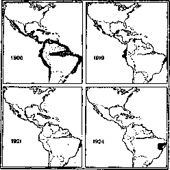
Figure 3. Distribution of yellow fever in Central and South America in 1900, 1919, 1921, and 1924. (Foci are blackened.) Therefore, taking into account the speed of travel on modern communication routes (airplanes) and the possibility of passive transport of the yellow fever mosquito from unfavorable regions (Africa) to favorable ports of the East that have never known yellow fever, modern prophylaxis puts forward the requirement to pre-arrange all threatened ports in such conditions that even the importation of a yellow fever patient or an infected vector could not cause an outbreak so common to America and Europe of the 19th century. This is provided for in the corresponding paragraphs of the international sanitary convention of 1926. Regarding the USSR, it must be remembered that the yellow fever mosquito is present in Batumi, Poti, and Sukhumi, from where it is sometimes carried by locally plying steamboats to upstream ports: Gudauta, Gagra, Tuapse (Lindtrop, 1925).
Related articles
Mentioned in
Cite this page
“Yellow Fever.” Soviet Medical Encyclopedia. English translation of Bolshaya Meditsinskaya Entsiklopediya, 1st ed. (Moscow, 1928–1936), ed. N. A. Semashko. https://sovietmedicalencyclopedia.pages.dev/article/yellow-fever/