Leukoma
Historical document, translated for reference. It reflects medical knowledge of the 1920s–30s and is not medical advice.
Summary
Leukoma is an opaque white opacity of the cornea resulting from scarring after inflammation or injury. This article describes the types, causes, anatomical features, clinical manifestations, diagnosis, prevention, and treatment of leukoma.
Encyclopedia article (1928–1936)
LEUKOMA (leukoma'), cataract, an opaque white opacity of the cornea resulting from scarring after inflammation or injury to it. In rare cases, congenital leukomas are observed, and their causes vary: on the one hand, inflammatory processes of the cornea in the embryo, on the other, apparently abnormalities in embryonic development. Depending on their size, partial and complete leukomas are distinguished (leukoma partiale et leukoma totale); when adhesions form with the iris, adherent leukomas (leukoma adhaerens, Fig. 1) result. Leukoma represents the most intense scarred opacity of the cornea; less intense opacities, depending on their transparency, are called nubeculae or spots (macula). Anatomically, all these opacities represent a layer of newly formed connective tissue of varying thickness, developed in place of the destroyed part of the cornea (Figs. 2 and 3); in leukoma tissue, newly formed vessels are also present. In leukomas, secondary changes may develop in the form of colloidal, hyaline, and amyloid degenerations, sometimes with lime deposits. Clinically, this manifests as yellow-colored spots of various shades against the main background of the leukoma. Leukomas may be at the same level as the surrounding cornea; on the other hand, a slight depression-facet may form in their place due to insufficient development of scar tissue; there may also be protrusion due to stretching of the scar (leukoma ectaticum) as a transition to corneal staphyloma or, more rarely, due to excessive development of scar tissue and thickening of the epithelium. With extensive leukomas, flattening (applanatio) of the cornea may occur. When evaluating leukomas, particular importance
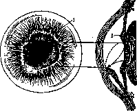
Figure 1. Leucoma adhaerens; l-iris.
is attached to their adhesions with the iris (anterior synechiae), which are recognized by displacement of the pupil toward the adhesion, change in its shape in the form of a pear (Fig. 1), and uneven depth of the anterior chamber; sometimes the dark color of the scar indicates its adhesion to the iris. Such adhesions may vary in size, from very thin threads to extensive adhesions involving the entire pupillary border of the iris. Leukomas cause a decrease in vision, the extent of which depends on the position of the leukoma in relation to the pupil and on the size and intensity of the leukoma itself. Light rays, passing through the opacity, instead of proper refraction, are scattered in all directions and do not give any clear image on the retina. These circles of light scattering worsen vision even in cases where the leukoma occupies only part of the pupillary area and the rest of the cornea is transparent. This light scattering also explains the peculiar sensation of blinding that patients with leukomas sometimes complain about. In addition, scars of the cornea usually result in irregular astigmatism with all its consequences for vision. This is particularly pronounced in ectatic leukomas. No less important for the eye are the indirect consequences of leukomas, especially those adherent to the iris. Of these consequences, the most important is increased intraocular pressure, with subsequent glaucoma-a frequent cause of blindness in the affected eye. Similar consequences of leukomas include strabismus, myopia, and nystagmus, which most often develop in cases where the leukoma has existed from early childhood.-The basis for differential diagnosis of leukoma from inflammatory opacities (infiltrates) of the cornea is: their bluish-white or purely white color, smooth shiny surface, and absence of the usual signs of irritation and inflammation of the eye, especially pericorneal injection.-Prevention of leukomas reduces to prevention of inflammations and injuries to the cornea. Since leukoma represents a scar, their clarification is impossible, but since along with scar tissue, remnants of infiltrate may also be present in leukomas, irritating therapy is usually prescribed to dissolve these remnants. This includes: yellow mercury ointment 1-3%, gray mercury ointment, ointment with potassium iodide, di-onin-3-10%, Lithium benzoicum-5-10%, Thiosinamin-5-10%, and others; massage through the eyelid with ointment or vibrational massage with the Maklakov or Pisberg apparatus, iontophoresis. Partial transplantation of transparent cornea to the site of the scar currently gives only a 10% satisfactory result. In cases where the leukoma occupies the pupillary area, iridectomy is performed for optical purposes. In leukomas adherent to the iris, peripheral iridectomy has, in addition to optical, also preventive antiglaucomatous significance. For cosmetic purposes, tattooing with ink or gold chloride is performed on leukomas.

Figure 2.
Figure 3.
Figure 2. Macula corneae. Figure 3. Leukoma corneae: 1-epithelium; 2-membr. Bowman; 3-stroma; 4-Descemet's membrane; 5-endothelium.
Related articles
Mentioned in
Cite this page
“Leukoma.” Soviet Medical Encyclopedia. English translation of Bolshaya Meditsinskaya Entsiklopediya, 1st ed. (Moscow, 1928–1936), ed. N. A. Semashko. https://sovietmedicalencyclopedia.pages.dev/article/leukoma/