Leukopathias
Historical document, translated for reference. It reflects medical knowledge of the 1920s–30s and is not medical advice.
Summary
Leukopathias are a form of achromia characterized by depigmented patches on the skin due to complete or partial disappearance of normal skin pigment. This article classifies leukopathias as congenital or acquired, discussing various forms including albinism, leukonychia, naevus anaemicus, and vitiligo, along with their hereditary patterns and causes.
Encyclopedia article (1928–1936)
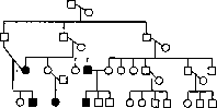
LEUKOPATHIAS (leukopathia), synonym achromia, a variety of achromia (see), the formation on the skin of depigmented spots as a result of the complete or partial disappearance of the normal skin pigment. The spots are either milky-white or rosy-white due to the transparency of the blood vessels. Leukopathias are distinguished as congenital (L. congenitalis) and acquired (L. acqutsita). - Congenital leukopathias. These include the phenomena of albinism (see) - general or partial. General albinism is inherited as a recessive autosomal trait (fig. 1). In this case, one must assume that in three consecutive generations, a heterozygous member of the family each time married a person who had the albinism factor. Partial albinism is inherited as a dominant trait. There are known cases when this anomaly appeared in several generations (e.g., in a case from New Zealand in 5 generations). Partial albinism in the form of a streak of white hair (pilosis circumscripta, canities) has been traced as a dominant trait up to the 6th generation. The milky-white coloration of the nails (leukonychia, leukopathia unguium) occurs either in the form of points, single or multiple, appearing at the lunula and gradually moving toward the edge of the nail, or in the form of complete whitening of the entire nail plate (leukonychia totalis). Spotted leukonychia develops due to mechanical damage to the nail bed; leukonychia totalis is very rare, is congenital, and is genotypically determined. This anomaly is inherited as a dominant trait; there is a known case of Bauer where the inheritance of this trait was noted in 4 generations (fig. 2).- Among congenital leukopathias, one must also distinguish naevus anaemicus and leukoderma acquisitum (Sutton). Naevus anaemicus is often encountered; it is characterized by the presence from the first years of life of white spots in varying numbers, shapes, and sizes; > A. 1 I L Ш ВДШ111 th ш a o Figure 2. ГП male O female Щ Leuconychia upon pressing on the periphery with a glass slide, the spots disappear, and after rubbing they appear even more sharply, maintaining their pale color among the reddened surrounding normal skin. On histological examination, in some cases a reduced number of vessels is found at the site of the spot, in others - their spasm. No other pathological changes are found. There are no indications of hereditary transmission. Naevus anaemicus often occurs in combination with a greater or lesser number of birthmarks and apparently represents their variety. Sutton's disease - white spots of varying sizes, round or oval in shape, located around pigmented birthmarks. Sutton, who described this disease in 1916, considered it a variety of vitiligo; subsequently it was classified as a group of pigmented birthmarks (Leszczynski). Probably this type of leukopathia, like naevus anaemicus, should be classified as paratypic diseases; it is not inherited.- The group of leukopathias also includes vitiligo (see) and syphilitic leukoderma.- Leukopathias after the regression of psoriasis, parapsoriasis, lichen planus, Gibert's pink lichen, seborrheic eczema, pityriasis versicolor, microsporia, ringworm and other erythemato-squamous dermatoses is explained by the fact that skin covered with scales is protected from the action of ultraviolet rays and does not tan. This form of leukopathia is called leukoderma solare (Meshchersky). Leukopathias that developed under the influence of chemical substances on the skin (chrysarobin, iodine, etc.) are called pseudoleukoderma (Voronom). A characteristic feature of the group of exogenous leukopathias is that they are temporary, last from several weeks to several months and gradually disappear parallel to the fading of the tan of the main background of the skin.
yan
Figure 1.
Related articles
Cite this page
“Leukopathias.” Soviet Medical Encyclopedia. English translation of Bolshaya Meditsinskaya Entsiklopediya, 1st ed. (Moscow, 1928–1936), ed. N. A. Semashko. https://sovietmedicalencyclopedia.pages.dev/article/leukopathias/