Aspheric Lenses
Historical document, translated for reference. It reflects medical knowledge of the 1920s–30s and is not medical advice.
Summary
This article from the 1928–1936 Soviet Great Medical Encyclopedia defines aspheric lenses as those designed to correct oblique astigmatism. It highlights their specific application in correcting aphakic eyes, often referred to as Gullstrand cataract lenses.
Encyclopedia article (1928–1936)
ASPHERIC LENSES, lenses that correct oblique astigmatism. An aspheric surface is a surface of revolution that deviates in a regular manner from a spherical surface.
Thus, for every point on an aspheric surface, from the center to the periphery, the radius of curvature is constantly changing.

In aspheric lenses, 'oblique astigmatism' is corrected. Aspheric lenses have become widely used for the correction of aphakic eyes under the name of Gullstrand 'cataract lenses.' The advantages of aspheric lenses are evident from the table (see figure), in which the left column (a) corresponds to
...to the image obtained with ordinary biconvex lenses (13.0 D), and the right (b) - with the help of Gullstrand lenses of the same power. The side numbers indicate in degrees the degree of rotation of the eye behind the lens. ASPHYXIA (from Greek a-negative particle, and sphygmos-pulse), absence of pulse, suffocation, a pathological state of the organism due to a lack of air, an excess of CO2, poisoning by gases, or a disturbance of respiratory function, characterized by sharp respiratory disorders, changes in blood circulation, and other phenomena, often ending in death from paralysis of the respiratory center. Asphyxia due to a lack of O2 can be caused either by its low content in the external environment (ascent to high altitudes) or by an obstacle to its passage through the respiratory tract (blockage of the respiratory tract, tumors, drowning, strangulation, etc.). Asphyxia due to an increased content of CO2 depends either on its accumulation in the external environment or on obstacles to its excretion from the organism. Asphyxia from internal causes can also be of various types. Diverse causes also determine the different character of asphyxia in the sense of its external manifestations: phenomena during a lack of O2 differ sharply from those during asphyxia from an excess of CO2. It should be noted that in pure form, asphyxia is rarely encountered; usually, it is of mixed origin. Asphyxia due to a lack of oxygen is most often encountered during ascent to high altitudes. In former times, the phenomena arising in the organism during this were linked to a decrease in atmospheric pressure. For the first time, P. Bert showed that animals can live even at a pressure of 80 mm, provided there is a sufficient amount of oxygen. Later, it was proven that the phenomena developing at mountain altitudes are of a somewhat different character, and that one cannot draw a complete analogy with those during a lack of O2. According to Mosso's data, asphyxia at altitudes depends not so much on a lack of oxygen as on the rapid excretion of carbonic acid; hence, insufficient irritation of the respiratory center, which regulates breathing depending on the CO2 content in the blood. Albitsky and others established that amounts of oxygen half the normal, i.e., 9.5-10%, do not cause any pathological phenomena. At 7-8%, an excited state, dyspnea, a drop in temperature by 1-2°, and sometimes hemoglobinuria appear. At 5-6%, animals die a day or two after the experiment, and only a few survive. With a decrease of O2 to 2.5%, complete asphyxia and inevitable death occur. Animals die calmly, without convulsions (the latter, apparently, are caused by carbonic acid). Claude Bernard noted even earlier that an animal adapts to a gradual decrease in oxygen; a bird that has experienced oxygen starvation survives a second time at such an oxygen content at which a control one dies. Levy and Zuntz showed that this habituation is explained by an intensification of breathing, but it is possible only up to a certain limit, namely, 35 mm Hg, which corresponds to 2.5% oxygen (Albitsky). Another compensatory phenomenon during oxygen depletion is the strengthening of cardiac activity, which serves as a very sensitive reaction. Thus, Croce-Spinelli observed in himself at a pressure of 705 mm an increase to 92 beats, at 560 mm-100 beats, at 510 mm-116 beats, and, finally, at 410 mm-135 beats. Despite a strong increase in heart contractions, blood pressure does not rise or increases insignificantly; subsequently, with increasing palpitations, it falls. While maintaining a normal composition of air, asphyxia can occur due to difficulty in the passage of air into the respiratory tract on the basis of mechanical causes (foreign body), infectious diseases of the respiratory tract (syphilis, diphtheria), or of a purely nervous character (spasm); in newborns, asphyxia is a consequence of premature respiratory movements of the unborn child and the entry of amniotic fluid into its respiratory tract. The initial respiratory tract in a human, who usually breathes through the nose and mouth, can hardly constitute a barrier to the access of air (excluding violent strangulation). However, in a child who usually breathes through the nose, asphyxia of this kind does not represent an exceptional phenomenon and is usually observed during sucking. Much more often, blockage of the larynx and trachea is encountered, which happens due to the lodging of solid food, a foreign body, during compression of the larynx by a tumor, enlarged thymus and thyroid glands, during edema of the glottis, during inflammatory diseases of the larynx, during angina Ludovici, sometimes during diphtheria due to blockage by diphtheritic membranes, but more often from edema of the glottis, during spasm on the basis of tetanus, poisoning by chlorine, ammonia, hydrogen sulfide, etc., during paralysis of the mm. crico-arytenoidei post., during strangulation. Blockage of individual large bronchi is less dangerous due to their branching; here, spasm or blockage by catarrhal mucus of many bronchioles can lead to asphyxia. Blockage of lung tissue is tolerated without particular harm if the process occurs slowly, because the remaining parts begin to work with compensatory intensification. However, even here, asphyxia is possible, for example, due to the filling of alveolar ducts with water in drowning victims, bilateral pneumonia, tuberculosis (in the latter case, compensatory phenomena stand out sharply). To this, one can also refer cases where access of air is hindered not directly, but indirectly, for example, due to compression of the chest, bilateral pneumothorax, empyema of the pleura, during exudative pleurisy, etc. Experimentally, asphyxia of this kind was studied relatively long ago. Thus, Preynoso as early as 1851 caused asphyxia in animals by constricting the throat. Senator (1854) hindered access of air by pouring oil into the lungs or bandaging the chest; Bert drowned various animals in water and came to the conclusion that the smaller the animal (the smaller its surface), the faster it dies without access of air (pigeons live without air 1 1/2 m., rats-2 m., cats-3 m., dogs-4 1/2 m.). In general, the younger the animal, the easier it copes with a lack of oxygen (a rat pup at the age of 12-15 hours lives without oxygen 30 min., a six-day-old-15 min., a 20-day-old-1 min. 35 sec.; an adult human-3-6 min., a newborn-10-15 min.). If blockage of the respiratory tract occurs slowly, then the picture of asphyxia is similar to that during oxygen starvation. In cases of acute asphyxia, excitement, convulsions, and an increase in blood pressure come to the fore; the respiratory rhythm may remain unaccelerated (if there is an obstacle to the exit of air). Asphyxia from carbonic acid. With a sufficient amount of oxygen, even relatively large amounts (up to 23%) of CO2 are tolerated by animals completely harmlessly (Albitsky). Only at 23-30% of its content does the animal fall into a drowsy state, does not react to stimuli, and breathing and heartbeat are strengthened. At 30-50%, drowsiness quickly passes into a comatose state, and death occurs. Blood pressure rises sharply from the action of CO2 due to the increase in heartbeat and due to the contraction of vessels. At very large amounts of CO2, blood pressure falls, a delay in breathing occurs, and even a spasm of the bronchial musculature of a reflex character with the participation of the n. trigemini and n. vagi. Metabolism during this kind of asphyxia increases sharply, especially protein metabolism; a large amount of urea and phosphoric salts appears in the urine, and hyperglycemia in the blood. Asphyxia from poisoning by other gases generally fits more or less into the general picture of asphyxia (see below). Separately stands the group of asphyxia from damage to nerves and respiratory muscles. This includes cases with mechanical injuries to the medulla oblongata, wounds of the vagus nerves, paralysis of the n. phrenici during multiple neuritis, during eclampsia, tetanus, during poisoning by strychnine and curare. Finally, a completely special group is represented by asphyxia from a disorder of internal respiration. They can occur either from the loss by tissue cells of the ability to assimilate O2 (poisoning by hydrocyanic acid) or due to a lowering of the blood's ability to carry a sufficient amount of O2 for tissue cells (for example, during large losses of blood or depletion of erythrocytes of hemoglobin). A large role in internal asphyxias is played by the sufficient buffering of the blood (the ability of salts to bind carbonic acid). The picture of asphyxia, depending on the origin, varies greatly; however, since in the majority of cases asphyxia of a mixed character is observed, some general signs can be given. Hogyes notes four stages in asphyxia: I-intensification of breathing with intensified inspiration (lack of O2); II-with intensified expiration (excess of CO2); sometimes accompanied by clonic convulsions; the III stage is characterized by a cessation of breathing, starting from a few seconds to a few minutes, which depends either on irritation of the n. vagi, or on a fall in the excitability of the respiratory center (lack of carbonic acid in the blood), or on a loss of excitability of the muscles; then comes the last (IV) stage (rare deep inspirations with passive expirations), lasting 3-8 min., but sometimes hours (Albitsky).
Cardiac activity is initially unchanged (excluding oxygen starvation), then the rhythm slows down, the volume of contractions increases due to irritation of the vagus nerve. After breathing stops, the heart continues to beat for some time. Thus, Mascka observed the heart beating in hanged persons for 4-5 minutes after breathing ceased, and Hofmann even for 8 minutes. Blood pressure rises and then falls only with severe slowing of cardiac activity. Among other phenomena, one can note intestinal peristalsis, erection, and increased secretion of sweat and urine (dilation of peripheral vessels and an increase in blood pressure). In the blood, leukopenia is observed due to the migration of leukocytes from peripheral vessels to internal organs (mainly the lungs), eosinophilia (in guinea pigs under the influence of carbon monoxide, according to Bogomolets); a drop in blood clotting, and an increase in viscosity. Hemoglycolysis drops sharply under the influence of CO2. In acute cases, hyperglycemia is noted, in subacute cases—glycosuria without hyperglycemia. The calcium content in the blood increases. The freezing point of blood decreases, O in the blood drops to 0–0.96%, CO2 rises to 56%. Gas exchange initially increases, as does nitrogen metabolism. The amount of urea and phosphoric acid salts increases in the urine. Among subjective sensations, darkening of the eyes, ringing in the ears, and then loss of consciousness are observed. Pathological-anatomical picture: significant congestive hyperemia (cyanosis) of the integuments and internal organs, absence of postmortem blood clotting, hemorrhages in the brain, on the mucous and serous membranes, and in the lungs.
N. Sprotishga. Asphyxia of the newborn, a state of breathlessness of the child, accompanied by a weakening of cardiac activity. Asphyxia of the newborn is mostly a consequence of paresis of the respiratory center, which occurs after its excessive irritation in the uterus. At its core always lies a disorder of placental gas exchange, a lack of O and an excess of CO2 in the child's blood, which irritates its respiratory center. The causes of asphyxia of the newborn are compression of the umbilical cord, its tight wrapping, tears, prolapse, partial detachment of the placenta; mechanical irritation during artificial extraction of the fetus (breech, version); often hemorrhages in the brain; on the part of the mother—a decrease of O in her blood during heart and lung diseases, during blood loss; finally, chronic and acute infections, toxicosis, e.g., eclampsia. In rare cases, the cause of asphyxia is congenital heart and lung deformities of the fetus. If, under the influence of one of these causes, the respiratory center of the fetus is irritated, then respiratory movements begin ahead of time, i.e., when air does not yet have access to the lungs; as a result, asphyxia occurs, or 'suffocation not brought to death,' in the words of Schultze. A consequence of premature respiratory movements is the penetration of amniotic fluid, mucus, blood, and sometimes meconium into the child's respiratory tract. Sometimes, even without a preliminary dyspneic phase, paralysis of the respiratory center can occur due to sharp venous congestion of the blood during violent and continuous contractions (tetanus uteri). Disturbance of cardiac activity, in the form of its slowing and irregularities, occurs due to the excitation of the vagus nerve center by sharply venous blood. A distinction is made between congenital asphyxia and acquired asphyxia, which arises after birth and is based on difficulty in breathing by the lungs due to some central or local causes. At autopsies of children who died from congenital asphyxia, one finds an overfilling of the right heart and large vessels with blood, congestion and edema of the brain, thoracic and abdominal organs, hemorrhages in the skin, pleura, pericardium, and sometimes aspiration masses in the bronchi. Two types of asphyxia are only different degrees of the same affliction. In asphyxia of the first degree, or cyanotic, the skin is cyanotic, muscle tone is preserved, as are the corneal and pharyngeal reflexes; heart sounds are slowed but distinct; asphyxia of the second degree, pale, or syncopal, is characterized by sharp waxy pallor of the integuments (except for slightly cyanotic lips), complete relaxation of the muscles, absence of reflexes, and sharp weakening of cardiac activity. Diagnosis of incipient fetal asphyxia should be made on the basis of contamination of amniotic fluid with meconium, violent convulsive movements of the fetus, and, mainly, on the basis of auscultation of the fetal heart, which in threatening cases should be performed with a watch in hand every 2-3 minutes. Slowing of heart activity in the intervals between contractions, and then tachycardia (excitation and paralysis of the vagus nerve), require acceleration, and sometimes immediate termination of the birth act. One should distinguish asphyxia from physiological apnea, when a child can, after being born, live for some time (up to 10 minutes) without breathing, at the expense of the excess O present in its blood (for example, during a cesarean section). The prognosis is favorable in cyanotic asphyxia and very serious in pale asphyxia (34% mortality, according to Demelin). Children die immediately or within a day from weakening of the respiratory center, or, finally, later from bronchopneumonia. Long (more than half an hour) non-appearance of breathing, complete and persistent absence of reflexes, increasing pallor, and bloody foam from the mouth and nose should be considered bad omens. However, resuscitation should be continued as long as a heartbeat is audible. Measures pursue three goals: 1) to clear the respiratory tract of mucus; 2) to stimulate respiratory movements; and 3) to increase the energy of blood circulation. One must act in precisely this order; everything necessary for resuscitation must be prepared in advance. In asphyxia, the umbilical cord is immediately cut and the respiratory tract is cleared of mucus. If in cyanotic asphyxia deep wiping with absorbent cotton is sufficient, then in pale asphyxia, mucus should be aspirated with a catheter with a bulb, carefully inserting it into the trachea behind the epiglottis. It is good to place the child at an incline, with the head lowered, for the drainage of mucus. Hanging by the legs is dangerous in cases of cerebral hemorrhages. Then, breathing is stimulated by skin irritation: patting, rubbing, sprinkling with ether, cold water, and finally, alternating baths (warm at 40° and room temperature), starting and ending with a warm bath. In pale asphyxia, one should not delay, and after 10-12 baths, one should move on to artificial respiration. The Schultze swinging method, well known to every doctor, is absolutely contraindicated in cases of bone fractures, as well as in premature infants and in cases of cerebral hemorrhages, as it increases bleeding. Performed by an inexperienced person, this method can be disastrous for the child. Ruptures of the liver, spleen, stretching of the spinal cord with hemorrhages (Knapp), etc., have been described. Even in Germany, they insistently demand its abandonment (Birk). The Sylvester method (Sylvester, 1863), which is calmer and allows for warming the child, can be fully recommended; for a child lying on its back with slightly raised shoulders: 1) raise the arms on both sides of the head, stretch and pull them for a few seconds (inhalation); 2) lower them and press them on both sides of the chest (exhalation); do this 10-15 times per minute (see figures 1 and 2). It is good to fix the child's legs, and also to pre-extend the tongue and hold it in that position for air access. The Prochownik method consists of hanging the child by the legs, with the head resting with the forehead on the table to straighten the trachea (see figure 3). The chest (lungs and heart) is rhythmically compressed by hand, whereby the aspirated fluid is squeezed out. A simple and good method for aspiration asphyxia, it may not be indifferent in the presence of cerebral hemorrhage. The Japanese method (Ogata), less known, consists of the following: the child in a lying position is held by one hand by the legs, the other supports the back, wrapping fingers around the neck. This hand slowly lifts and bends the head and upper part of the torso toward the legs until the face

Figure 1. Sylvester I. Inhalation.
touches the back of the feet (exhalation). Then extension, again flexion, and so on 8-10 times per minute. The Sokolov method is essentially close to the latter, but the child lies on the table with the head hanging down. The doctor, standing

Figure 2. Sylvester II. Exhalation.
on the right, with the left hand bends the head to the chest, and with the right simultaneously bends the child's knees to the abdomen (exhalation). Then the legs are extended, and the head is tilted back (inhalation). Rhythmic tongue extensions
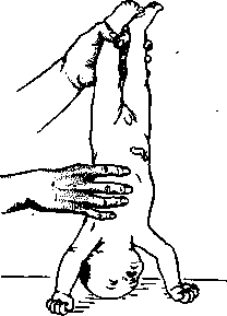
Figure 3. Prochownik's method. (Laborde) have not become widespread among us, just as insufflations of air or O into the lungs have not. They require certain instrumentation (Ribemont's insufflator, English tracheal catheters, Tiegel's apparatus) and skill in inserting the catheter into the larynx. With unskilled use of this method, ruptures of the lung tissue and emphysema have been observed. Injections of adrenaline into the heart (1:1000-1 cc), recommended in Germany, may be tried in cases of severe pale asphyxia, however, only with the simultaneous application of other measures. Judging the effectiveness of this method is therefore difficult. During all these manipulations, the child should be constantly warmed (hot diapers, baths, heating pads), and cardiac activity should be supported. Subsequent careful observation is necessary, as the activity of the respiratory center may weaken again, and the child, left to their own devices, may perish.
Related articles
Mentioned in
Cite this page
“Aspheric Lenses.” Soviet Medical Encyclopedia. English translation of Bolshaya Meditsinskaya Entsiklopediya, 1st ed. (Moscow, 1928–1936), ed. N. A. Semashko. https://sovietmedicalencyclopedia.pages.dev/article/aspheric-lenses/