Intracranial Pressure
Historical document, translated for reference. It reflects medical knowledge of the 1920s–30s and is not medical advice.
Summary
Intracranial pressure refers to the pressure of cerebrospinal fluid produced by the choroid plexuses and filling the ventricles of the brain, its cisterns, and subarachnoid spaces. This article discusses measurement methods, normal and pathological values, clinical symptoms, and associated conditions like hydrocephalus.
Encyclopedia article (1928–1936)
Intracranial pressure, the pressure of cerebrospinal fluid produced by the choroid plexuses and filling the ventricles of the brain, its cisterns, and subarachnoid spaces of the brain and spinal cord. The existence of intracranial pressure was known long ago, both based on experiments on animals and on clinical observations; it was also known that in a number of pathological processes it can significantly increase. With the aim of reducing pathologically elevated intracranial pressure, Quincke in 1891 proposed spinal puncture. Thanks to this method, clinical practice gained the ability not only to examine cerebrospinal fluid but also to objectively determine intracranial pressure, which consists of elastic and hydrostatic pressure. To measure pressure, the needle introduced into the subarachnoid space is connected with a rubber tube to a thin glass tube, which is held vertically; in it the fluid begins to rise until equilibrium is reached between atmospheric and intracranial pressure. With lumbar puncture in the sitting position, pressure, due to the fact that the column of cerebrospinal fluid in an adult averages 60 cm (distance from crown to puncture site), will be significantly higher than in the lying position. In view of the fact that the specific gravity of cerebrospinal fluid is 1.005-1.007, the obtained height of the column can be considered as the pressure of a water column; in the sitting position it can reach 400-410 mm (Kronig, Kozhevnikov), in the lying position - 125-150 mm (Quincke). This pressure is not constant and it undergoes fluctuations depending not only on pathological but also physiological factors. Of these latter, one must first point to the influence of pulse and respiration. If one looks at the tube connected to the subarachnoid space, it is easy to notice that the level of fluid in it oscillates almost continuously, with some oscillations of smaller amplitude and more frequent, others - of larger amplitude and less frequent. The first coincide with the systole of the heart (with each pulse wave the pressure increases by 1-4 mm); the latter depend on respiration: with each inhalation (due to the outflow of blood from the brain) pressure falls and with each exhalation it increases; these fluctuations reach 20 mm. Cough, screaming, straining, emotions accompanied by a rush of blood to the head cause a significant increase in intracranial pressure. A particularly significant increase in pressure is observed in certain pathological processes: 1) in tumors of the brain itself, as well as its membranes and skull, 2) in hemorrhages into the brain or its membranes, 3) in purulent and serous inflammatory processes of the soft membranes, which, on the one hand, are often accompanied by increased secretion of cerebrospinal fluid by the choroid plexuses, on the other hand - decreased absorption of fluid. If these changes are persistent, then due to constantly significantly elevated pressure, hydrocephalus develops. With the integrity of the communication between the ventricles and the subarachnoid spaces, but with decreased absorption, communicating internal hydrocephalus develops, and with the closure of the Magendie (foramen Magendie) and Luschka (Luschka) openings or the Sylvian aqueduct (aquaeductus Sylvii) or the Monro openings (foramina Monroi), internal obstructive hydrocephalus develops. Pathoanatomically, in the corpse, elevated intracranial pressure manifests itself in the fact that when the skull is opened, the dura mater is found to be tense and hyperemic, the brain appears enlarged in volume and closely adheres to the membranes; the convolutions of the brain are flattened, the sulci are smoothed out. In the presence of hydrocephalus - the corresponding picture. A significant increase in intracranial pressure is accompanied by a number of clinical symptoms of a general cerebral nature: headaches, vomiting, stagnant papillae with further transition to atrophy. If the increase in pressure occurs
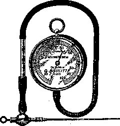
in small children, then initially bulging of the fontanelles is noted, then divergence of the sutures and progressive increase in head size, sometimes reaching a significant degree (70 cm or more). In most cases, this is a consequence of cerebrospinal meningitis. To measure pressure, Claude uses a special manometer, manufactured in Paris by engineer Bullet (see figure).
Related articles
Mentioned in
Cite this page
“Intracranial Pressure.” Soviet Medical Encyclopedia. English translation of Bolshaya Meditsinskaya Entsiklopediya, 1st ed. (Moscow, 1928–1936), ed. N. A. Semashko. https://sovietmedicalencyclopedia.pages.dev/article/intracranial-pressure/