Dermatophytes
Historical document, translated for reference. It reflects medical knowledge of the 1920s–30s and is not medical advice.
Summary
This article from the 1928–1936 Soviet Great Medical Encyclopedia discusses dermatophytes (dermatomycetes), pathogenic fungi that parasitize the upper layers of the skin, hair, and nails. It covers historical discoveries, laboratory cultivation methods, morphological classification, and associated clinical conditions.
Encyclopedia article (1928–1936)
DERMATOMYCETES (from the Greek derma - skin and mykes - fungus), syn. dermatophytes (phyton - plant), pathogenic fungi parasitizing predominantly the upper
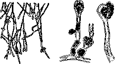
Figure 1.
layers of the skin and its appendages—hair and nails. Of these, the most widespread and well-known are: Achorion (the causative agent of favus, see Figure 1), Trichophyton (causative agent of ringworm, see Figure 2), Microsporum (parasite of microsporia, see Figure 3), Epidermophyton (causative agent of eczema marginatum, see Figure 4), Microsporon furfur—causative agent of pityriasis versicolor, Microsporon minutissimum—of erythrasma, and finally Trichosporon (see Figure 5), which affects only the hair (piedra). Some authors (Unna) propose to classify the latter three groups as saprophytes in view of the fact that they never penetrate deeper than the superficial layers of the skin. All dermatomycetes have a mycelium that settles in the skin and reproduce by forming conidia. Other stages of development that could give them a more detailed characterization from a systematic point of view are absent in them, as a result of which they are still classified among the so-called imperfect fungi—Fungi imperfecti. According to the newest classification proposed by Ota and Langeron

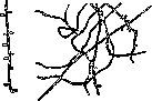
FIG. 2.
Figure 3.
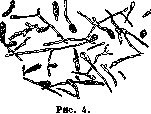
(Ota, Langeron), dermatomycetes belong to the group of hyphomycetes. The first of this group to be discovered was the causative agent of favus—Achorion. In 1839, Schönlein first proved the "vegetative nature" of the scutulum in favus. Independently of Schönlein, Gruby in 1841 discovered and described this same fungus, and in 1845 Remak succeeded in obtaining a pure culture of Achorion and proving its pathogenic role on himself. Three years after the discovery of Achorion, Gruby described the causative agents of two other clinical forms—trichophytosis and microsporia—and immediately thereafter, within the next decade, followed a series of discoveries of new representatives of dermatomycetes in both humans and animals. Believing that the nutrient medium is a source of error in identifying the fungus, Sabouraud at the London Congress of Dermatologists in 1894 proposed for the cultivation of dermatomycetes the so-called international test medium (milieu d'épreuve) with ingredients not only of a definite composition but also from a specific Paris firm: Maltose brute 4.0; Pepton gelose 1.0; Agar 1.8; Aqua 1,000.0. Growth at a temperature of 15-28°. During the World War, Grütz successfully substituted German manufactured products for the French maltose and peptone. Grütz's medium: Pepton Knoll 5.0; Nervina-Malz 80.0; 1.8% agar—1,000.0. Finkelstein suggests, to avoid overgrowth, treating the initial material (hair, scales) before inoculation with a 2% antiformin solution for 2-5 minutes followed by washing in sterile physiological sodium chloride solution. Cultivate
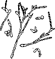
Figure 5.
at a temperature of 35-36°. In view of the fact that sugar media promote rapid aging and degeneration of initial cultures, Sabouraud proposed a special medium for preserving the latter, consisting of 1% peptone-agar without carbohydrate content. The classification of dermatomycetes proposed by Sabouraud has generally survived to the present time. All pathogenic fungi, according to Sabouraud, are subdivided into 3 genera: Microsporum, Trichophyton, and Achorion. Microsporum in turn is divided into 2 groups: human Microsporum (Microsporon Audouini) and Microsporum parasitizing animals, with the most widespread representative Microsporon lanosum (see table, Fig. 1) or Microsporon canis. Trichophyton, depending on the character of the arrangement of fungal elements, is divided into Endothrix, Ectothrix (see table, Figs. 2, 5, and 6), and Neo-endothrix (see table, Fig. 3). Spores of fungi belonging to the Endothrix group are located inside the hair; in Ectothrix, spores and hyphae are located predominantly around the hair; to Neo-endothrix belong species of fungi that have a long period of penetration into the hair, and therefore their elements are encountered both outside and inside the hair. Ectothrix-type parasites are encountered chiefly in animals, while human parasites mostly belong to the Endothrix type. Achorion is also divided into 2 main groups: human Achorion (Achor. Schönleini; see separate table, Fig. 4) and animal Achorion (Achor. quinckeanum, Achor. gallinae, Achor. gypseum, etc.). Some subspecies (Achor. quinckeanum and Achor. gypseum) occupy an intermediate position, combining the properties of both Achorion and Trichophyton. In addition, experiments on animals have shown that, for example, a guinea pig that has undergone some fungal disease reacts with increased sensitivity not only toward the species with which it was infected, but also against other species, more or less pathogenic. Thus, the question still remains disputed whether all microspora and trichophytons represent true species or whether one is dealing merely with varieties of the same species. The question of the production of toxins by dermatomycetes is resolved in the affirmative; as early as 1902, Plato and Neisser succeeded by cultivating the fungus on sugar broth in obtaining a filtrate (trichophytin) that causes both a local and a general reaction, especially in cases of past deep trichophytosis.
S. Fried. Dermatomycoses, or dermatophytosis, skin diseases caused by the settling of fungi (Mycetes) in the skin. Three main groups of dermatomycoses are distinguished. The first group includes the most superficial dermatomycoses, in which fungi settle only in the cornified layer of the epidermis and do not cause a distinct reaction on the part of the skin proper. These diseases are (almost) non-contagious, although fungi are encountered in great abundance in scales taken from the lesions. Unna called these forms saprophytias. These include pityriasis versico[lor...]
tt
Should read
Printed
read Fig. 1
Fig. 6
Fig. 4
Fig. 3 Fig. 2
Fig. 5
Fig. 5
Fig. 1 Figure 3
Fig. 4
Fig. 6
Fig. 2


s? i
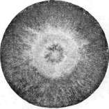
Figure I, Micr. lanosum (30-day culture). Figure 2. Trich. cerebriforme (2-month culture). Figure 3. Trich. Neo-endothrix. Figure 4. Achorion Schönleini (45-day
culture). Figure 5. Trichophyton ectothrix. Figure 6. Trichophyton endothrix. To the article Dermatophytes. [lor] and erythrasma. In dermatophytosis of the second group, the fungi settle not only in the stratum corneum, but also in other structures of horny substance (hair, nails). These forms are for the most part highly contagious and therefore more frequently encountered on exposed parts of the body. Here the process is accompanied by a distinct (usually subacute) inflammatory reaction, which sometimes (e.g., in kerion and parasitic sycosis) may reach the degree of deep purulent inflammation. This group is united under the name of epidermophytoses (Darier), which include favus, microsporia, and trichophytia, as well as epidermophytia and oidiomycosis. The latter two forms are distinguished by lesser contagiousness, absence of appendage involvement of the skin, and a tendency of the process to localize in skin folds. Epidermophytoses also include the so-called tropical mycoses (carate, tokelau, etc.), which are found only in tropical countries. Clinically, superficial dermatophytoses are characterized by the formation of sharply demarcated, mostly regular-shaped lesions with more acute inflammatory phenomena (in epidermophytoses of smooth skin) along the periphery. The third group includes dermatophytoses in which the fungi settle and multiply in the deeper parts of the skin proper. These diseases mostly have a chronic course, expressed by phenomena of granulomatous inflammation. They spread through the lymphatic and blood vessels and are frequently complicated by diseases of the lymph glands, bones, internal organs, etc. These forms are called deep dermatophytoses or true dermatophytoses (Darier). These include blastomycosis, sporotrichosis, hemisporosis, cladiosis, and others. The clinical and histological picture of these forms bears great similarity to that of tuberculosis or syphilis, and their exact diagnosis is established only through careful bacteriological examination. Furthermore, this category usually includes a number of similar diseases caused by pathogenic mold fungi. These diseases more often take the form of chronic inflammatory tumors (mycetomas). Such is, for example, Madura foot, which is found in tropical countries (very rarely in Europe). Finally, skin diseases caused by trichomycetes can be classified among deep dermatophytoses. According to their morphological and biological properties in botanical classification, the latter occupy an intermediate position between fungi and bacteria. These include actinomycosis, some forms of Madura foot, and streptotrichoses. Many forms of dermatophytoses are also observed in animals, and the causative agents of others are found in a saprophytic state (sporotrichons, etc.). Clinical observations and experiments on animals have revealed that in dermatophytoses (even superficial ones, e.g., in superficial trichophytosis), phenomena of immunity-allergy develop in the organism, and that a special role in this is played by the skin, toward which certain fungi (e.g., trichophyton fungi) exhibit a special dermatotropism (formation of skin lesions only, even upon their intracardiac administration). Connected with the phenomena of immunity is the extreme rarity of reinfections in dermatophytoses, the sharp inflammatory reaction in deep trichophytoses, and allergic rashes, often apparently of hematogenous character (trichophytids, etc.). For the development of saprophytias, epidermophytosis, and erythrasma, local conditions of the skin (mainly moisture?) apparently have essential significance. Epidermophytoses are mostly very contagious, but usually constitute the lot of childhood, partly curing themselves (trichophytosis and microsporia) independently by the period of puberty. For the treatment of dermatophytoses, exfoliating agents (parasitifuga) are used, most often in combination with disinfecting substances (parasiticida). In lesions of the hair (on the head), preliminary depilation is inevitable; deep dermatophytoses mostly respond well to treatment with potassium iodide internally + X-rays.
N. Chernogubov.
Related articles
Mentioned in
Cite this page
“Dermatophytes.” Soviet Medical Encyclopedia. English translation of Bolshaya Meditsinskaya Entsiklopediya, 1st ed. (Moscow, 1928–1936), ed. N. A. Semashko. https://sovietmedicalencyclopedia.pages.dev/article/dermatophytes/