Favus
Historical document, translated for reference. It reflects medical knowledge of the 1920s–30s and is not medical advice.
Summary
Favus is one of the oldest known skin diseases, caused by various species of the fungus Achorion. The article details the different species that infect humans and animals, their characteristics, and the immunological responses they provoke.
Encyclopedia article (1928–1936)
FAVUS (syn. favus, tinea lupinosa, tinea vera, porrigo lupinosa, dermatomycosis favosa), one of the oldest diseases, known even to ancient medicine. The true cause of F. remained unclear until the causative agent of F. was discovered almost simultaneously by three researchers: in Germany by Remak (Remak, 1837), who found fungal elements in the scutula but did not recognize their etiological role, and by Schonlein (Schonlein, 1839), who established the mycological nature of the disease, and in France by Gruby (Gruby, 1841), who independently of the first two came to the same conclusion. The causative agent of F. was named by Remak Achorion Schonleini. Grawitz (1885) was the first to obtain a culture of the fungus. At present, several species of Achorion are distinguished; among them, Achorion Schonleini is the only cause of human favus; transmitted from person to person, it is the classic causative agent of favus of the scalp. Opposed to it are several species of Achorion, causative agents of favus of animals. When transmitted to humans, they cause lesions of the smooth skin in the latter; these species include: A. quinckeanum, A. gypseum, A. violaceum, A. gallinarum, Oospora canina (see Dermatophytes) and others. Characteristics of the causative agent of favus in humans and animals. 1. Achorion Schonleini as the causative agent of favus of the human scalp is found almost everywhere. For growing Achorion, besides the usual fungal media (milieu d'epreuve, de conservation, beer wort), Sabouraud recommends strongly peptonized media (10%) and a medium with honey (8%); the latter media, ensuring typical growth, simultaneously prevent pleomorphism (degeneration) of cultures. Many authors (Sabouraud) grow cultures at 37° in the first days; this is also recommended by Yu. Finkel'shtein when using anti-formalin treatment. The culture of Achorion (on agar) is very typical; it belongs to the type of smooth cultures (without aerial hyphae); it rises sharply above the surface of the medium, is sharply contoured, its surface is waxy, dry, and in its mass it is empty, spongy-porous. Pleomorphism of cultures under appropriate media occurs rarely. The microscopic picture of the culture is best observed in a hanging drop or by Plaut in situ; in it, arthrospores are observed; from other fruiting organs, individual authors (on special media) have seen rudimentary spindles; very characteristic are candelabra-like thickenings of the mycelium and combs, as well as chlamydospores (in old cultures). A. Schonleini only as an exception infects animals. 2. A. formoseum (1927) was isolated by Hasegawa on Formosa (18 out of 27 cases of favus of the human scalp), and later also in Mongolia; mycologically it is a variant of A. Schonleini, but does not form vesicles in the hair. To Achorion of animal origin belong: 3. A. quinckeanum (discovered by Quincke in 1885)--causative agent of favus of mice; it is also spontaneously observed in cats, dogs, rats, canaries. In humans it affects only the smooth skin. It is much more contagious than A. Schonleini; sometimes it causes epidemics. It usually does not penetrate the hair; the only such case was described by Plaut. Its culture grows quickly; some describe the culture of this fungus as white-furry, others as an uneven white snow-like coating with radial depressions and a reddish tint (in old cultures). Microscopically, lateral chlamydospores (aleuriospores) and spindles (fuseaux) are found, which brings it closer to Microsporon. 4. A. gypseum (discovered by Bosch in 1907)--causative agent of favus of horses, dogs, cats, rats; the culture grows quickly, soon giving pleomorphism. The colony is light yellowish-brown with a central button and radial grooves, as if sprinkled with powder (hence the name); under the microscope, many spindles are visible. In humans it is found on the smooth skin, occasionally giving scutula; it penetrates the hair and gives the same picture as in A. Schonleini; it often forms Kerion. 5. A. violaceum (Bloch, 1911) is spontaneously observed in mice; it can cause an epidemic (Bloch). It grows slowly, best on 4% glycerin-agar, but also on simple agar; it easily degenerates; externally it resembles Trich. violaceum; it gives scutula microscopically similar to those in A. Schonleini. 6. A. gallinae (Megnin, Sabrazes; 1881)--causative agent of chicken favus. Spontaneously not found in humans, but infection from animals (cats, canaries) is possible. Its culture at low t° is white, above 30°-pink; in the center of the colony there is a button-like elevation, brain-like convolutions, radial grooves. Under the microscope, many spindles are visible. 7. Oospora canina (Sabrazes, 1893)--causative agent of favus of dogs; it is not often found, spontaneously not found in humans, it forms scutula (on the outer surface of the ears); on media it gives a white dense fluff covered with a thin powder; it produces pigment diffusing into the medium. 8. A. sejisei (Cazalbou, 1913) was isolated from a horse. By culture it resembles A. gypseum; under the microscope-many spindles, colored in light brown. 9. A. passerinum (Fischer, 1928) is close to A. gallinae (without pink color). Experimental data. The most susceptible to Achorion, especially to the species quinckeanum, turned out to be the guinea pig (Bloch). The process after cutaneous inoculation according to Bloch in it proceeds very typically and regularly: incubation is 4-6 days, the process lasts the same time, in general after 12-15 days reverse development begins, and the disease spontaneously ends after 3-4 weeks. In rabbits, as well as experimentally in humans (Bloch, Sutter, Fischer), the process proceeds less regularly. Inoculation is also achieved in other animals; it is interesting to note that in the mouse, which is the host of this fungus, inoculation does not always succeed. A. Schonleini is less pathogenic for animals. Experiments were made on cats, dogs, rabbits, guinea pigs; even in the latter, the most susceptible of the mentioned animals, the process is not typical: scutula are absent or they are rudimentary. Hematogenous infection was achieved only with A. quinckeanum: when feeding with culture, scutula on the head were obtained (Unna). Sabouraud notes that infection of animals with intracutaneous introduction of pieces of scutula (central part) is easier than with inoculation of cultures. Immunity. By the research of many authors, it has been established that the body of an animal and human that has had favus infection becomes immune both to re-infection with live fungi and to their poisonous products (endotoxin, favin). By experimental reinoculations, authors were able to obtain all transitions from allergy to complete clinical and mycological immunity; the latter, according to some, occurs only when infected with very virulent strains, like A. quinckeanum, according to others-only with 'cutaneous' first infection (Bloch). Sutter in humans and Zeves in animals saw positive results only with multiple inoculations; in individual cases, positive results were also obtained by intracardiac and subcutaneous inoculations (Jadassohn). The body also passes into an allergic state when introducing fungal endotoxins-favin, trichophytin. The most active of favins can be considered those obtained from A. quinckeanum according to the exotoxin type (Bloch-Massini method) and according to the endotoxin type (Thardshimanjanz). Favin prepared from A. Schonleini is much weaker. The most sensitive reaction is intradermal. In favus caused by A. Schonleini, as a rule, there are no general phenomena, the skin reaction is insignificant. Stein (Stein) in 63 cases of favus of the scalp did not get a single positive reaction, and in 2 cases of the body, caused by animal Achorion, all were positive. Bloch in experimental infection of humans with A. quinckeanum obtained reactions on the 7-8th day after infection. When infected with A. quinckeanum, the toxin always gives a positive skin reaction, and sometimes also a general one. The latter is always obtained with deep inflammatory lesions. Cases of favids, achoriids are the most vivid expression of the allergic ability of the body. Neither allergy nor immunity have in fungal infections strict species specificity (group immunity), but still favus patients with the Schonleini causative agent react more sharply to favin and rarely to trichophytin, and patients with favus caused by animal Achorion react very weakly to favin. Thus, experiments on animals further confirm the already emphasized remoteness of animal Achorion from Schonleini, with which they are united only by the ability to form scutula. In general, toxin-allergy in favus has no great practical diagnostic value, and has no therapeutic value at all. The sensitivity of animals to the toxin is even much weaker; there are no general or focal phenomena; only the intracutaneous reaction may have some significance; in guinea pigs an inflammatory papule appears, disappearing after 1-2 days. Active immunization with killed cultures of the fungus or their toxin, as well as passive-by transferring serum or extract of organs of immune animals-do not achieve the purpose.
Along with the formation of allergy, there goes, though only partially, in deep mycoses in humans, the appearance of agglutinating, precipitating, and complement-fixing properties. The result of the reaction depends on the method of antigen preparation and on the virulence of the fungal strain included in the antigen. Montpellier-Boutin obtained a negative result with the serum of a patient with F., after injecting favin-positive; specificity is group. Seroreactions have not acquired diagnostic significance. The study of the problem of the transition of the body to an allergic state without primary infection is also devoted to the experiments of Martenstein. Martenstein's toxic antibodies, obtained by infusing a suspension of live spores with serum or crushed skin of sensitized guinea pigs, were found in experiments with A. quinckeanum so far only in guinea pigs. The laboratory diagnosis of favus is made on the basis of microscopy (without staining) of material collected from the patient (hair, scutula; nail scrapings), treated with 10%-20% caustic soda with careful heating (on a slide over a flame burner); with this method, there is a loosening and clarification of the material (tissues, fungi). If necessary for sowing (from smooth skin, nails), the material is subjected to preliminary treatment (according to Yu. Finkelstein) to destroy accompanying microbes: 2-3 minutes in 2% antiformine, washing in physiological NaCl solution, then sowing on Sabouraud agar (agar 1.8, maltose or glucose 4.0, peptone 1.0, water 100.0); the media are placed in an incubator, since the microbes-competitors dying from the specified treatment do not suppress the growth of F. fungi at 37°C, as is observed without antiformine treatment. The species of the grown fungal culture (after 5-6 days) is determined by its macro- and microscopic characteristics.
E. Sigalova. Clinical manifestations of favus. Favus of the head. The onset of the disease is characterized by the appearance of vesicles or small pustules on reddened skin. Gradually, the picture of favus develops, which is characterized by the following: the skin of the head is covered with peculiar yellowish-gray formations called scutula (scutuli favi) due to their resemblance to honeycombs. The scutula are distinguished by their round shape, have raised edges with a saucer-shaped depression in the center, in the middle of which a small hole in the form of a pinprick is noticeable, from which a hair protrudes. The average size of a scutulum is about 1/2 cm, but sometimes they form confluent plates with festooned outlines at the periphery. The scutula are firmly fused with the skin, and even with careful separation, an abrasion with slight bleeding appears. The skin around the scutula appears reddened and infiltrated, and the redness gradually transitions to normal color. The hairs in F. become dull, as if sprinkled with flour, but retain their normal length. The diseased hairs become thin, dry, less elastic, and therefore often stick out in different directions, giving the impression of being uncombed. They often turn prematurely gray, even in children. A consequence of F. is atrophy of the skin and complete baldness in those places where the pathological process existed for a long time. The scar changes appear continuous in the central parts and diverging in the form of cords at the periphery. The skin in the bald areas is smooth, { shiny, completely devoid of hair or with preserved individual thin hairs; it is thinned and gathers in folds resembling cigarette paper. At the border with healthy skin, there is a noticeable depression, which gives the impression of deep scars. F. usually is located on the vertex, from where it extends to the frontal, temporal, and occipital regions; therefore, the oldest changes are found on the vertex. Rarely does the disease extend beyond the hairy part of the head, and usually a border of unaffected skin and hair remains at the edge (Fig. 3). Subjective sensations in F. are insignificant: slight itching, a feeling of awkwardness and heaviness from the accumulation of crusts, tightness due to the gluing of hair. F. does not always manifest in the form of scutula; sometimes due to the addition of secondary infection or complication by pediculosis, atypical scutula are formed, but grayish-green loose crusts resembling impetigo. This variety of F. has been named impetiginous. It is not difficult to recognize it, since around the crusts there are hairs characteristic of F., and with prolonged existence, scarred areas also appear (Fig. 2). Sometimes F. manifests in a scaling or pityrioid variety, named so for its resemblance to seborrhea of the head. On the scalp, there is continuous scaling with white scales tightly adhering to the skin, starting from the vertex and spreading to almost the entire head. When it is removed, as well as when the scutula are removed, abraded skin is exposed; at the periphery it is inflamed and infiltrated, the hairs are changed, and subsequently atrophic changes appear (Fig. 1). Much less frequently, F. of the head occurs, similar to deep trichophytosis—a trichophytoid form. On the head in different areas, foci are scattered, covered with crusts gluing the hairs, sitting on inflamed, infiltrated skin. The hairs are dull, grayish, not cut short as in trichophytosis, not as strikingly noticeable as the hairs in the usual form of F., due to their small number corresponding to the size of the foci. After healing, numerous limited scars
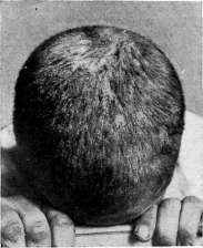
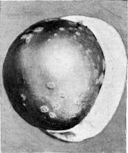
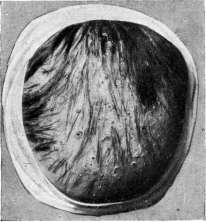
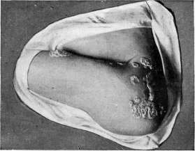
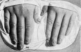
Figure 1. Scaling (pityrioid) form of favus. Figure 2. Scutulo-impetiginous form of favus; hairs epilated after radiotherapy; in the center—skin atrophy. Figure 3. Favus of the head: scutular form with atrophy in the frontal-parietal area. Figure 4. Favus of smooth skin; vesiculo-pustular
form. Figure 5. Favus of the nails. scatters scattered all over the head. This variety of F. is most difficult to diagnose, as it has neither scutula nor continuous atrophy; the changed hairs, due to their small number, are not so noticeable. The course of F. of the head is chronic, the disease lasts for years, and sometimes even decades. Its onset is more often sought in childhood, but it can first appear in mature age and even in the elderly. The disease proceeds as a local process, not accompanied by general phenomena, if one does not count certain subjective sensations from the scalp. In a number of cases, the glands in the back of the neck swell, reaching the size of a small hazelnut, without inflammatory phenomena on the skin. Children suffering from F. often have a sickly appearance, are pale, lethargic, and poorly nourished. The clinical diagnosis of F. in all cases must be confirmed microscopically. In the clinical recognition of F., one should keep in mind: a) eczema, for which the presence of an exudative process is characteristic; the signs of the latter can be masked where there are crusts, but around the hairy part, behind the ears, it is easy to detect the presence of vesicles, excoriations, weeping; under the crusts, the skin is also excoriated and weeping in places. In addition, eczema never leaves scars. The hairs can also be slightly cloudy in eczema, which can be misleading in diagnosis. b) Impetigo, in which crusts accumulate on the crown, just as in F.; but in impetigo there are no cloudy, diseased hairs, the crusts are loose, dark in color, easily separate from the skin, which appears under them either slightly reddened or only slightly scaly. Impetigo usually does not leave scars. c) Deep folliculitis, which can end in scars and thus resemble F.; but in them, along with scars, there are also more recent manifestations, which represent a pustule sitting in the mouth of the follicle; subsequently they dry into a crust. The hairs show no changes, but fall out as a result of the deep inflammatory process ending in scarring. d) Deep trichophytosis; but in it the hairs fall out quickly and cloudy hairs are not noticeable, in the foci there is much purulent exudate, drying into loose crusts that glue the hairs together until they 1 have fallen out. Scars are either absent or appear as sitting separately and shallow; trichophytosis, in addition, proceeds acutely, within days or weeks. In the atrophy stage, F. can resemble other atrophic processes, namely lupus erythematosus and atrophic alopecia (pseudopelade of Brocq); the latter disease resembles atrophy in F. so much that Darrier calls it pseudofavous atrophy. But unlike F., around the scarred areas in atrophic alopecia, no changes are visible on the skin except the slightest scaling, and sometimes weak inflammatory discoloration. In lupus erythematosus, the scarred area is surrounded by densely adherent white scales and a zone of stagnant redness at the periphery. In both diseases, there are no changes in the hairs except for their falling out during the atrophy stage. In all suspicious cases of F., the microscope finally decides the matter. Much less frequently than F. of the head, F. of smooth skin occurs, and usually it accompanies the lesion on the head; its clinical picture is not always the same. More often there is the form erythematosa, which manifests as small, from 72 to 2 cm in diameter, reddish-colored spots with sharp contours and irregular outlines. The skin on them scales in the form of small-plate white scales. The spots are located on the face, neck, shoulders, and the upper parts of the trunk behind and in front. Sometimes along with this, the patient with F. also has a few scutula on the smooth skin - on the knees, shoulders, or forearms. Another variety of F. of smooth skin is scutular; in it, typical F. formations - scutula - appear separately or in groups simultaneously with F. of the head (more often) or more rarely only on the smooth skin. The size of the scutula ranges from 1/a to 1 cm in diameter; they are also grayish-yellow in color, dry, somewhat embedded in the skin and have a depression in the center from which a downy hair protrudes. The skin around is somewhat inflamed. Unlike F. of the head, on smooth skin after removal of the scutula and healing of favus, no atrophy remains. Even rarer can be found a third variety of F. on smooth skin - vesicular; in it, groups of small vesicles and pustules appear on a reddened base, among which the smallest scutula can sometimes be noticed. The groups are few in number and are scattered most often on the extremities. The causative agent of the vesicular form of F. is more often A. quinckeanum (fig. 4). Extremely rarely, F. of smooth skin is a widespread disease, expressed by confluent scutula covering continuously the upper and lower extremities, trunk, head, and even face. Such generalized F. is a severe disease, accompanied by nutritional decline and phenomena of depression from the psyche: patients are lethargic, depressed, and not very sociable. Subjective sensations in F. of smooth skin are either completely absent or expressed only by slight itching. Only in widespread F. are intense itching and pains observed when the scutula are torn off. The course of F. of smooth skin depends on the condition of the lesions on the head; if there is a constant possibility of infection from falling crusts and transfer by nails, F. of smooth skin can last a long time, but after healing the process on the head, it quickly yields to treatment. - As a rare disease, cases of F. of mucous membranes have been described: oral cavity, esophagus or even the entire gastrointestinal tract. On the mucous membrane there are small violations of the integrity of the cover with pale yellow spots - colonies of the F. fungus. As extremely rare cases, lesions of F. of internal organs have been described: spleen, liver, lungs, bones, etc. F. of the nails [onychomycosis favosa (fig. 5)] - see Onychomycosis. Patho-anatomical changes of the skin, affected by favus, consist of the following: the scutulum represents a collection of fungal elements, epidermal cells, and fatty detritus. In the central parts, the scutulum is composed of numerous spores (conidia), at the periphery - of an interwoven felt of mycelial threads, partly well-staining and partly dead, representing only a fine-grained mass. The scutulum is surrounded by a ring of leukocytes, then there is a zone of exudate with remnants of destroyed epithelial cells. Besides the scutulum, the fungus can be found among the epithelial cells up to the Malpighian layer; especially abundant is the accumulation of fungus in the horny layer; sometimes Achorion penetrates the epidermis and penetrates into the connective tissue (Fridman). In the depth of the follicle, densely packed with fungal elements, Achorion penetrates the hair through the inner root sheath and reaches the hair bulb, never penetrating into the hair papilla. In the connective tissue, an infiltrate is observed, consisting of plasma cells, lymphocytes, and fibroblasts. Halle and Darrier saw the formation of a tubercle with giant cells. In the atrophy stage, the papillary layer is absent, the sebaceous glands are destroyed, cysts are formed from the dilated excretory ducts of the sweat glands, which are also almost completely absent. In far-advanced cases, the elastic tissue is destroyed. When examining nail lesions histologically, the fungus is located in the thickness of the nail plate, not penetrating into the dermal parts under the nail. The treatment of favus is closely connected with the removal of hairs - epilation, and where it has occurred, the process ends. Only the removal of diseased hairs, and with them the parasite, gives the parasiticidal agent the possibility to penetrate deeper into the hair follicle and exert its action. If the hairs are not removed, then no, even strongly acting disinfectant, can cure the patient with F., because the dense cuticle of the hair prevents the penetration of ointments to the deep parts of the follicle, where the fungus nests, and this is the failure of therapy with ointments, alcohol solutions or vapor substances. The best results are given by epilation with X-rays, since during X-ray therapy the hair thins out and falls out especially easily; the hair does not break off above the exit from the skin, but is removed together with the root part. It is much more difficult to achieve good epilation by removing hairs with tweezers or by using various irritating substances, which cause inflammatory phenomena and thereby some loosening of the hair. Buschke's method - removal of hairs with thallium acetate - has not found wide application due to the complications that thallium gives and the insufficiently satisfactory epilation. X-ray therapy is therefore the most reliable method of treating F. of the head. Before sending the patient for irradiation, the scalp is freed from crusts, scales, and scutula, for which it is smeared for two to three days with vaseline oil with 2% carbolic acid or 2% salicylic ointment. The day before irradiation, the head is well washed with ordinary or green soap and is subjected to treatment in this cleansed form. The hair on the head should be cut no shorter than 1/2 inch.
At the State Röntgenological and Radiological Institute in Leningrad, the following method of epilation is usually used: the head is divided into four fields: frontal (1st field), occipital (2nd), left lateral (3rd), right lateral (4th), with four sessions of irradiation (dosage and technique—see Röntgenotherapy). During the following 12 days, the head is cleared of the forming crusts by washing and with weakly disinfecting ointments or keratolytic ointments: 2% ointment of white precipitate of mercury, 2% carbolic ointment, or 1-2% salicylic ointment. To avoid skin irritation, it is better not to apply any treatment for the last 2-3 days before hair loss, i.e., starting from the 10th day after irradiation, and to limit oneself to daily head washing. On the 12th-14th day, hair loss usually occurs; this is the most important period of treatment, as the complete removal of hair determines the success of the entire therapy. Spontaneously falling hairs are first removed by hand, pulling them, and then the remaining ones are epilated with tweezers in such a way that the scalp is completely clean, freed of a single hair. It is permissible to preserve a border of healthy hair at the edge of their growth if they are difficult to remove there. Usually, with proper dosage and carefully performed Röntgenotherapy, hairs are epilated easily, removed along with the root part, and the success of treatment is ensured. If hairs for some reason are removed with difficulty and break off above the skin, it is necessary in the future to carefully monitor their regrowth and to epilate them again. After hair removal, if there are no signs of dermatitis on the scalp, ointment therapy begins. Various remedies are proposed—5% naphthalene ointment, Wilkinson's ointment, 3-5% chrysarobin ointment, etc. Saburo's method: for three days, T-ra Jodi (5%) is rubbed into the skin, on the 4th day the head is washed with green soap and then for the next three days the following ointment is rubbed in: Ac. pyro-gall. 5.0; ol. Rusci 20.0; Vasel. 100.0. Saburo also advises applying the scalp with an ointment made of Hydrarg. sulfur. 0.5; Sulfur, pp. 25.0; Vaselini 100.0. Ointment treatment continues for an average of 2-3 weeks after epilation, when the scalp becomes completely clean and it can be left without treatment, using only daily washing. On the 4th week after epilation, the growth of vellus hairs begins, and after 2½-3 months after Röntgenotherapy, the head is already covered with a rather dense layer of hair. The end of treatment, and along with it, the cure, can be judged by various methods: by repeated, multiple microscopic examinations of the pathogen over 4-6 months or even by culture and by observing clinical signs—redness of the skin around the growing hair, the formation of a small pustule around it, or scaling. These clinical signs are of great importance, as they allow one to unmistakably decide on the beginning of a relapse. The treatment of favus of smooth skin is significantly less complicated. Smearing with ointments containing keratolytic and disinfecting agents (5% salicylic, 2% pyrogallic, 5% sulfur or Wilkinson's ointment) quickly makes it possible to eliminate the disease. o. Podyvotskaya. The distribution of favus is by no means uniform in different countries. It occurs very frequently in Japan and China, in the latter especially in connection with frequent head shaving already in childhood. Individual cases of favus are described in America; according to statistics of the American Dermatological Society, out of 205,000 skin diseases registered from 1877 to 1892, there were 707 cases of favus. Favus is more common in Mexico. Of European countries, favus is rarely encountered in England (according to Crocker, 1 case per 2,000 skin diseases), in France (in Paris in 1920-21, only 37 patients with favus were treated, of which 27 were from other countries, although Saburo believes that in the southern departments of France favus occurs endemically among the native population), in Germany (for example, Buschke in one of the largest hospitals in Berlin observed over 9 years only one case, registered between 1920-1923). Among the most affected European countries is Poland, where, according to Altmann, in 1924 a special expedition to combat favus, organized by American Joint Distribution, treated 10,000 people in one year. According to Kumer, the situation is no better in Bessarabia and Romania. Favus was also a relatively common disease in Italy: according to Passini, in Venice in 1905-08, 141 cases of favus were registered, in the dermatological clinic of Milan from 1912 to 1922—575 cases of favus. In pre-revolutionary Russia, according to conscription data in 1891, among 862,024 recruits, 744 were exempted due to favus, in 1892 among 710,267—727; from 1893 to 1895 among 2,437,184, 1,953 were exempted (Petersen). In Russia, favus affected almost the entire territory of the country. According to pre-war registration data in 77 provinces, cases of favus were noted, with the largest number observed in Bessarabian, Kazan, Mogilev, Orenburg, Ufa, and Erivan provinces, i.e., favus was most common in provinces with Muslim and Jewish populations—peoples in the worst social conditions, deprived even of primitive sanitary-hygienic skills. In Kiev before the imperialist war, about 200 patients with favus sought treatment per year, which constituted 28.5% of all skin diseases for the period from 1909 to 1913 (Lurie, Rabinovich), and among fungal diseases, favus constituted 53%. The absence in pre-revolutionary Russia of specialized institutions for the treatment of fungal diseases, accessible medical assistance for the poorest layers of the population, and widely carried out preventive measures led to far from complete registration of favus morbidity. The opening in the early years of Soviet healthcare of dermatovenereological dispensaries and somewhat later special favus houses (dispensaries), conducting dispensary examination of each registered patient with favus and systematic examinations of schoolchildren, contributed to a more complete coverage of existing patients and gave in some cities a high registration of favus, which was then replaced—due to the implementation and improvement of social conditions—with a sharp decrease in morbidity. For example, in favus dispensaries organized in 1923 in Kiev and Odessa, patients with favus turned to: Cities 1923 1924 1925 1926 1927 1928 Odessa... 675 430 Kiev ... 715 100 1 1 448 393 In the USSR, favus is most common in Ukraine, Belarus, the Tatar Republic, Uzbekistan, and Azerbaijan. The sharp decrease in favus morbidity in the USSR compared to the pre-revolutionary period can be judged by the following data: while in 1872-1885 favus constituted 1.5% of all skin diseases registered at the Military Medical Academy, at present in Leningrad and Moscow it occurs only in individual cases. Favus is most common in childhood; most patients get sick before the age of 10; the youngest age is 28, even 22 days (Saburo). According to data from the Odessa Dermatovenereological Institute for 1917-27 (Mgebrov), out of 100 patients with parasitic diseases of the hair, 1.7% fell ill before 1 year of age, 26% from 1 to 5 years, 40.9% from 6 to 10 years, 24.55% from 11 to 15 years, 3% from 16 to 20 years, 2.8% from 21 to 30 years, 1.5% from 31 to 51 years. According to Kiev data (Lurie), favus up to 15 years of age was found in 84.7%, in adults after 15 years—in 15.3%; apparently, the onset of the disease in these 15.3% should be attributed to a younger age. Among children, favus is more common in boys, among adults—in women. According to data from the Kiev Favus Dispensary, favus was found in 47.7% of boys and 36.7% of girls, in 6.7% of men and 8.9% of women. Apparently, this difference in childhood is explained by more frequent haircuts and the possibility of infection during them in boys. Favus is registered somewhat more often in women than in men, apparently due to greater contact of women with sick children. The existing opinion about the greater susceptibility of Jews and Muslims to favus is explained by the fact that in pre-revolutionary Russia, the Jewish and Muslim working population was in the worst social and living conditions. The increased morbidity of favus among them was a reflection of the overcrowding, cramped conditions, and poverty of national minorities. Improvement of these conditions is accompanied by a sharp decrease in patients with favus. Thus, according to data from the Kiev Favus Dispensary, which began identifying patients with favus in 1924, the following was registered: Years Jews Russians and Ukrainians I 1924 456 368 1925 277 213 1926 185 235 1927 204 128 1928 111 4
Thus, the total number of registered cases of F. from 1924 to 1929 decreased from 715 to 364 (by 50.9%), and this decrease is observed in all national groups of patients. This decrease is particularly evident in 1928, while the slight increase in the following year 1929 should be attributed to population growth and the completeness of patient registration coverage. Under poor sanitary-hygienic conditions, certain professions may to some extent be a predisposing factor for infection with F. For example, during an examination in 1924 of schoolchildren in Radomysl, among children with F, in 44% of cases there was a possibility of bringing the disease into the family by parents who, due to the nature of their work, had to come into contact with animal skin; however, poor sanitary-hygienic conditions undoubtedly also played a role in the introduction of infection. In addition, the custom of covering the head (among Jews and Muslims) plays no small role in the spread of F: the skin under the hat is always moist, deprived of the action of air and therefore more susceptible to infection. The most important predisposing factor for infection with F is undoubtedly prolonged proximity to an affected individual. The contagiousness of F is significantly weaker than that of ringworm. In the Kyiv favus dispensary for 1928-30, 83 families were found in which cases of the disease were noted among several members (see table). From this table it is clear that the possibility of infection with prolonged cohabitation with a patient is significant; on average, 56.8% of family members of patients with F were found to be ill; in 25 families, the mother and children were ill, in 2 families the father and children, in 2 families the grandmother and grandchildren. Closer and longer contact of the mother with children is a predisposing factor for infection of children; in most cases, children slept in the same bed with the mother. Cases of F infection were observed in poorly organized, overcrowded children's shelters during the imperialist war. School epidemics of F were not observed in the USSR; in individual cases of illness among schoolchildren, when investigating sources of infection, it always turned out to be someone from the family. Indirect infection with F is extremely rare; thus, infections through compresses, baths, bedding, and pillow feathers have been described; some only admit the possibility of infection through a hairbrush. Prevention. The social nature of favus determines the system of measures carried out in the USSR in the fight against F. The national policy of the Soviet power, the rapid growth of the economic and sanitary-hygienic level of the working people, the elimination of illiteracy, the Soviet system of education, the development of school sanitary supervision, the increase in the number of kindergartens and nurseries, systematic examinations of children in them—all this served as the basis for the successful implementation of preventive measures carried out both by general dermatovenereological dispensaries and by special favus dispensaries opened in the most affected areas. The tasks of these dispensaries are: 1) to observe all patients with F from the moment of diagnosis until the final cure of the patient; 2) to treat patients; 3) to carry out follow-up treatment after X-ray therapy; 4) to identify foci and, first of all, to identify families of the ill; 5) to examine schools, boarding schools, etc.; 6) to register and account for all patients; 7) to develop scientific and public issues related to the organization of the fight against F; 8) to involve the working and rural public in the implementation of preventive measures. Examination of families of the ill is one of the most important aspects of work, as it makes it possible to identify sources of F among older persons who are often the source of infection for children. Identification of sources and their disinfection is the main condition for the elimination of endemic foci. Due to the infectious nature of F, it is necessary by legislative means to introduce mandatory notification of the relevant health authorities about each detected case. Along with improving the economic well-being of the population, overcoming the housing crisis, raising the cultural level of the masses, and deepening sanitary education, it is necessary to improve knowledge about F among doctors, especially school doctors; for the protection of children's groups (nurseries, kindergartens, schools), patients with F found among them can be returned to the groups only after complete recovery and under the supervision of a dispensary or knowledgeable doctor. It is necessary to monitor children's contact with animals, especially cats. In the treatment of favus, enhanced supervision and isolation are necessary, especially during hair loss; specially equipped washing rooms should be set up in favus dispensaries; falling hair must be burned; during treatment, it is necessary to wear a cap; the clothing of patients and premises must be disinfected. The care personnel must wear special clothing (robes that button in the back); the hands of the personnel, especially the nails, must be thoroughly washed with disinfectant solutions, warm water and soap, and Lurie
Related articles
Mentioned in
Cite this page
“Favus.” Soviet Medical Encyclopedia. English translation of Bolshaya Meditsinskaya Entsiklopediya, 1st ed. (Moscow, 1928–1936), ed. N. A. Semashko. https://sovietmedicalencyclopedia.pages.dev/article/favus/