Human Nerves
Historical document, translated for reference. It reflects medical knowledge of the 1920s–30s and is not medical advice.
Summary
This article provides detailed anatomical illustrations and descriptions of the human nervous system, focusing on the sympathetic nervous system, cervical region, and various nerve plexuses. The diagrams show the topography of nerves in different body regions including the neck, head, pelvis, and buttocks.
Encyclopedia article (1928–1936)
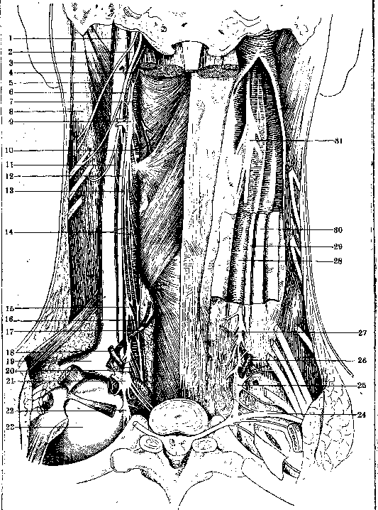
Figure 29. Sympathetic nervous system of the cervical region (posterior view): 1- internal jugular vein; 2- vagus nerve; 3- digastric muscle; 4- meningeal artery; 5- accessory nerve of Willis; 6- hypoglossal nerve; 7- sympathetic trunk; 8- sternocleidomastoid muscle; 9- superior cervical ganglion; 10- superior laryngeal nerve; 11- descending branch; 12- descending branch of the hypoglossal nerve; 13- vagus nerve; 14- sympathetic trunk; 15- superior cardiac nerve; 16- middle cardiac nerve; 17- inferior thyroid artery; 18- vertebral artery; 19- subclavian vein; 20- inferior cardiac nerve; 21- inferior cervical ganglion; 22- first thoracic ganglion; 23- pleura; 24- first dorsal nerve; 25- inferior cervical ganglion; 26- vertebral artery; 27- inferior thyroid artery; 28- common carotid artery; 29- vagus nerve; 30- internal jugular vein; 31- superior cervical ganglion
;
Figure 30. Topography of superficial organs of the neck (sternocleidomastoid muscle and clavicle removed): 1- thyroid gland; 2- thoracic duct; 3- trachea; 4- internal mammary artery; 5- subclavian vein; 6- subclavian artery; 7- clavicle; 8- lymph nodes and lymphatic trunk; 9- inferior thyroid artery; 10- brachial plexus; 11- middle sympathetic ganglion; 12- common carotid artery; 13- internal jugular vein; 14- vagus nerve; 15- sternocleidomastoid muscle
Figure 31. Topography of organs of the neck (internal jugular vein removed): 1- superior thyroid artery; 2- inferior thyroid artery; 3- ansa Vieusseni; 4- thoracic duct; 5- pleurotracheovascular ligament; 6- common carotid artery; 7 and 14- phrenic nerve; 8- subclavian vein; 9- subclavian artery; 10- clavicle; 11- pleura; 12- eighth anastomotic branch of the brachial sympathetic plexus; 13- middle cervical ganglion; 15- vagus nerve
Figure 32. Topography of organs of the neck (common carotid artery removed): 1- middle cervical ganglion; 2- inferior thyroid artery; 3- supreme intercostal artery; 4- ansa Vieusseni; 5- recurrent nerve; 6- cardiac sympathetic nerve; 7- phrenic nerve; 8- vagus nerve; 9- common carotid artery; 10- subclavian artery; 11- trachea; 12- internal mammary artery; 13- pleuroscalene ligament; 14- inferior cervical ganglion
Figure 33. Topography of organs of the neck (most vessels removed): 1- inferior cervical ganglion; 2- recurrent nerve; 3- cardiac sympathetic nerve; 4- vagus nerve; 5- phrenic nerve; 6- first thoracic ganglion; 7- vertebral nerve
Figure 34. Horizontal section through the human head at the level of the odontoid process: 1- spinal nerve; 2- transverse process of the first vertebra; 3- parotid gland; 4- medial pterygoid muscle; 5- masseter muscle; 6- mental nerve; 7- tongue; 8- external maxillary artery; 9- facial vein; 10- sympathetic trunk; 11- internal carotid artery; 12- vagus nerve; 13- hypoglossal nerve; 14- internal jugular vein; 15- external carotid artery; 16- ascending pharyngeal artery; 17- accessory nerve of Willis; 18- occipital artery; 19- digastric muscle
Figure 35. Horizontal section through the neck at the level of the vocal cords. The center of the figure shows one of the cervical vertebrae with the spinal cord from the dorsal side and the spinal nerve and vessels from the side in the transverse canal. Dorsal to the vertebra, on both sides of the spinous process and nuchal ligament, lie the dorsal muscles of the neck. Ventral to the vertebra lie the deepest muscles of the neck (longus capitis and colli muscles), the prevertebral fascia of the neck, the retropharyngeal fascia, the pharynx, and the cartilages of the respiratory tract with the vocal cords. The lateral parts are occupied by the cervical vascular-nervous bundle, the lateral muscles of the neck, the superficial muscles of the neck, and their fasciae. 1- sternocleidomastoid muscle; 2- internal jugular vein; 3- cervical sympathetic trunk; 4- vagus nerve; 5- descending branch of the hypoglossal nerve; 6- cervical sympathetic trunk; 7 and 10- arteries and veins of the superior thyroid; 8- one of the nerves of the brachial plexus; 9- artery, vein, and nerve of the vertebral; 11- common carotid artery; 12- vagus nerve; 13- internal jugular vein
Figure 36. Section through the neck at the level of the upper edge of the respiratory tract: 1- scalenus muscle; 2- one of the nerves of the brachial plexus; 3- cervical sympathetic nerve; 4- inferior thyroid artery; 5- inferior thyroid artery; 6- sternocleidomastoid muscle; 7- vagus nerve; 8- jugular vein; 9- cardiac sympathetic nerve; 10- inferior thyroid artery; 11- recurrent nerve; 12- thyroid gland; 13- esophagus; 14- recurrent nerve; 15- vagus nerve; 16- internal jugular vein; 17- inferior thyroid artery; 18- common carotid artery; 19- inferior thyroid artery; 20- cervical sympathetic nerve; 21- artery and vein of the vertebral
Figure 37. Section through the neck at the level of the epiglottic cartilage: 1 and 23- spinal nerve; 2 and 22- jugular vein; 3- vagus nerve; 4- lymph gland; 5- descending branch of the hypoglossal nerve; 6- internal carotid artery; 7- glossopharyngeal nerve; 8- external carotid artery; 9 and 11- lingual artery; 10- parotid gland; 12- facial vein; 13- superior cervical ganglion; 14- glossopharyngeal nerve; 15- external carotid artery; 16- internal carotid artery; 17- temporal vein; 18- descending branch of the hypoglossal nerve; 19- vagus nerve; 20- superior laryngeal nerve; 21- cervical plexus; 24- vertebral artery
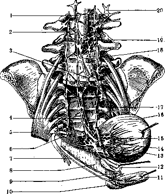
Figure 38. Diagram of the right iliac-sacral plexus from the front: 1- lateral cutaneous branch; 2- iliohypogastric nerve and with it the lateral cutaneous branch; 3- genitofemoral nerve; 4- ilioinguinal nerve; 5- branches for the major and minor psoas muscles; 6- lateral femoral cutaneous nerve; 7- external spermatic nerve; 8- lumboinguinal nerve; 9- branch for the iliacus muscle; 10- femoral nerve; 11- branch for the major psoas and iliacus muscles; 12- accessory obturator nerve; 13- obturator nerve; 14- superior gluteal nerve; 15- peroneal nerve; 16- inferior gluteal nerve; 17- tibial nerve; 18- branch for the internal obturator and gemelli muscles; 19- branch for the quadratus femoris muscle; 20- branch for the adductor magnus, semimembranosus, semitendinosus, and long head of the biceps muscles; 21- posterior femoral cutaneous nerve; 22- inferior medial cluneal nerve; 23- perineal and dorsal nerves of the penis (clitoris); 24- inferior hemorrhoidal nerves; 25- branch for the levator ani muscle; 26- branch for the coccygeus muscle; 27 and 28- anococcygeal nerves; 29- branch for the piriformis muscle; 30, 32, 34, 36 and 38- branches for the intertransversarii muscles; 31, 33, 35 and 37- branches for the quadratus lumborum muscle; Th12-Lm- lumbar plexus; L4-S3- sacral plexus; S2-S4- pudendal plexus; S5-Co- coccygeal plexus. Figure 39. Schematic representation of a preparation of the sympathetic nervous system of the abdominal cavity and pelvis of a man from the right side. In the upper part of the preparation on the left, three intercostal spaces are shown, in which the intercostal arteries branching from the thoracic aorta are shown, and below them are the intercostal nerves connecting with the nodes of the border trunk of the thoracic sympathetic nervous system through connecting branches. Between the border trunk and the thoracic aorta are the large and small splanchnic nerves. In the upper right part of the preparation under the diaphragm, the stomach is shown with its dorsal surface, on which its nerve plexus is left. Further, the duodenum and transverse colon are cut. All other intestines are turned to the left, and the right kidney with the ureter and peritoneum are separated to show the sympathetic nervous system and the nerves of the iliac-sacral plexus. 1- lumbar ganglion of the sympathetic trunk; 2- renal plexus; 3- iliohypogastric nerve; 4- sacral femoral cutaneous nerve; 5- interganglionic branch; 6- anastomosis of the hypogastric plexus with the lumbar aortic plexus; 7- sacral ganglion of the sympathetic trunk; 8- sacral plexus; 9- spermatic plexus; 10- deferential plexus; 11- inferior mesenteric plexus; 12- superior mesenteric plexus; 13- solar plexus. (For illustration to the article Human Nerves)
Fig. 38
Figure 39.
Figure 40. Main nerves of the heart. On the preparation, the respiratory tract, main bronchial branches, both carotid arteries, and the nerves lying on the sides of the respiratory tract are left; below, the aortic arch with its large vessels is left, the superior vena cava is cut rather low, and both the subclavian and internal jugular veins are removed; finally, the pericardial sac is opened and its flaps are spread apart. 1- left recurrent nerve; 2- right recurrent nerve; 3- right vagus nerve; 4- tracheal branches; 5- cardiac ganglia and plexus; 6- right pulmonary plexus; 7- right phrenic nerve; 8 and 9- branches of the cardiac plexus; 10- anterior pulmonary plexus; 11- left phrenic nerve; 12- inferior cardiac nerve; 13- middle cardiac nerve; 14- superior cardiac nerve; 15- left vagus nerve
Figure 41. Nerves of the gluteal region: 1- posterior gluteal muscle; 2- posterior superior iliac spine; 3- superior gluteal nerve; 4- piriformis muscle; 5- sciatic nerve; 6- pudendal nerve; 7- posterior sacrospinous ligament; 8- posterior sacrotuberous ligament; 9- superior gemellus muscle; 10- inferior gemellus muscle; 11- semimembranosus muscle; 12- biceps femoris and semitendinosus muscles; 13- adductor magnus muscle; 14- quadratus femoris muscle; 15- bursa of the piriformis muscle; 16- bursa of the pectineus muscle; 17- lesser trochanter (major psoas muscle); 18- bursa of the external obturator muscle; 19- middle, anterior, and posterior gluteal muscles; 20- trochanteric bursa of the gluteus minimus muscle; 21- middle iliac spine; 22- nerve for the tensor fasciae latae muscle; 23- nerve for the gluteus minimus muscle; 24- nerve for the gluteus medius muscle; 25- long posterior sacroiliac ligament; 26- short posterior sacroiliac ligament. (For illustration to the article Human Nerves.)
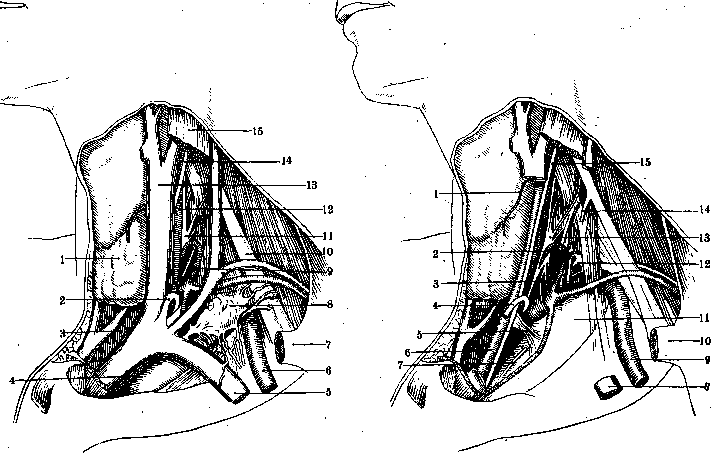
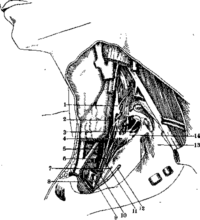
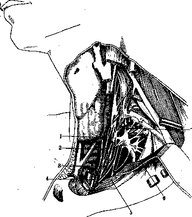
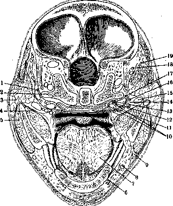
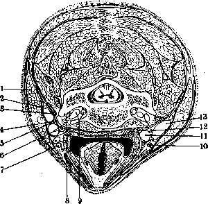
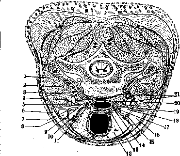
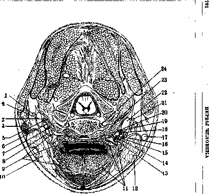
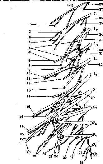
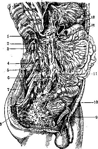
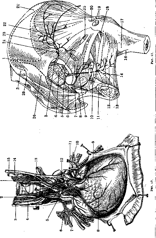
Figure 42. Right vagus nerve: 1-20 p. spinalis; 2-p. hypoglossus; 3-p. vagus; 4-p. cervicalis secundus; 5-gangl. cervicale sup. trunci sympathici; 6-gangl. cervicale inf. trunci sympathici; 7-truncus sympathicus; 8-plexus oesophageus; 9-vena azygos; 10-a. splanchnicus major; 11-gangl. semilunare dextrum; 12-plexus Solaris; 13-rami gastrici; 14-plexus pulmonalis; 15-ramus cardiacus medius; 16-n. recurrens; 17-nn. cardiaci sup.; 18-n. laryngeus sup. (ramus ext.); 19-r. pharyngeus; 20-n. laryngeus sup.
Figure 43. Left vagus nerve: 1-p. lingualis; 2-p. glosso-pharyngeus; 3-p. laryngeus sup.; 4-rami pharyngei; 5-n. recurrens; 6-r. cardiacus med.; 7-rr. pulmonales; 8-r. hepaticus; 9-n. splanchnicus major; 10-plexus oesophageus; 11-truncus sympathicus; 12-gangl. cervicale inf.; 13 and 14-plexus brachialis; 15-gangl. cervicale sup.; 16-n. vagus.
Figure 44.
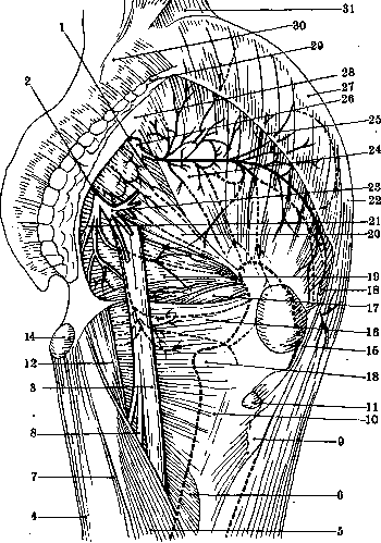
Figure 44. Nerves of the gluteal region: 1-foramen suprapiriforme; 2-foramen infrapiriforme; 3-n. ischiadicus; 4-m. adductor magnus; 5-m. biceps femoris, caput longum; 6-m. adductor magnus; 7-m. semitendinosus; 8-r. muscularis flexorius; 9 and 20-m. glutaeus max.; 10-m. adductor minimus; 11-bursa glutaeo-femoralis; 12-m. semimembranosus; 13-trochanter minor; 14-bursa ischiadica m. glutaei max.; 15-bursa trochanterica m. glutaei max.; 16-m. quadratus femoris; 17-m. obturator ext.; 18-n. for m. tensor fasciae latae; 19-m. obturator int. cum gemellis; 20-m. piriformis; 21-n. pudendus; 22-spina iliaca superior ant.; 23-n. glutaeus inf.; 24-m. glutaeus med.; 25-tractus ilio-tibialis (Maissiati) (pars supratrochanterica); 26-n. glutaeus superior; 27-spina iliaca media; 28-lig. sacro-tuberosum; 29-spina iliaca superior posterior; 30-m. quadratus lumborum.
Figure 45. Lumbar plexus and its branches: 1-p. ilio-hypogastricus; 2-p. ilio-inguinalis; 3-p. cutaneus femoris lateralis; 4-p. genito-femoralis; 5-p. femoralis; 6-p. obturatorius; 7-p. plexus sacralis; 8-gangl. sacrale trunci sympathici; 9-ramus communicans; 10-gangl. lumbale trunci sympathici; 11-ramus communicans.
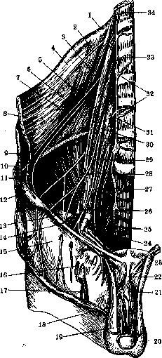
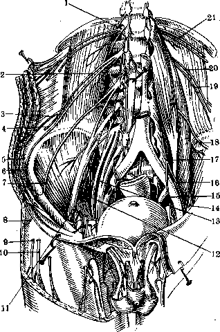
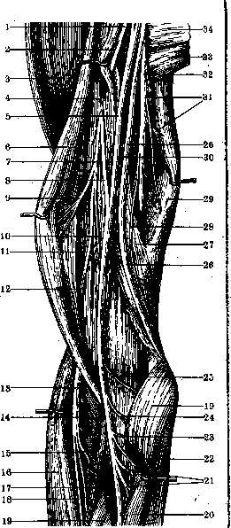
Figure 46. Drawing of the right half of the abdominal wall, pelvis and upper thigh region; from the specimen the parietal peritoneum has been removed, and some fibers of the quadratus lumborum muscle and the psoas major muscle have been split. Only the skin and subcutaneous tissue of the thigh and the dorsal part of the penis have been removed from the outside. 1-costa XII; 2-p. intercostalis XII; 3-m. quadratus lumborum; 4-aponeurosis m. transversi abdominis; 5-m. psoas major; 6-n. ilio-hypogastricus; 7-n. ilio-inguinalis; 8-m. obliquus abdominis ext.; 9-m. obliquus abdominis int.; 10-n. cutaneus femoris lat.; 11-ramus cutaneus lat. n. ilio-hypogastrici; 12-m. iliacus; 13-fascia ilio-pectinea; 14-lig. inguinale (Pouparti); 15-fascia lata; 16-fascia cribrosa; 17-v. saphena; 18-funiculus spermaticus; 19-tunica vaginalis communis; 20-scrotum; 21-n. dorsalis penis; 22-lig. suspensorium penis; 23-m. pyramidalis; 24-ductus deferens; 25-a. and v. iliaca ext.; 26-n. femoralis; 27-n. obturatorius; 28-n. lumbo-inguinalis; 29-n. spermaticus ext.; 30-n. genito-femoralis; 31-ramus communicans; 32-ganglia trunci sympathici; 33-truncus sympathicus; 34-vertebra thoracalis XII. Figure 47. Lumbar plexus and its branches. Drawing of the posterior wall of the abdominal cavity, pelvis and upper thigh region. From the specimen all internal organs except the bladder and part of the rectum have been removed, the leaf of parietal peritoneum has been removed, on the right side the psoas major muscle has been removed; under such conditions on the right side all nerves of the lumbar plexus are visible from their exit from the intervertebral foramina; on the left side it is visible how these nerves leave the psoas major muscle. 1 and 21-p. intercostalis XII; 2 and 20-p. ilio-hypogastricus; 3-ramus muscularis; 4 and 8-ramus cutaneus lat.; 5-m. abdominis; 6 and 19-p. ilio-inguinalis; 7 and 18-p. cutaneus femoris lat.; 9 and 10-rami cutanei femorales lat.; 11 and 14-p. femoralis; 12-ramus musculi psoatis; 13-plexus sacralis; 15-n. spermaticus ext.; 16-n. lumbo-inguinalis; 17-n. genito-femoralis. (For illustration in article Human Nerves.)
Figure 48. Diagram of irradiation of gastric pains to spinal centers and irradiation of vomiting reflexes to medulla oblongata centers: 1-IX thoracic segment; 2-p. splanchnicus; 3-truncus sympathicus; 4-plexus coeliacus; 5-peripheral nerve; 6-p. vagus; 7-gangl. nodosum; 8-gangl. jugulare. Figure 49. Diagram of innervation of human sexual organs: 1-upper lumbar part of spinal cord; 2-lower sacral part of spinal cord; 3-m. sphincter urethrae; 4-plexus ductus deferentis; 5-plexus hypogastricus; 6-plexus aorticus; 7-gangl. mesentericum inf.
Figure 50-
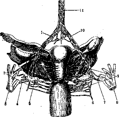
Figure 51. Figure 50. Innervation of uterus: 1 and 10-plexus hypogastricus; 2 and 5-plexus Frankenhauseri; 3 and 6-plexus pudendus; 4 and 7-p. erigens; 5-gangl. Frankenhauseri; 6-vaginal branches; 11-plexus aorticus. Fig. 51. Innervation of urinary bladder: 1-aorta abdominalis; 2-truncus sympathicus dexter; 3-plexus hypogastricus sup.; 4-plexus hypogastricus inf. dexter; 5-n. ischiadicus; 6-nn. erigentes; 7-n. pudendus; 8-m. levator ani; 9-m. transversus perinei profundus; 10-m. ischio-cavernosus sin.; 11-m. bulbo-cavernosus; 12-prostata; 13-plexus vesicalis; 14-nn. vesicales; 15-ureter dexter; 16-vesica urinaria; 17-plexus hypogastricus inf. sin.; 18-ramus ant. n. lumbalis IV; 19-plexus aorticus; 20-plexus renalis sin. ?44
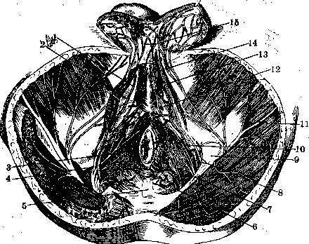
Figure 52. Nerves and arteriesg of male perineum: 1-n. pudenda communis; 2-diaphragma urogenitale; 3-m. sphincter ani ext.; 4 and 7-lig. sacro-tuberosum; 5-os coccygis; 6-a. haemorrhoidalis int.; 8 and 10-n. pudenda communis; 9-n. pudendus; 11-tuberositas ossis ischii; 12-n. cutaneus femoris post.; 13-a. bulbosa; 14-m. ischio-cavernosus; 15-a. scrotalis post.; 16-nn. scrotales post.
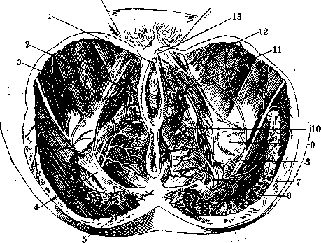
Figure 53. Nerves and arteries of female perineum: 1-glans clitoridis; 2-bulbus vestibuli; 3-glandula Bartolini; 4-lig. sacro-tuberosum; 5-os coccygis; 6-m. glutaeus max.; 7-n. pudendus; 8-n. cutaneus femoris post.; 9-tuberositas ossis ischii; 10-a. transversa perinei; 11-a. bulbosa; 12-m. ischio-cavernosus; 13-n. dorsalis clitoridis. NERVES OF HUMAN 74
Figure 54. Anastomosis of n. musculo-cutanei with n. medianus: 1-branch to m. brachialis; 2-anastomosis with n. medianus; 3-p. cutaneus antibrachii lat.; 4-p. radialis; 5-p. cutaneus antibrachii dors.; 6-anastomosis; 7-ramus volaris n. cutanei antibrachii medialis; 8-n. medianus; 9-branch to m. biceps brachii; 10-p. cutaneus antibrachii medialis; 11-branch to m. coraco-brachialis; 12-n. musculo-cutaneus;
Figure 55. Anastomosis of n. musculo-cutanei with n. medianus: 1 and 7-n. musculo-cutaneus; 2-ramus muscularis; 3-m. deltoideus; 4-m. coraco-brachialis; 5-n. medianus; 6-m. biceps brachii; 7-anastomosis with n. medianus; 8-ramus muscularis for m. biceps; 9-ramus muscularis for m. brachialis; 10-m. brachialis; 11-p. cutaneus antibrachii lat.; 12-n. radialis; 13-m. supinator; 14-ramus profundus n. radialis; 15-m. brachioradialis; 16-a. radialis; 17-ramus superficialis n. radialis; 18-m. pronator teres; 19-m. flexor carpi radialis; 20-branch for m. flexor digitorum sublimis; 21-for m. flexor carpi rad.; 22-for m. pronator teres; 23-for m. palmaris longus and flexor sublimis and indicis; 24-ramus muscularis for m. pronator teres; 25-n. ulnaris; 26-m. triceps (caput mediale); 27-ramus collateralis ulnaris; 28-m. triceps (caput longum); 29-a. brachialis; 30-rami musculares for caput longum m. triceps; 31-n. radialis; 32-m. teres major; 33-m. latissimus dorsi.
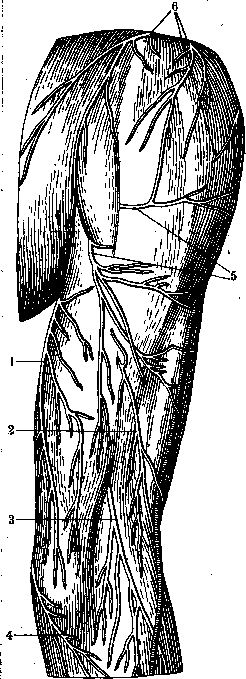
Figure 56. Distribution of cutaneous nerves of the arm: 1-p. cutaneus brachii medialis; 2-p. cutaneus brachii post.; 3-n. cutaneus antibrachii dorsalis; 4-ramus ulnaris n. cutanei antibrachii medialis; 5-n. cutaneus brachii lat.; 6-nn. supraclaviculares.
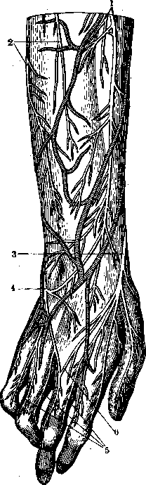
Figure 57. Distribution of cutaneous nerves on the dorsum of forearm and hand: 1-p. cutaneus antibrachii dorsalis; 2-ramus ulnaris p. cutanei antibrachii medialis; 3-ramus superficialis n. radialis; 4-ramus dorsalis manus n. ulnaris; 5-nn. digitales dorsales; 6-ramus anastomoticus ulnaris. "i
Figure 58.
Figure 59.
Figure 58. Branches of p. cutaneus antibrachii lat., radialis superficialis, ulnaris and medianus on the hand: on the left side-usual relationships, on the right side-deviations.
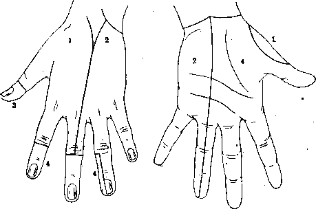
i p. cutaneus antibrachii lat. -------p. radialis superficialis - . - P. ulnaris . . . p. medianus Figure 59. Zones of innervation; 1-p. radialis; 2-p. ulnaris; 3 and 4 - p. medianus. Figure 60. Distribution of p. cutanei humeri interni and rami cutanei humeri anterioris: 1-nn. intercosto-brachiales; 2-n. supraclavicularis; 3-n. cutaneus antibr. medialis; 4-n. ulnaris; 5 and 5-ramus volaris and ramus ulnaris%i. cutanei antibr. medialis; 7-v. basilica; 8-v. cephalica; 9-v. mediana cubiti; 20-n. cutaneus antibr. lat.; Л-rami cutanei brachii ant. n. cutanei antibr. medialis; 12-n. cutaneus brachii medialis. Figure 61. Distribution of cutaneous nerves on the forearm:
' 1 - p. cutaneus antibrachii dorsalis; 2-p. cutaneus antibrachii lat.; 3-ramus superficialis n. radialis; 4-ramus palmaris n. mediani; 5-ramus cutaneus palmaris n. ulnaris; 6-n. cutaneus antibrachii medialis (ramus ulnaris and ramus volaris). Figure 62. Nerves of the right index finger from the lateral side: l-n. digitalis dorsalis; 2-aponeurosis palmaris; 3-n. digitali- volaris proprius; 4-cutaneous branch; 5-a. digitalis volaris propria; 0-branches '
длй}ф;орсальноп стороны; 7-branch for the lateral surface of the finger; «--anastomosis between the dorsal and volar nerves. Figure 63. Anastomoses between p. digitalis dorsalis n. radialis and p. digitalis volaris proprius n. mediani: . 1 --p. digitalis dorsalis n. radialis; 2 -anastomoses; 3-p. digitalis volaris proprius n. mediani. (For illustration in Human Nerves.)
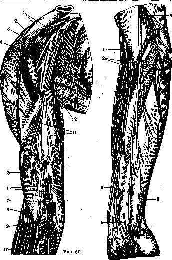
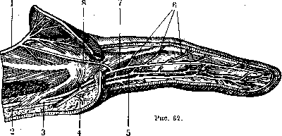
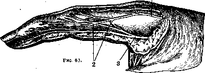
Figure 61.
Figure 64. Position of p. radialis on the arm: J-n. radialis; 2-vena collat. ulnaris sup.; з and 7-p. collateralis ulnaris p. radialis; 4-branch for caput lat. m. tricipltis; 5-n. cutaneus antibr. dorsalis; б-branch for caput mediale m. tricip.n m. anconaeus; £-n. ulnaris; 9-caput longum m. tricip.; 10-caput mediale m. triq/JS / ?-n. cutaneus antibrachii medialis; 12-n. cutaneus humeri post.; 13-n. cutaneus humeri internus; /4-^branch for caput longum m. tricipims; 15-m. latissimus dorsi. Figure 65. Anastomosis of p. mediani with p. ulnaris:
/-tendon of m. bicip.; 2-p. medianus; 3-m. brachio-radialis; 4-branch for m. pronator teres; 5-n. radialis superfic; в and 8- a. radialis; 7-branch for m. flexor pollicis long.; 9-m. flex. poll, long.; lO-m. abd. poll, long.; 11- m. pronator quadratus; 12-membrana interossea; 13-ramus interosseus (med.); 14-ramus anast. n. mediani cum n. ulnari; 15- m. flexor dig. prof.; 16-m. flexor carpi uln.; 17-n. ulnaris; 18-nerve for m. flex. dig. subl.; 19- nerve for m. flexor carpi rad.; 20-nerve for m. palmaris long.; 21-m. pronator teres (caput ulnare); 22-nerve for m. pronator teres; 23-m. brachialis internus. Figure 66. Terminal branches of nerves of the hand: 1-m. flexor pollicis longus; 2-tendon of m. flexoris carpi radialis; 3-tendon of m. brachio-radialis; 4-m. pronator quadratus; 5-n. medianus; 6-tendon of m. abductoris pollicis long.; 7-vagina tendineum mm. flexorum communlum; 8 and 25-Ug. carpi transversum; 9-m. abductor pollicis brevis; 10-m. opponens pollicis; 11- ramus muscularis n. mediani; 12- m. flexor pollicis brevis (caput superfic); 13-nn. digitales volares communes n. mediani; 14-m. adductor pollicis; 15-mm. lumbricales; 16 and J8-^-nn. digit, vo.lares proprli; 17-branches for the palmar-lateral side of the fingers; 19-cutaneous branches; 20-p. digitalis volaris communis; 21 -ramus anastomoticus; 22-rami musculares; 23-m. abductor digiti V; 24-ramus profundus; 26-m. flexor digit! V brevis; 27-branch to the joint; 28-ramus dorsalis; 29-n. ulnaris; 30-m. flexor digitorum profundus; 31-m. flexor digitorum sublimis; 32- m. flexor carpi ulnaris. (For illustration in Human Nerves.)
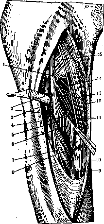
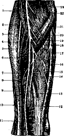
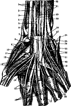
Figure 64-
HUMAN NERVES
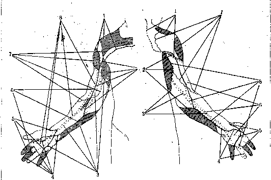
Figure 67.
f
Figure 68.
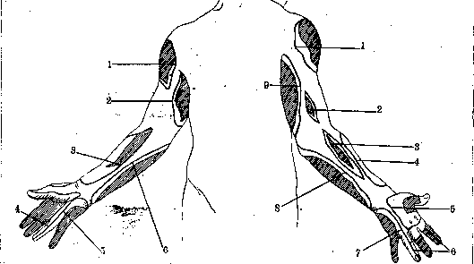
Figure 69.
Figure 70.
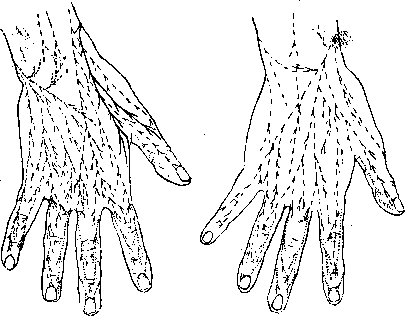
Figures 67 and 68. Diagrams of autonomous maximal areas of distribution of cutaneous nerves of the hand from the palmar and dorsal surfaces. Figure 67: 1- nn. supraclaviculares; 2-p. cutaneus humeri internps; 3-p. cutaneus antibracbii medialis; 4-n. ulnaris; 5-n. medianus; 6-n. cutaneus antibrachii lat,.; 7-n. cutaneus humeri lat.; 8-n. radialis. Figure 68. I-nn. supraclaviculares; 2-n. cutaneus humeri jnternus; other designations are the same as in fig. 67. Figures 69 and 70. Diagram of distribution of defects in cutaneous sensitivity on the palmar and dorsal sides with interruption of individual nerve trunks; Figure 69. 1-p. cutaneus humeri lat.; 2-p. cutaneus humeri - internus + intercosto-brachialis; 3-n. cutaneus antibrachii lat.; 4-n. medianus; 5-n. ulnaris; 6-m cutaneus antibracbii medialis. Figure 70. 1-p. cutaneus humeri lat.; 2-n. cutaneus humeri post.; 3-n,. cutaneus antibrachii dorsalis; 4-n. cutaneus antibrachii lat.; 5-n. radialis superficialis; 6-n. medianus; 7-n. ulnaris; 8-n. cutaneus antibrachii medialis; 9-n. cutaneus brachii internus 4- intercosto-brachialis. (According to Foerster.)
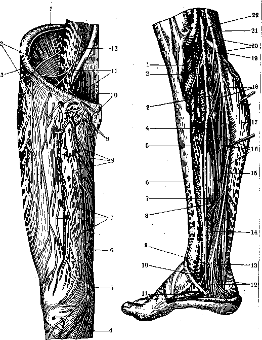
f"Pnc. 71. Distribution of cutaneous nerves on the anterior surface of the thigh: 1 and »-p. ilio-inguinalis; 2-p. cutaneus femoris lat.; з-га- -mus cutaneus lat. p. ilio-hypogastrici; 4-p. saphenus; 5-ramus infrapatellaris n. sapheni; .' 6-ramus cutaneus n. obturatorii; 7 and s-rami cutanei femoris ant.; 2 0-n. spermaticus ext.; , 11-n. lumbo-inguinalis; 2 2-n.genito-femoralis. Figure 72. Branches of p. tibialis within: 1- m. popliteus; 2-й. interosseus cruris; 3-branch for m. tibialis post.; 4-a. peronea; 5-a. tibialis post.; 6-m. flexor digit. longus; 7-m. tibialis post.; 8- branch for a. tibialis post, and m. tib. post.; 9-n. plantaris lat.; 10-n. plantaris medialis; 11-m. abductor hallucis; 22-rami calcanei mediales; 13- ramus articularis; 2 4-branch for syndesmosis tibio-fibularis; 25-m. flexor hallucis longus; 16-rami musculares for m. flexor hallucis longus; 17-m. soleus; 18-rami muse, for m. soleus; 19-m. plantaris; 20- rami muse, for m. popliteus; 21- n. tibialis; 22-a. poplitea. Figure 73. Distribution of cutaneous nerves on the posterior surface of the thigh: 1-nn. clunium medii; 2-rami perineales; 3- small branches of n. cutanei femoris post.; 4-ramus cutaneus n. obturatorii; 5-ramus cutaneus crurrs medialis n. sapheni; б-branches of p. cutanei surae lat.; 7-branches of p. cutanei femoris lat.; 8-n. cutaneus femoris post.; 9-nn. clunium inf.; 10-branches ',..
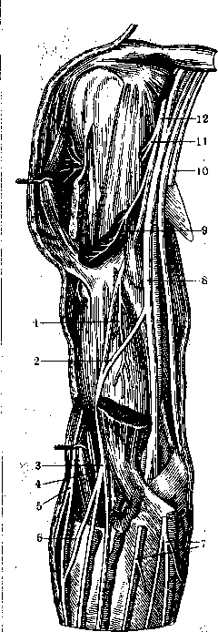
p. cutanei femoris lat.; 11-nn. clunium sup. Figure 74. Branches of p. femoralis: 1 and 25-p. cutaneus femoris lat.; 2-ramus muscularis for m. iliacus; 3- rami musculares for m. sartorius; 4-rami muse, for m. quadrioeps femoris; 5-rami cutanei ant.; 6-m. sartorius; 7 and 9-p. saphenus; 8-hiatus tendineus (adductorius); io and 11- rami cutanei n. obturatorii; 12- m. adductor longus; 13-v. saphena magna; 14-\. femoralis; 15-branch for m. pectineus; 16-a. femoralis; 17-ramus muscularis n. femoralis for m. pectineus; 18-n. spermaticus ext.; 19 and 23-rami musculares for m. iliacus; 20-m. psoas major; 21-n. femoralis; 22-a. iliaca communis; 24-m. iliacus. Figure 75. Branches of p. tibialis from behind: 1-m. semitendinosus; 2-m. semimembranosus; 3-m. adductor magnus; 4-a. and v. poplitea; 5-n. tibialis; 6-fossa poplitea; 7-branch for a. poplitea; 8-rami musculares; 9-ramus articularis; io, 14 and 20-m: gastrocnemius; il-m. popliteus; 22- arcus tendineus ra. solei; 13-m. soleus; 25-fascia cruris; 16-n. suralis; 2 7-ramus anastomoticus peronaeus; 18- n. cutaneus surae medialis; 19 and 24-p. peronaeus communis; 21- m. plantaris; 22- rami musculares; 23-n. cutaneus surae lat.; 25- ramus articularis; .26-m. biceps femoris (caput breve); 27--ramus muscularis; 28- m. biceps femoris (caput longum); 29-n. ischiadicus. (For illustration in Human Nerves.)
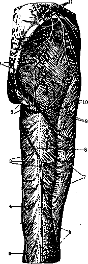
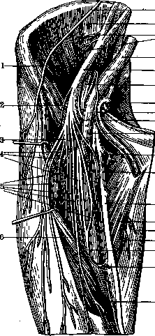
Figure 73.
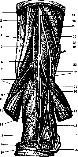
Figure 75. ?в7
HUMAN NERVES
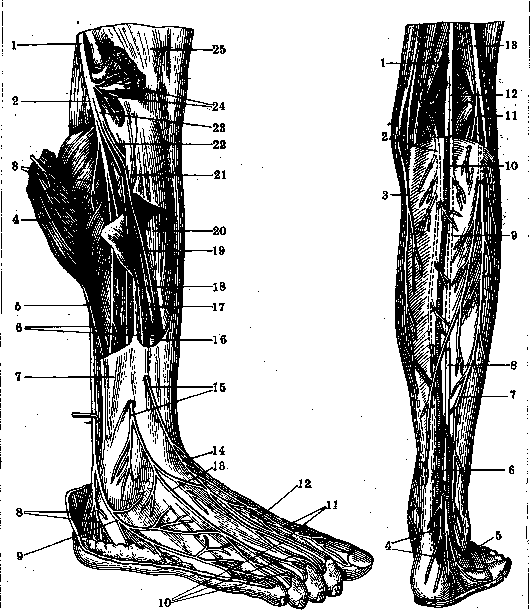
Figure 76.
Figure 77.
Figure 76. Distribution of p. suralis and p. peronaei superficialis: I-p. peronaeus communis; 2, в-vs. 15-p. peronaeus superfic; 3-rami musculares; 4-та. peronaeus longus and brevis; 5-septum iriter-i mqseulare ant. (fibulare); 7-fascia cruris; s^rami calcanei lat.; S-n. cutaneus dorsalis lat.; 10-nn. digitales dorsales pedis; 11-nn. digitales dorsales hallucis lat. and digiti II .'medialis; 12-terminal branch of p. -peronaei profundi; 13-p. cutaneus dorsalis intermedius; J4-n. cutaneus dorsalis medialis; 16 and 21-m. ext. digitorum longus; 17-m. peronaeus tertius; IS-m. extensor hallucis longus; ' 19 and 23-p. peronaeus profundus; 20-rri. tibialis ant.; 22- ramus muscularis; 24-rami musculares; 25-m. peronaeus longus. Figure 77. Cutaneous nerves of the posterior surface of the leg: 1-p. tibialis; 2-p. cutaneus femoris post.; J--ramus cutaneus cruris medialis; 4-rami calcanei lat.; 5 - n. cutaneus dorsalis lat.; 6 - n. suralis; 7 - ramus anastomoticus peronaens; 8, 9 ж 12-n. cutaneus surae medialis; 10 - v. saphena parva; 11- n. cutaneus surae lat.; 13 - n. peronaeus communis. 769
HUMAN NERVES
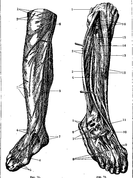
Figure 78. Cutaneous nerves of the anterior inner surface of the leg; 1-rami cutanei femoris anteriores; 2 and 3-branches of n. cutanei surae lateralis; 4-n. peronaeus superficialis; 5-nn. digitales dorsales; 6-terminal branch of n. peronaei profundi; 7-rami calcanei medii n. tibialis; 8-n. saphenus; 9-ramus infrapatellaris n. sapheni. Figure 79. Deep anastomoses between n. suralis and n. peronaeus profundus: 1-n. peronaeus communis; 2-rami musculares; 3-m. extensor digitorum longus; 4-m. extensor hallucis longus; 5-rami calcanei laterales; 6-n. cutaneus dorsalis lateralis; 7-m. extensor digitorum brevis; 8-nn. digitales dorsales pedis; 9-nn. digitales dorsales hallucis lateralis et digiti secundi medialis; 10-m. extensor hallucis brevis; 11 and 15-ramus muscularis; 12 and 16-rami musculares; 13-m. tibialis anterior; 14-n. peronaeus profundus. ..- '. B.M.E. Vol. XX. 21
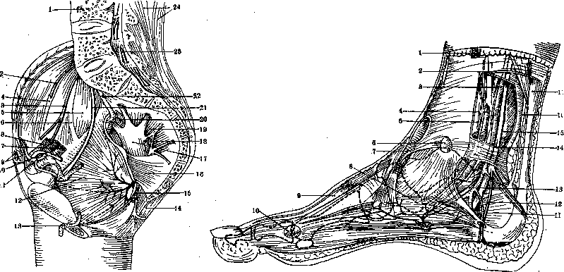
Figure 80.
Figure 8
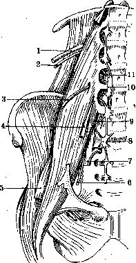
Figure 80. Nerves of the pelvic region: 1-vertebra lumbalis IV; 2-n. ilio-inguinalis; 3-n. genito-femoralis; 4-lig. inguinale; 5-m. psoas major; 6-m. psoas minor; 7-vasa iliaca ext.; 8-vasa spermatica int.; 9-m. rectus and pyramidalis abdominis; 10-ductus deferens; 11-ramus epigastrico-obturatorius; 12-m. obturator int.; 13-v. dorsalis penis; 14-m. glutaeus maximus; 15-vasa pudenda int.; 16-m. coccygeus; 17-n. ischiadicus; 18-vasa glutaea inferiora; 19-m. piriformis; 20-vasa glutaea superiora; 21-n. obturatorius; 22-ramus lumbo-sacralis; 23-n. sacralis I; 24-m. sacro-spinalis. Figure 81. Vessels and nerves of the medial part of the foot: 1-v. saphena magna and n. saphenus; 2-fascia cruris; 3-a. tibialis post.; 4-m. flexor digitorum longus; 5-m. tibialis post.; 6-bursa subcutanea malleoli med.; 7-lig. cruciatum pedis; 8-m. abductor hallucis; 9-m. extensor hallucis longus; 10-bursa subcutanea metatarso-phalangea I; 11-m. flexor digitorum longus; 12-n. calcaneus medialis; 13-n. plantaris medialis; 14-lig. laciniatum; 15-m. flexor hallucis longus; 16-m. plantaris; 17-tendo calcaneus (Achillis).
'
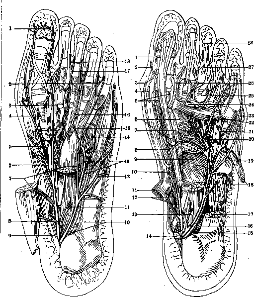
Figure 82.
Figure 83.
Figure 82. Nerves on the plantar surface of the foot: 1-aa. digitales plantares propriae; 2-aa. digitales plantares communes; 3-m. lumbricalis I; 4-n. articularis metatarso-phalangeus hallucis; 5-r. digitalis medialis hallucis proprius; 6-a. plantaris medialis; 7-m. flexor digitorum brevis; 8-n. plantaris medialis; 9-m. abductor hallucis; 10-m. abductor digiti V, pars proximalis cum nervo; 11-caput plantare m. plantaris brevis (caro quadrata) cum nervo; 12-n. digitalis lat. proprius digiti V; 13-r. profundus (muscularis) n. plantaris lat. et vasa plantaria lat.; 14-n. articularis metatarso-phalangeus V; 15-anastomosis between nn. plantares lateralis et medialis; 16-m. lumbricalis III; 17-n. digitalis communis; 18-n. digitalis proprius. Figure 83. Deep nerves and vessels on the plantar surface of the foot: 1-tendo flexoris digitorum long.; 2-a. digitalis medialis propria hallucis; 3-bursa subcutanea metatarso-phalangea I; 4-bursa subabductoria hallucis; 5-ossa sesamoidea; 6 and 9-m. abductor hallucis, caput obliquus; 7-m. flexor hallucis brevis; 8-motor anastomosis between nn. plantares medialis et lat.; 10-a. plantaris medialis; 11 and 14-m. abductor hallucis; 12-m. tibialis post.; 13-caput plantare m. plantaris brevis (caro quadrata) cum nervo; 15-n. for m. abductor digiti V; 16-n. plantaris lat.; 17 and 18-m. abductor digiti V; 19-r. profundus (muscularis) n. plantaris lat.; 20-arcus plantaris; 21-n. articularis metatarso-phalangeus V; 22-m. adductor hallucis (caput transversum); 23-bursa subcutanea metatarso-phalangea V; 24-lig. capitulorum transversum; 25-capitulum ossis metatarsalis III; 26-aa. digitales plantares communes; 27-m. flexor digitorum brevis; 27-m. flexor digitorum longus. Figure 84. Diagram of autonomous and maximal areas of distribution of cutaneous nerves of the anterior surface of the leg: 1-n. lumbo-inguinalis; 2-n. spermaticus ext.; 3-n. cruralis totalis; 4-n. tibialis + cutaneus surae medialis; 5-n. peronaeus + cutaneus surae lateralis; 6-n. ischiadicus; 7-n. cutaneus femoris lateralis. Figure 85. Diagram of areas of distribution of defects in cutaneous sensitivity of the anterior surface of the leg with interruption of individual nerve trunks: 1-n. lumbo-inguinalis; 2-n. cutaneus femorus internus; 3-nn. cutanei femoris ant.; 4-n. saphenus; 5-n. tibialis; 6-n. peronaeus communis; 7-n. ischiadicus; 8-n. cutaneus femoris lat.; 9-n. spermaticus ext.; 10-n. ilio-hypogastricus. Figure 86. Diagram of autonomous and maximal areas of distribution of cutaneous nerves of the medial surface of the leg: 1-n. obturatorius; 2-n. ischiadicus; 3-n. peronaeus + cutaneus surae lat.; 4-n. tibialis + cutaneus surae medialis; 5-n. peronaeus; 6-n. cruralis. Figure 87. Diagram of distribution of defects in cutaneous sensitivity on the medial surface of the skin of the leg with
interruption of individual nerve trunks: 1-n. cutaneus femoris int.; 2-n. ischiadicus; 3-n. tibialis; 4-n. peronaeus communis; 5-n. saphenus; 6-nn. cutanei ant. The autonomous areas of the corresponding nerves are indicated by hatching. (For illustration in article Human Nerves.)
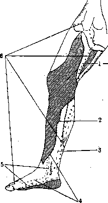
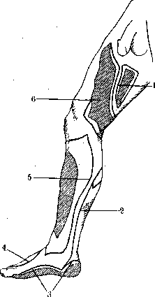
Figure 84.
Figure 85. Figure 86. Figure 87. Figure 88. Diagram of autonomous and maximal areas of distribution of cutaneous nerves of the leg from the posterior side: 1-n. cutaneus clunium sup.; 2-n. ilio-hypogastricus; 3-n. cutaneus femoris lat.; 4-n. ischiadicus; 5-n. cruralis; 6-n. peronaeus + cutaneus surae lat.; 7-n. tibialis + n. cutaneus surae medialis; 8-n. cutaneus femoris post. Figure 89. Diagram of areas of distribution of defects in cutaneous sensitivity on the posterior surface of the leg with interruption of the nerve trunk: 1-n. cutaneus femoris lat.; 2-n. cutaneus femoris post.; 3-n. cutaneus surae medialis; 4-n. peronaeus communis; 5-n. ischiadicus; 6-n. tibialis; 7-n. saphenus; 8-nn. cutanei femoris ant.; 9-n. cutaneus femoris int. Figure 90. Diagram of autonomous and maximal areas of distribution of cutaneous nerves of the leg on the lateral surface: 1-n. ilio-hypogastricus; 2-n. cutaneus femoris lat.; 3-n. ischiadicus; 4-n. cruralis; 5-n. peronaeus + cutaneus surae lat.; 6-n. tibialis + n. cutaneus surae medialis; 7-n. cutaneus femoris post. Figure 91. Diagram of areas of distribution of defects in cutaneous sensitivity on the lateral surface of the leg with interruption of the nerve trunk: 1-n. cutaneus femoris lat.; 2-n. ischiadicus; 3-n. suralis; 4-n. peronaeus communis; 5-n. cutaneus femoris post. The autonomous areas of the corresponding nerves are indicated by hatching. (For illustration in article Human Nerves.)
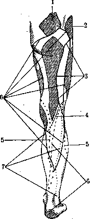
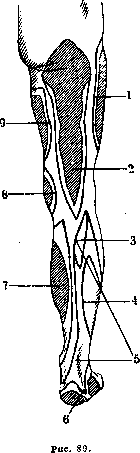
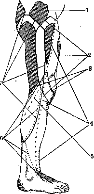
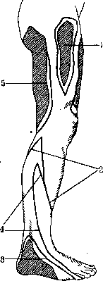
Figure 90.
Figure 91.
Related articles
Mentioned in
Cite this page
“Human Nerves.” Soviet Medical Encyclopedia. English translation of Bolshaya Meditsinskaya Entsiklopediya, 1st ed. (Moscow, 1928–1936), ed. N. A. Semashko. https://sovietmedicalencyclopedia.pages.dev/article/human-nerves/