Ampoule and Ampullitis
Historical document, translated for reference. It reflects medical knowledge of the 1920s–30s and is not medical advice.
Summary
This entry provides a historical overview of the medical ampoule, detailing its introduction, manufacturing, and usage for sterile drug storage. It also includes a brief clinical description of ampullitis, an inflammation of the ampulla of the vas deferens.
Encyclopedia article (1928–1936)
AMPOULE (from Latin ampulla, diminutive of amphora—a vessel among the Romans), a glass vessel with a wide base and a narrow neck. A medicinal ampoule serves for the storage of dosed sterilized solutions, sera, as well as powders and liquids that change when acted upon by air. Introduced in 1886 by the Parisian pharmacist Limousin, ampoules have gained wide application in medical practice due to the following advantages: 1) the substance sealed in ampoules is completely isolated from the surrounding air, it does not change and remains sterile until the ampoule is opened; 2) ampoules contain a precisely dosed quantity of the preparation; and 3) they are more portable than vials. The appearance of ampoules varies (see Figure 1); the volume is from 1 to 250 cubic cm (the usual volume of ampoules containing solutions for injection is 1–3 cubic cm). Since many preparations decompose in the presence of even negligible traces of alkalis, glass containing no free alkali is used for the manufacture of ampoules. Immediately before filling, the ampoules are thoroughly washed first with a 1% solution of hydrochloric acid, and then with distilled water. The washed ampoules are sterilized (see Sterilization), dried, and filled, usually through a graduated burette, the lower end of which is connected either to a glass capillary tube or to a syringe needle. In factories, mass filling of ampoules is used with the help of various devices built on the principle of air evacuation. Filled ampoules are immediately carefully sealed and sterilized! To open an ampoule, a notch is made with a file on its neck, closer to the base, and the neck is broken off; solutions are drawn up by immersing the syringe needle into the ampoule. Liquid or powder not used immediately upon opening a medical ampoule is unfit for further use. Recently, double ampoules, iso-ampoules according to Hay-Oleyn (see Figure 2), have been produced in Germany: identical in volume, the upper and lower ampoules are separated by an obliquely soldered glass tube C, sealed slightly above the base and at the top. A weighed quantity of the medicinal substance is placed into the lower reservoir A, after which the air is evacuated from the ampoule, and it is sealed; a solvent liquid (sterilized water, physiological saline, oil, etc.) is poured into the upper reservoir B; this part is also sealed. Sealed on both sides, the ampoule is sterilized. Upon opening, a notch is made on the neck E, and the cap B is broken off; with a finger, the tube C is moved in the direction opposite to the inclination—the tube breaks off without splinters, and the liquid pours into the lower reservoir. After complete dissolution, a notch is made on the neck F, a part of the upper ampoule is broken off, and the solution is drawn from the lower one into a syringe. Advantages of iso-ampoules: 1) the medic
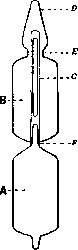
Figure 2.
inal substance is stored in a vacuum separately from the solvent and 2) the possibility of using a freshly prepared sterile solution.
L. Medvedkova. AMPULLITIS, a disease of the ampulla of the vas deferens, most often combined with inflammation of the epididymis, significantly less often encountered as an independent disease. Symptoms: severe pain in the anus and during ejaculation, the presence of blood or pus in the ejaculate, general malaise, and fever. Sometimes partial inflammation of the pelvic peritoneum is added to ampullitis. Upon examination through the rectum, the inflamed ampulla is palpated as a dense, painful cord to the left or right above the prostate, and sometimes as an elongated, doughy tumor. In differential diagnosis, it is often difficult to distinguish ampullitis from inflammation of the seminal vesicle. Treatment in acute cases is reduced to rest, hot micro-enemas, and injections of autovaccine; subsequently, diathermy and massage are indicated.
Related articles
Cite this page
“Ampoule and Ampullitis.” Soviet Medical Encyclopedia. English translation of Bolshaya Meditsinskaya Entsiklopediya, 1st ed. (Moscow, 1928–1936), ed. N. A. Semashko. https://sovietmedicalencyclopedia.pages.dev/article/ampoule-and-ampullitis/