Infarction
Historical document, translated for reference. It reflects medical knowledge of the 1920s–30s and is not medical advice.
Summary
Infarction refers to tissue death in an organ caused by blocked blood flow, typically in end arteries. The article distinguishes between anemic (white) and hemorrhagic (red) infarctions, describing their formation, characteristics, and outcomes.
Encyclopedia article (1928–1936)
INFARCTION, infarctus (from Latin infarcire - to stuff, to pack), "stuffing" of tissues and organs with some material foreign to them, for example, salts, extravasated blood, products of decay, etc. After the view expressed by Virchow that hemorrhagic infarction is the result of embolism, the term I. began to be applied to all wedge-shaped necrotic changes in organs caused by emboli, even when, as in the case of anemic infarction, no such stuffing of tissues occurred. The terms proposed to designate anemic I., such as "anemic sequestrum," "wedge-shaped necrosis," etc., did not gain acceptance in the literature. At present, the word I. almost exclusively implies those foci of necrosis in organs that are the result of a cessation of blood flow in so-called end arteries, regardless of what caused this cessation (thrombosis, embolism, spasm of the vessel); only for certain types of deposits of salts and pigments in the kidney is the term I. still used in the former sense of "stuffing." - The doctrine of I. was developed mainly through the works of Virchow (1856), Cohnheim (1872), Litten (1880), Grawitz (1891) (Virchow, Cohnheim, Litten, Grawitz) and others. Cohnheim clarified this question in particular, being the first to reproduce I. experimentally and the first to introduce the concept of end arteries (Endarterien), i.e., arteries that, up to their branching into capillaries, have no arterial anastomoses, or such in which the existing anastomoses prove insufficient due to loss of their elasticity or depending on weakening of heart activity (functionally-end arteries). When such arteries are blocked, blood circulation cannot be completely restored,
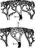
Scheme of embolic closure of vessels in the presence of arterial collaterals (above) and in so-called end arteries (below): 1-embolus; 2-network of capillaries.
and the area corresponding to the branches of the blocked artery undergoes necrosis (see figure). End arteries are present in most organs: lungs, heart, brain, spleen, kidneys, etc. I. represents necrosis of tissues deprived of blood supply. I. usually have a cone-shaped (on cross section - triangular) shape, with the apex of the cone directed into the organ and corresponding to the site of vessel occlusion, while the base is located on the surface of the organ. The coagulative necrosis of tissues occurring in I. gives them a certain density and causes some increase in volume of the affected area. I. of the brain usually have a soft consistency, which is why they are called softening of the brain (red or white), but at the very beginning of their formation they also appear denser compared to the surrounding tissues. Two main types of I. are distinguished: 1) anemic (ischemic), or white I., and 2) hemorrhagic, or red I. Anemic I. forms when the collateral capillaries of the end artery are poorly developed and its occlusion leads to complete anemia of the area irrigated by it [see separate table (pp. 791-792), fig. 6]. On cross section, anemic I. have a yellowish-white color, and the cross-section surface appears dry; they are usually surrounded by a red rim, depending on hemorrhage from the capillary network surrounding the I. If the end artery has a well-developed network of capillary anastomoses, then its vessels are filled with blood from neighboring capillaries, and the blood, by way of diapedesis, impregnates the entire area of I.; a red, or hemorrhagic I. results [see separate table (pp. 791-792), fig. 5]. The most characteristic location for the formation of red I. is the lungs. Despite the fact that in the lungs there is a dual blood supply (a. pulmonalis and rami bronchiales aortae) and that occlusion of branches of one of these systems can be compensated by the other, still it is precisely in the lungs more often than in other organs that I. are observed and, as a rule, hemorrhagic ones. (In the spleen and kidneys, anemic ones are more common; in the brain and heart - both types equally; in the testes and intestines - almost exclusively hemorrhagic.) Experiments have shown that simple occlusion of branches of the pulmonary artery is not sufficient to produce I., and that they form mainly when there is venous stasis, as occurs, for example, in certain types of heart defects. Unlike ordinary hemorrhages encountered in the lung and having a soft consistency and moist appearance of the cross-section surface, I. of the lung appears dense and dry in appearance, which depends on the fact that its basis, as in anemic I., is coagulative necrosis. The fate of I. varies: small I. may be resorbed without a trace, especially I. of the lung, but for the most part the dead mass of I. undergoes organization, and on the site of the former infarction a retracted scar forms (after red infarcts, scars may be pigmented). Sometimes liquefaction of I. occurs (so-called colliguative necrosis) and a cyst forms in its place. In some cases, the dead mass of I. is partially or completely petrified, and then calcareous concretions are found in the tissue of the scar. If the vessel occlusion was caused by an embolus containing bacteria, then suppuration of the corresponding area occurs - septic I., and sometimes it is difficult to determine whether one is dealing with a suppurated infarct or simply an abscess. - Deposits of salts or pigments in the tubules, vessels, and interstitial substance of the kidneys are also called I. These deposits can occur both by the hematogenous route and as a result of the secretory activity of the kidneys. Uric acid, calcareous, and bilirubin I. are distinguished; some authors also speak of hemosiderin and silver I. The so-called uric acid infarct deserves special attention.
Related articles
Mentioned in
Cite this page
“Infarction.” Soviet Medical Encyclopedia. English translation of Bolshaya Meditsinskaya Entsiklopediya, 1st ed. (Moscow, 1928–1936), ed. N. A. Semashko. https://sovietmedicalencyclopedia.pages.dev/article/infarction/