Gaucher Disease
Historical document, translated for reference. It reflects medical knowledge of the 1920s–30s and is not medical advice.
Summary
This article from the 1928–1936 Soviet medical encyclopedia details Gaucher disease, a systemic disorder affecting the spleen, liver, lymph nodes, and bone marrow. It describes the pathological anatomy, clinical symptoms, and differential diagnosis of the condition, which is now known to be caused by a genetic deficiency.
Encyclopedia article (1928–1936)
GAUCHE DISEASE, or Gaucher splenomegaly (morbus Gaucher), is named after the author who first described this form of disease in 1882 as a special type of epithelioma of the spleen. At present it is known that Gaucher's disease is not an epithelioma but a systemic disease primarily of the spleen, liver, lymph glands, and bone marrow, in which the pathological process involves, according to Pick, only the reticular, and according to other authors, the entire reticuloendothelial apparatus. The essence of Gaucher's disease lies in the fact that the cellular elements of the mentioned apparatus absorb and accumulate in themselves a peculiar substance. The latest chemical investigations by Epstein and Lieb (Epstein, Lieb) have shown that this microscopically and optically completely indifferent substance, in its main mass, consists of kerasin (C4H91NOs) belonging to cerebrosides (sphingolipids). Due to this change in the macrophage system, the affected organs appear enlarged, penetrated by light, large (up to 80 μm) rounded polyhedral, so-called "Gaucher cells." Their nucleus is small and pyknotic, and the protoplasm is finely reticular or fibrillar or homogeneous. - Pathological anatomy. The spleen is most affected in Gaucher's disease, and it can reach giant sizes (8 kg). It is then dense, on section gray-red, brick-colored, or chocolate-colored. The cut surface is variegated, as if "dusted" with grayish or grayish-yellow small foci and stripes (accumulations of Gaucher cells). Sometimes infarcts are encountered, often cavities filled with blood and Gaucher cells. In the order of cicatrization, both may be replaced by fibrous tissue. More often, however, fibrous nodes form in place of necroses in the cellular masses of Gaucher cells. Hemosiderin may be found in the scars. It is often contained in the endothelium of the splenic sinuses and sometimes diffusely impregnates Gaucher cells. In the connective tissue cells of the trabeculae, a proteinogenic brown pigment is often encountered, which does not give a reaction for iron. - The liver is also always enlarged (up to 4,800 g), but relatively less than the spleen. Here, too, grayish and yellowish spots and stripes are scattered over the red-brown or chocolate-colored surface. They correspond to accumulations of Gaucher cells, located primarily inside the capillaries. The proliferation of connective tissue along the Glisson capsule gives the liver a cirrhotic appearance. Brown pigmentation (as in the spleen) completes the picture. - Lymph glands are affected in the same way, but to a lesser degree. Extrathoracic and extrabdominal glands are involved in the process only exceptionally. - Bone marrow involvement usually recedes into the background, but rare cases are observed in which the disease is localized primarily in the bones; in this case, the spongy substance of all bones of the skeleton is diffusely penetrated by the cellular mass of Gaucher cells, and in the tubular bones it is located as gray and yellow nodes among the dark-red bone marrow. A definite tendency of Gaucher cell foci to fibrous-sclerotic transformation is noted. All this leads to bone deformation and spontaneous fractures; due to the flattening of individual vertebrae, a hump may form. - Such a clearly expressed involvement of the skeletal system in some cases of Gaucher's disease forced Pick to distinguish this form under the name of the skeletal form of Gaucher's disease (Skeletal form). In typical cases, the process does not extend beyond the mentioned organs. However, cases are observed in which the thyroid gland, lungs, kidneys, and other organs are involved. The etiology of the disease apparently lies within the change of germ plasm, a mutation (see), inherited by the recessive type. Pathogenetically, the disease represents an anomaly of metabolism. Since kerasin belongs to lipoids, it is possible that its production is the result of a deviation of lipoid metabolism in an abnormal direction. - Clinical picture. Gaucher's disease is a congenital constitutional disease; in part of the cases (up to 35%) it is familial. It occurs very rarely. So far, only about 50 reliable cases have been published. Women suffer twice as often as men. Gaucher's disease can fully manifest itself already in infants or in early childhood, but often it proceeds "ultrachronically." Symptoms increase gradually, and only after many decades does the picture of the disease reach its full development. Symptoms of splenomegaly and hepatomegaly predominate. Their enormous size, especially the first, cause perisplenitis and perihepatitis, leading to abdominal pain. Ascites is generally absent. In the skeletal form, symptoms of bone involvement predominate, while splenomegaly and hepatomegaly recede into the background. The yellow-brown coloration of the skin on exposed parts of the body (face, neck, hands) is already early noticeable. The mucous membranes are not affected. This pigmentation has nothing in common with jaundice and is a partial manifestation of hemochromatosis (see), which always accompanies Gaucher's disease. Leukopenia is also discovered early, either in the form of neutropenia or lymphopenia; subsequently, moderate hypochromic anemia joins it. In far-advanced cases, signs of hemorrhagic diathesis (bleeding from the nose, gums, stomach, uterus, etc.) are observed, accompanied by thrombocytopenia. Among complications, tuberculosis of the lungs and peritoneum is encountered most often. - Gaucher's disease has to be differentiated from various "splenomegalies." In childhood, especially with splenhepatomegaly of the Niemann-Pick type. In all cases, microscopic examination of a biopsy of a lymph gland or a splenic punctate helps. The finding of typical Gaucher cells in them, which do not give a reaction to lipoids, unlike morphologically similar Niemann-Pick cells, ensures the diagnosis. - Prognosis is relatively favorable. Death most often occurs from intercurrent diseases. In isolated cases, a favorable effect from splenectomy has been observed. Theoretically, however, such an operative intervention is not justified.
E. Gerzenberg.
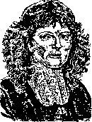
GRAAF, Regnerus (Regnerus de Graaf, 1641-1673), one of the outstanding Dutch anatomists of the 17th century, is known for his research on the anatomy of the sexual organs. In the book "De virorum organis generationi inservientibus" (Lugduni, 1668) he first described the seminiferous tubules as "vessels that produce seed" and gave a simple method for their detection by shaking the testicle in water after removal of the tunica albuginea. In the subsequent work of Graaf "De mulierum organis generationi inservientibus tractatus novus, demonstrans homines et animalia coetera omnia quae vivipara dicuntur, haud minus quam ovipara, ab ovo originem ducere" (Lugduni, 1672) vesicular formations in the ovary are described, which later received the name Graafian vesicles, which he took for eggs. G. then attempted to trace the development of the egg in a rabbit that had come out of the ovary, by performing systematic dissections at certain times after mating, and gave images of a series of eggs from the 3rd day to the appearance of an embryo in them. G. was the first to propose the term ovary instead of the former testes muliebres. The name G. is also associated with the introduction into anatomical practice of instruments for injection: the syringe (which he called a siphon) and the cannula ("De clysteribus, de usu siphonis in anatomia, defensio partium genitalium", Amstel, 1677). G. belongs to the first work on the nature of pancreatic juice ("Disputatio medica de natura et usu succi pancreatici", Lugduni, 1663). G. proposed a method of creating salivary and pancreatic fistulas on a dog.
Related articles
Mentioned in
Cite this page
“Gaucher Disease.” Soviet Medical Encyclopedia. English translation of Bolshaya Meditsinskaya Entsiklopediya, 1st ed. (Moscow, 1928–1936), ed. N. A. Semashko. https://sovietmedicalencyclopedia.pages.dev/article/gaucher-disease/