Anaplasma
Historical document, translated for reference. It reflects medical knowledge of the 1920s–30s and is not medical advice.
Summary
This article describes Anaplasma as coccoid inclusions found in erythrocytes, historically associated with the disease anaplasmosis in cattle. It discusses the debate over whether these organisms are independent pathogens or degenerative cellular changes, and details the clinical symptoms, transmission, and pathology of the disease as understood in the early 20th century.
Encyclopedia article (1928–1936)
ANAPLASMA, Anaplasma (from the Greek plasma—image), coccoid inclusions in erythrocytes, with a diameter of 0.1 to 0.5 μ, which stain red with Giemsa stain and give the impression of nuclei without any traces of a protoplasmic body. They were first described as the causative agents of anaplasmosis. In view of the fact that Anaplasma were often encountered in the blood simultaneously with Babesia (see), the opinion was expressed that they are a stage in the development of the latter. Other authors doubt that Anaplasma are independent microorganisms and believe that they are degenerative changes of erythrocytes of the Jolly body type. It is not
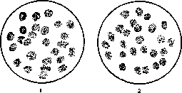
1—Anaplasma centrale; 2—Anaplasma marginale.
excluded that Anaplasma are indeed only a characteristic degeneration of erythrocytes accompanying an infection with another, as yet unknown, pathogen. Similar formations have also been described in humans, horses, dogs, goats, and sheep. Anaplasmosis (synonyms: cattle malaria, pernicious anemia of cattle) is a specific febrile disease of cattle transmitted through tick bites. The disease was first described by Theiler in the Cape Colony (Africa); it is also found in southern Africa, North and South America, and Australia; it was also described by Dzhunkovsky near Ganja in Azerbaijan. When animals are infected with blood containing Anaplasma, the incubation period is 16–47 days. When infected through tick bites, the disease manifests after 60–80 days. The febrile period lasts 7–12 days. Simultaneously, a number of symptoms are observed in the animals: trembling, loss of strength, edema in the lower part of the abdomen, and rapidly increasing anemia; the number of red blood cells drops to 1.5 million and lower. There are no other changes in the blood. This disease is often encountered simultaneously with other diseases of cattle, such as piroplasmosis, trypanosomiasis, and spirochetosis. Upon autopsy of dead animals, an infiltrate is found in the lower part of the abdomen and in the sternal region, transudate in the pleura, hemorrhages on the surface of the heart, an enlarged yellow liver, and an enlarged spleen, sometimes with small hemorrhages on the surface. Diagnosis of this disease presents great difficulties in terms of distinguishing it from piroplasmosis. There is no specific treatment. Schamber and Smith successfully used treatment with injections of mercuric chloride and quinine. Mortality reaches 50% and more. Immunity to anaplasmosis, even in animals that have recovered, is not noted. Young animals tolerate the disease more easily. Combating this disease is difficult and must be directed toward the destruction of ticks (see) and freeing animals from them by means of tick baths.
Related articles
Mentioned in
Cite this page
“Anaplasma.” Soviet Medical Encyclopedia. English translation of Bolshaya Meditsinskaya Entsiklopediya, 1st ed. (Moscow, 1928–1936), ed. N. A. Semashko. https://sovietmedicalencyclopedia.pages.dev/article/anaplasma/