Hemorrhoids
Historical document, translated for reference. It reflects medical knowledge of the 1920s–30s and is not medical advice.
Summary
This article from the 1928–1936 Soviet Great Medical Encyclopedia defines hemorrhoids as varicose dilations of the veins of the lower rectum. It details the anatomical distinction between internal and external hemorrhoidal nodes and their respective venous systems.
Encyclopedia article (1928–1936)
HEMORRHOIDS, haemorrhois (from Greek haima - blood and rheo - to flow; Russian folk name: pochechuy), varicose dilation of the veins of the lower rectum (varices haemorrhoidales). Hemorrhoidal dilations quite often lead to bleeding, which gave this disease its name. Internal and external hemorrhoidal nodes are distinguished, depending on the involvement of one or another venous plexus of the rectum. External nodes are formed from the lower venous plexus (plexus haemorrhoidalis inferior), located directly under the skin of the anus; these nodes are often called subcutaneous (varices haemorrhoidales subcutaneae). This venous plexus belongs to the branches of the vena haemorrhoidalis inferior (see Figure 1, a), which drains into the vena pudenda and ultimately into the vena cava. Internal nodes are found in the area of distribution of the upper venous plexus (plexus haemorrhoidalis superior), located under the mucous membrane of the rectum, in the area of the folds of the latter (columnae rectales Morgagni); these varicose nodes are called submucosal (varices haemorrhoidales submucosae). Internal nodes drain through the vena mesenterica inferior into the vena portae. Between the lower and upper venous plexuses, there are numerous anastomoses connecting the portal vein system with the inferior vena cava; they dilate during obstructed outflow in one of the systems (e.g., in cirrhosis of the liver), which leads to an increase in hemorrhoidal venous plexuses. A stronger filling of these venous plexuses of the rectum in old age can be considered an almost physiological phenomenon. If these venous dilations reach a more significant size, protrude from the anus, and cause a number of disorders, then one speaks of hemorrhoids. In any case of hemorrhoids, one is dealing with true varicose dilations of the veins of the lower rectum, similar to how there are varicose dilations of the veins on the lower extremities and in the area of the spermatic cord. The opinion of Reinbach and some other researchers, who considered hemorrhoids a vascular tumor, a cavernoma, i.e., a neoplasm, can be considered [erroneous].
Figure 1. Veins and their plexuses in the lower rectum: a-vv.
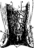
haemorrhoidalis inferior; b-plexus haemorrhoidalis superior; c-columna rectalis Morgagni; d-vena haemorrhoidalis superior; e-cross-section of the mucous membrane (according to Corning).
finally refuted; at the present time, no one doubts that hemorrhoids are a varicose process. As for the predisposing and etiological factors leading to the formation of hemorrhoids, a whole series of them is known. Local anatomical conditions already favor the formation of hemorrhoids. The venous hemorrhoidal plexuses are located in the lower part of the trunk, the outflow from here is obstructed, these veins lack valves, which favors the stagnation of blood in the nodes, and subsequently their dilation. Furthermore, during the normal act of defecation, and especially during constipation, there is a straining of the abdominal muscles and an increase in pressure in the veins; the venous plexuses of the rectum and the hemorrhoidal nodes swell sharply and gradually expand. These factors act in the majority of people, which explains the frequency of hemorrhoids. To these factors are added a number of other moments leading to stagnation in the veins of the small pelvis, for example, pregnancy, uterine retroflexion, tumors. Furthermore, the profession and lifestyle undoubtedly influence the development of hemorrhoids. It has long been believed that a sedentary lifestyle, lack of movement, and excessive eating and drinking contribute to the development of hemorrhoids. However, accurate statistical data do not always confirm these deep-seated views. The material of the Schaak clinic, just processed by Beckerman in 333 cases of patients operated on for hemorrhoids, gives 45.2% of patients who performed their work standing, and 35.1% sitting. Approximately the same figures for the incidence of hemorrhoids for persons working predominantly standing or sitting are also given by the statistics of Quenu and Rotter. Constitutional, racial, and hereditary factors also have significance for hemorrhoids. Jews suffer from hemorrhoids particularly often. The heredity of hemorrhoids in the aforementioned Schaak material was noted in 14% of all cases. Most often, hemorrhoids manifest in mature age, between 25 and 35 years. According to all statistics, men fall ill much more often than women (approximately in a ratio of 5:3). In childhood, hemorrhoids are observed extremely rarely. Pathological anatomy. Hemorrhoidal nodes are varicose-dilated veins. By injecting these vessels, it has been proven that they have various rounded, spindle-shaped, bunch-like, loop-like protrusions. The walls of these veins are thickened due to the development of connective tissue; in some places, the vessel wall, on the contrary, is sharply thinned, so that the nodes can rupture and sometimes cause profuse bleeding. The mucous membrane of the lower rectum covering the nodes is often sharply altered; it is thickened, hyperemic, and in a state of catarrhal inflammation. The microscopic picture of hemorrhoidal nodes is diverse. The lumens of the vessels have different diameters, the walls are thickened, the endothelial cells are swollen, the cells of the muscular layer are atrophied, and the elastic fibers are degenerated and reduced in quantity. In some places, the elastic tissue disappears completely due to the development of connective tissue, i.e., a picture is obtained similar to the changes observed in varicose veins of the lower extremities, but only in hemorrhoidal veins does the sclerosis of the walls not reach such a high degree. Many of the dilated veins and nodes are thrombosed. Subsequently, hemorrhoidal nodes undergo changes: the thrombi organize; sometimes an inflammatory process is added; abrasions and scratches forming on the mucosa of the anus serve as gateways for infection, phenomena of thrombophlebitis occur; the matter can reach suppuration and the formation of ulcerative surfaces on the mucosa. Then, in some cases, prolapse of large internal nodes outside the sphincter is observed; such nodes swell sharply, become blue-purple, and so-called "strangulation" of hemorrhoids occurs; nutrition in such strangulated nodes is sharply impaired, and they undergo partial necrosis. Very thinned nodes can burst and, as already mentioned, lead to significant bleeding. On the other hand, the inflammatorily altered mucosa of the lower rectum with small ulcerative surfaces covered with granulations can also give rise to small hemorrhages. These minor hemorrhages, if they are repeated often over a more or less prolonged time, can lead to secondary anemia. Clinical course of hemorrhoids and symptomatology. In the initial stages, hemorrhoids manifest little; subsequently, patients develop unpleasant sensations in the anal region, a feeling of discomfort, fullness, and heat, and a frequent sensation of itching and burning. In the further picture of the disease, one should distinguish symptoms depending on external and internal hemorrhoidal nodes. External hemorrhoidal nodes show through the skin of the anal opening in the form of bluish formations the size of a pea or a hazelnut; they often form a wreath around the anus. During straining, these nodes swell; they can be emptied by the pressure of a finger. Thrombosed nodes, on the contrary, are rather firm and do not empty. With inflammatory phenomena with thrombophlebitis, the nodes become extremely painful. Severe pain occurs during this time during defecation, especially with constipation. Internal hemorrhoidal nodes usually cause patients great trouble. They form in the lower part of the rectum, in the region of the columnae Morgagni (see figure 1), representing rounded, mucosa-covered protrusions, ranging in size from a hazelnut to a walnut. These nodes, located around the intestine, also form a wreath; to the touch, they are soft, like folds of the mucous membrane. The mucosa covering them is often inflammatorily altered and hyperemic. At first, these nodes are hidden and do not manifest themselves, but having reached a certain size, they cause an unpleasant feeling of heat, burning, and fullness in the rectum, interfere with sitting, and cause pain during bowel movements. Internal nodes almost always cause bleeding. Blood appears during a stool, staining the feces and toilet paper. These minor hemorrhages occur more often from the ulcerated surface of the inflammatorily altered mucosa. More severe hemorrhages are not uncommon, when pure bright red blood pours out from the anus (after a stool or during it) in a significant quantity. After such bleeding, occurring from a damaged or burst vein, patients sometimes feel a certain relief, which is why some patients look forward to such hemorrhages, which sometimes repeat periodically. Frequent hemorrhages lead to a disturbance of the general condition of the patients and (in many cases) to severe anemia. This anemia can be the most serious consequence of hemorrhoids. The second unpleasant feature of internal nodes is their tendency to prolapse. The submucosal layer of the lower rectum is very mobile on the muscular layer; during straining and the passage of a column of feces, the mucosa with the hemorrhoidal nodes easily descends downwards through the sphincter and protrudes from the anus. At first, these prolapsed nodes retract back into the intestine on their own after a stool. In later stages, the nodes remain outside (see figure 2) and must be reduced back by the patient himself after each bowel movement, which is very unpleasant and painful. With the swelling of the prolapsed nodes and the contraction of the sphincter, strangulation of the hemorrhoidal nodes can occur. In such a case, the anal opening is surrounded by a wreath of blue-purple shiny nodes of significant size, covered with altered edematous mucosa. These formations


are extremely painful; the manual reduction of the nodes is impossible, attacks of severe pain and tenesmus occur, the so-called "hemorrhoidal attack." Patients in a severe condition must lie down. The nodes become hard, thrombose, and in the coming days they may become gangrenous and even fall off completely. The remaining ulcers scar over, and thus, in successful cases, something like a self-cure can occur. In more severe and unfavorable cases, more extensive suppuration with subsequent formation of fistulas (see Intestine) can be added to the thrombophlebitis and gangrene of the nodes, or, which presents a direct danger to
life, suppurating thrombi can lead to general blood poisoning, to pyemia. Such severe complications are observed rarely. In addition to the complications listed, internal nodes can cause significant catarrhal inflammation of the rectal mucosa. This catarrh is expressed by the discharge of mucus during a bowel movement. In more severe and neglected cases, mucus is discharged spontaneously without a bowel movement, bothering patients, soiling underwear, and causing weeping eczema in the area around the anus. Diagnosis. Recognizing hemorrhoids does not present difficulties. The patient's history alone, with complaints of pain and unpleasant sensations and bleeding during bowel movements, with an indication of the presence of nodes, or "lumps," in the anal area, makes one suspect hemorrhoids. External examination, which is best performed with the patient lying on their side, spreading the buttocks, easily confirms the diagnosis if external nodes are present. When the patient strains, the picture becomes sharper, the bluish-translucent nodes engorge, and with a somewhat relaxed sphincter, internal nodes may also appear. The latter can be made more visible by placing a suction Bier cup against the anus, as suggested by Strauss. A warm sitz bath also promotes the prolapse of internal nodes. Although the indicated examination in most cases clarifies the diagnosis of hemorrhoids, one still cannot limit oneself to an external examination alone. In order not to overlook more serious diseases of the rectum, a digital examination is mandatory in every case. Such a digital examination allows for the exclusion of rectal cancer, which initially gives symptoms very similar to hemorrhoids. Unfortunately, such confusion still occurs in practice. With such an examination, even an initial stage of cancer, which gives a clear sensation of an ulcer with dense, nodular edges, cannot escape attention. Rectal polyps, which give symptoms similar to internal hemorrhoidal nodes, are also determined by digital examination. External nodes can sometimes be confused, especially in women, with anal condylomas, but the latter have a characteristic pointed appearance; furthermore, the presence of gonorrhea and the spread of smaller condylomas toward the genital organs allow for a correct diagnosis. In doubtful cases, it is also useful to examine the rectum with special specula; well-lubricated with Vaseline or oil, they are carefully inserted through the anus, then the blades of the speculum are spread, and the mucosa of the lower part of the rectum becomes accessible for direct visual inspection. For a more detailed examination of the rectum in its upper sections, a special examination with a rectoscope (rectoscopy) is required. Prognosis. The prognosis for hemorrhoids is generally favorable. If there are no special complications, hemorrhoids can exist for years, only occasionally bothering patients. In more severe cases, especially those accompanied by frequent bleeding, or those complicated by inflammatory processes, the prognosis is more serious. Sharp, progressive anemia can threaten life, and frequent prolapse of nodes, their strangulation, and inflammatory thrombophlebitis can make life unbearable and, in any case, sharply reduce the patient's ability to work. In these cases, energetic treatment is required, in most cases operative.
Treatment. In the treatment of hemorrhoids, one should first pay attention to the predisposing factors present in the given case and, if possible, eliminate them first; if there is a tendency toward constipation, it is mandatory to eliminate the latter and regulate the diet. Patients are advised to take mild laxatives at night: 1-2 teaspoons of compound licorice powder (Pulv. Liquiritiae compos.), rhubarb tablets, or Cascara sagrada. After this, there is a soft stool in the morning that does not irritate the hemorrhoidal nodes. If this is not enough, then ordinary or oil enemas are resorted to. It is advised to wash the anal area with cool water in the morning and evening. One should avoid coarse, irritating food (cabbage, coarse vegetables), spicy seasonings (pepper, mustard), and alcohol abuse. In mild cases of hemorrhoids and in a whole series of moderate ones, these measures are quite sufficient so that the hemorrhoids do not bother the patients. For pain, suppositories made of cocoa butter with the addition of narcotic substances (Morphii mur. 0.01 or Extr. Belladonnae 0.015, Butyr. cacao q. s. ut f. suppos.) or ready-made Anusol suppositories available for sale are helpful. For complicated cases of hemorrhoids with constant prolapse of nodes, with frequent inflammatory phenomena (hemorrhoidal attacks), and especially with recurring bleeding, the above-described conservative treatment is insufficient, and one must resort to surgical intervention (in most cases radically curing the hemorrhoids). Bleeding that affects the general condition and leads to anemia is an absolute indication for surgical intervention.
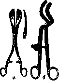

Operative treatment. At present, there are several methods for operating on hemorrhoids. For all methods, careful preparation of the patient is required. The rectum must be well emptied; a few days before the operation, patients receive a laxative 2-3 times. An enema is given only the day before, so that the remaining water does not interfere during the operation. On the day of the operation and in the following days, opium is given so that there is no bowel movement in the first days after the surgical intervention. Anesthesia during the operation for hemorrhoids can be varied. Many surgeons perform the vast majority of operations for hemorrhoids under local anesthesia. The area of the anus and the lower part of the rectum can easily be completely anesthetized with simple infiltration anesthesia. Others prefer general chloroform or ether anesthesia, spinal or parasacral anesthesia. The most commonly used operations for hemorrhoids include methods of cauterizing the nodes and the method of applying ligatures. The excision method is used less frequently. Methods of cauterization. In a patient lying on their back with legs raised, bent at the knees, and spread apart, the sphincter is first dilated with two fingers, after which the internal nodes prolapse somewhat. Now the nodes are grasped one by one with forceps, and under these forceps, special forceps, Langenbeck clamps (see Figure 3, a), are applied, over which the cauterization of the nodes is actually performed (with a Paquelin thermocautery); these clamps are equipped with heat-insulating bone plates on their wide branches. As a result of such cauterization, a wound remains under the scab. It is even more convenient to perform cauterization with Dombrovsky's forceps (see Figure 3, c). Their advantage lies in the fact that they cover the entire circumference of the anus; therefore, a radical removal of all nodes is performed without any blood loss. (Figure 4 shows the operation for hemorrhoids with Dombrovsky's forceps; the nodes of one half have already been cauterized, on the other, clamps are still hanging, grasping the hemorrhoidal nodes.) After removing the clamps, a rubber drainage tube wrapped in iodoform gauze is inserted into the rectum. The tube serves to drain gases. Recently, many surgeons have abandoned the insertion of drainage tubes without detriment to the patients.
[patients]. During subsequent dressings, ointment dressings with vaseline are widely used. In Shaak's clinic, 85% of all operations for hemorrhoids were performed by the method of cauterization with Dombrovsky's forceps (according to Shaak) with very good long-term results. Rokitsky's forceps are designed similarly to Dombrovsky's forceps. Ligation method. The application of ligatures is also a very simple, safe method used in many surgical clinics and departments. Preparation of the patient and dilation of the sphincter are the same as in the previous method. Then the nodes are grasped, pierced at the base with a needle and thread, and tied tightly. The constricted node becomes necrotic and falls off. For external nodes covered with skin, it is recommended to make a circular incision in the skin before applying the ligature to better tighten the node (Mirotvortsev spoke at the congress of physicians in Saratov in 1927 in favor of simple ligation of hemorrhoidal nodes with a silk ligature; the majority of surgeons present agreed with the speaker, considering the simple and safe ligation method to be the method of choice). Whitehead's excision method consists of the complete removal of all dilated hemorrhoidal veins together with a section of the altered mucous membrane. Circular incisions are made, through which all nodes are sharply excised. The operation is very bloody. In conclusion, a suture is applied, neatly connecting the mucosa to the skin. This operation is undoubtedly radical, but its negative aspects lie in a more complex technique, which is also often associated with significant blood loss. Furthermore, after Whitehead's operation, various complications were observed more frequently in the form of cicatricial stricture of the anus, rectal prolapse, and fecal incontinence. In view of this, this operation is currently rarely used, and only a few surgeons are its adherents. Besides the described methods, there are several other techniques that also lead to the destruction of hemorrhoidal nodes. This includes the method of forceful dilation of the anus, which was widely used in France. In our country, Subbotin was a proponent of this simple method. Dilation was performed with a special Trelat speculum-dilator. Such simple dilation and compression of hemorrhoidal nodes in a number of cases can lead to the emptying of dilated veins and to a cure, but still, the method cannot be considered radical, and for severe cases with complications, it is inapplicable. Furthermore, for a long time, the injection of chemical substances into hemorrhoidal nodes has been used for their destruction. Several drops of carbolic glycerin (Acidi carbolici liquef. 1.0, Glycerini 2.0-3.0) are injected into each node with a syringe. Voelcker recommends this method, which is common in America, and suggests injecting 1 drop of pure carbolic acid. Boas has recently again strongly recommended the injection method for the treatment of hemorrhoids; he uses 96% alcohol and injects 0.5-1 cubic cm of alcohol into the node under local anesthesia. His method has not found widespread acceptance among surgeons. The method is not radical and possesses no advantages over the generally accepted simple and safe methods of cauterization and ligation.
Related articles
Mentioned in
Cite this page
“Hemorrhoids.” Soviet Medical Encyclopedia. English translation of Bolshaya Meditsinskaya Entsiklopediya, 1st ed. (Moscow, 1928–1936), ed. N. A. Semashko. https://sovietmedicalencyclopedia.pages.dev/article/hemorrhoids/