Leishmaniases
Historical document, translated for reference. It reflects medical knowledge of the 1920s–30s and is not medical advice.
Summary
This introductory article from the first edition of the Great Medical Encyclopedia covers the classification and history of leishmaniases, focusing primarily on cutaneous leishmaniasis (oriental sore), its etiology, history of discovery, and parasite morphology.
Encyclopedia article (1928–1936)
LEISHMANIASIS. Contents: Cutaneous leishmaniasis.................574 American forest leishmaniasis.......588 Kala-azar.....................588 Canine leishmaniasis................604 Leishmaniasis is a disease of warm countries that affects humans and certain domestic animals. The disease is caused by a specific parasite and proceeds either in the form of skin lesions or as a febrile disease with damage to internal organs, and only in exceptional cases is a combination of these two forms of the disease observed. Cutaneous leishmaniasis is a disease characteristic exclusively of a hot climate; it has the appearance of a pimple that subsequently turns into an ulcer. Etiology. The question of the causative agent of oriental sore received its final resolution relatively recently. The first attempts to find the causative agent of the disease date back to 1868, when Smith described helminth eggs of Distoma in the discharge of ulcers; apparently, he mistook hyaline clumps, which are frequently encountered in cutaneous leishmaniasis, for eggs. Cunningham described special formations on frozen sections from an ulcer, which he considered to be parasites (family Monadina). Firth confirmed Cunningham's observations and gave these parasites the name Sporozoa furunculosa. Undoubtedly, both authors mistook tissue cells in whose protoplasm the actual parasites (Leishmania) were located for parasites. Subsequent research followed the path of searching for the causative agent among bacteria, with the most diverse bacteria being described: micrococci, streptococci, fungi of the species Streptothrix, capsular micrococci, which apparently represented, judging by their size, not bacteria, but leishmania. Borovsky (1898) in 20 cases of cutaneous leishmaniasis discovered special bodies, ranging in size from 0.5 to 2–3 µ, in sections as well as in smears of ulcers. Studies of the juice from papules and ulcers in a hanging drop showed the presence of a multitude of motile bodies. In smears, they had a thin process, the size of the diameter of the body itself, often with a spherical thickening at the end. Sometimes there were 2 or 3 such processes. In these formations, a nucleus lying in the center or on the periphery of the body could be detected. Undoubtedly, he was dealing with the true causative agent of the disease—Leishmania tropica. Shulgin (1902) on a small material (3 patients) confirmed Borovsky's observation. Martsinovsky jointly with Bogrov (1903) in one case of Persian sore discovered special parasites, which were described under the name Ovoplasma orientale. Simultaneously with this work, a work by Wright was published, who also in 1 case discovered the same parasites and described them under the name Helcosoma tropicum. These studies finally established that the causative agent of oriental sore is a parasite belonging to Protozoa and now known under the name Leishmania tropica (in honor of Leishman, who first described a similar parasite in kala-azar). Wenyon (1926) establishes the following place for this parasite among protozoa: phylum Plasmodroma, order Protomonadinae, family Trypanosomidae, genus Leishmania. Parasites are found in large numbers in smears from granulations lining the bottom of the ulcer. In the juice from ulcers in a hanging drop, individual bodies show weak amoeboid movements. The bodies are round, oval, or rice-grain-shaped and have a size of 2 to 4 µ. When stained with Giemsa, their protoplasm, which stains a pale blue color, contains... Figure 1. Lipofuscin in nerve cells of the sympathetic ganglion. Figure 2. Spleen in kala-azar: 1—atrophy of the follicle; 2—thickened trabecula; 3—pulp poor in cells; reticuloendothelial cells stuffed with leishmania are visible; 4—erythrocytes. Figure 3. Bone marrow smear in leishmaniasis: 1—large reticuloendothelial cell stuffed with leishmania (2); 3—erythroblast; 4—erythrocyte (other bone marrow elements not depicted). Figure 4. Liver in leishmaniasis: 1—enlarged intertrabecular spaces in which hypertrophied Kupffer cells stuffed with a mass of leishmania are visible; 2—hepatic trabeculae; 3—endothelium. Figure 5. Splenic pulp: 1—sharp depletion of the pulp in cells; swelling of the reticuloendothelial stroma; leishmania in reticuloendothelial cells; 2—sinuses with free cells of the reticuloendothelium. Figure 6. Splenic pulp: 1—atrophy of the pulp; almost exclusively reticuloendothelial cells (pale) are visible; 2—enlarged sinuses in which swelling of the endothelium, free macrophages, and erythrocytes are observed. Figure 7. Leishmania in culture. Figure 8. Smear from ulcer granulations in leishmaniasis. (To the illustr. art. Leishmaniases, Lipofuscin.) 9 >.
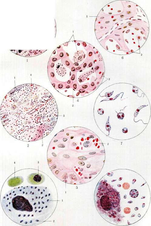
To the article Leishmaniasis, Lipofuscin. GOZNAK. a large round or slightly oval nucleus, opposite to which lies a smaller accumulation of chromatin, known by the name of blepharoplast or kinetonucleus. From the blepharoplast towards the nucleus extends a short filament—rhizoplast. The blepharoplast is of a round or rod-like shape. The protoplasm of the dead parasites stains a uniform reddish color. Leishmania tropica belongs to the group of intracellular parasites. On stained preparations from the ulcer granulations (see separate plate, figure 8), even at low magnification, one can usually notice round mononuclear cells whose protoplasm is stuffed with small granules staining in the color of the nucleus. At high magnification, the aforementioned granules represent chromatin masses of leishmania. Up to 70 parasites and more are found in a cell. Individual giant cells are encountered in which they are sometimes numbered by the hundreds. Occasionally these parasites are also encountered in polynuclear leukocytes; this made it possible to draw a conclusion about the possibility of their entering the bloodstream, which was subsequently confirmed. Entering the intercellular space upon cell destruction, the parasites begin to multiply by direct division and are captured by new cells. During reproduction, the nucleus shifts to the broader end of the parasite, and the blepharoplast is located at the narrower one. It takes the form of a rod standing perpendicular to the long axis of the parasite, and begins to divide in half. The body of the parasite increases somewhat in volume. A little later, the nucleus also divides into 2 parts, and finally the entire parasite breaks up into 2 new individuals. Sometimes division is multiple. In individual cases, formations known by the name of "gangue" are encountered on preparations, which represent the process of multiple division or, more likely, parasites squeezed tightly into a single heap. In sections from a young ulcer, enormous numbers of intracellular parasites are sometimes encountered, with which the entire field of view is speckled; they lie in special mononuclear epithelioid cells, and sometimes in giant ones. Culture of the parasite. Nicolle (first obtained a culture of leishmania on blood agar (NNN) (agar 14.0, sodium chloride 6.0, and distilled water 900.0. To 10 cm3 of agar in a test tube is added 1 cm3 of rabbit water; inoculation is made into the water of condensation, which must be tinted with hemoglobin). Multiplication of the parasites is observed already from the 2nd-3rd day. Upon the first appearance of parasites in culture, one can usually note 2 types of them: some have pale-staining protoplasm and a large loose nucleus, others stain an intense blue color, with a nucleus of comparatively smaller dimensions. Upon the transformation of parasites into flagellate forms, the difference in staining disappears. In cultures, the parasites have an elongated body, in the middle of which lies a round or oval nucleus, and at the anterior, narrower end—a blepharoplast, from which a long flagellum departs (see separate plate, figure 7). The parasites in culture possess lively movement. In old cultures, small blackish grains of velutin can usually be noted in the protoplasm of the parasites. Cultures withstand many generations. By inoculation of parasites from the ulcer, as well as from culture, it was possible to obtain the disease in certain animals, namely, in monkeys Macacus rhesus and others, dogs, mice, and guinea pigs. The question of the routes of infection of cutaneous leishmaniasis cannot yet be considered finally resolved. It must be considered most probable that blood-sucking insects play a role in the spread of the disease. The very name of the disease in some localities is associated with insect bites: "pasha-khurda" in Turkmenia (meaning mosquito bite), "cuncir" in Africa (derived from the word cunci—flea), and the like. Many of the blood-sucking insects with greater or lesser grounds were indeed suspected of transmitting the disease both on the basis of experiments and on the basis of epidemiological data. Patton and others found the appearance of flagellate forms of the parasite Leishmania in bugs which they infected with the help of L. tropica and L. Donovani; Adie found flagellate forms of the parasite in the salivary glands of infected bugs. Chatton and Blanc discovered flagellate forms of leishmania in bugs that fed on gecko lizards. All these experimental data, however, do not give the right to assume that bugs play a role in the epidemiology of cutaneous leishmaniasis, since these insects are extremely widespread, while the foci of cutaneous leishmaniasis are rather sharply limited. Experiments with fleas (Wenyon) and with lice (Patton) also gave negative results. Many observations were also made in relation to mosquitoes. Thus, Wenyon described the development of flagellate forms of leishmania in infected mosquitoes of the genus Aedes aegypti; but the results of the experiments were negative. Similar observations existed in relation to blood-sucking flies; in particular, Gachet experimented with flies Hippobosca canina, and he describes the appearance of leishmaniotic pustules in two people at the site of the bite by these flies. Finally, sandflies Phlebotomus were suspected in the transmission of the infection, in which flagellate forms of parasites of the type Herpetomonas were repeatedly discovered by many authors in the digestive tract. Thus, Acton in Mesopotamia found 6% of infected Phlebotomus; Patton in India—10%. Mackie discovered them in the same place in Phlebotomus minutus, and Laveran and Franchini—in Phlebotomus papatasi (France). The latter authors, with the help of cultures of parasites obtained from these insects, caused a general infection in mice and guinea pigs, and in dogs—skin lesions resembling ordinary leishmaniasis. Experiments with Phleb. minutus (bites and injections of emulsion) on man, monkey, and mice gave negative results for Sergent, Lemaire, and Senevet. In Africa, the following observations were made (Sergent, Parrot, Donatien, Beguet): 94 Phlebot. papatasi were caught in military hospitals, and after 3-4 days they were sent 600 km to Algeria, to a locality where leishmaniasis is not encountered; 71 Phlebo. arrived alive; no parasites were found in the emulsion from them, but upon rubbing this emulsion into the scarified skin of the forearm of an Algerian, a papule with a large number of L. tropica parasites appeared at this place after 84 days. Thus, the possibility of transmitting this disease via sandflies seemed to be proven, but this is still far from resolving all questions of epidemiology. Usually, Phlebotomus are always encountered in foci of cutaneous leishmaniasis; nevertheless, it can be thought that alongside them there may be some other transmitters of the disease. Reservoirs of the virus. In places of endemic spread of cutaneous leishmaniasis, disease in dogs is very frequently encountered (see below—Canine leishmaniasis), in which the disease proceeds either in the form of general leishmaniasis or in the form of skin lesions. Comparatively rarely, cutaneous leishmaniasis is encountered in cats. Cutaneous leishmaniasis in camels has also been described by Nicolle. Sergent and Nicolle discovered leishmania in geckos (Tarentola mauritanica) in 15.7%; it is difficult to say whether the parasites of the aforementioned animals are identical with human parasites, but morphologically they are extremely similar. Laveran infected geckos with the parasite L. tropica, and Chatton and Blanc also obtained a positive result in 3 experiments with geckos. In favor of the role of the gecko as a reservoir of the virus, one can cite the observations of Parrot, who fed 8 Phlebotomus on 6 specimens of geckos. The Phlebotomus flew out, and 3 months later 3 people in this house fell ill with cutaneous leishmaniasis. Nicolle, Blanc, and Laveran injected the blood of geckos with a natural infection into 2 humans and monkeys, as well as culture into 3 humans and a monkey. In all cases, the result was negative. Thus, there are no sufficient grounds to assert that geckos are a reservoir of the virus for man. Diseases of camels with leishmaniasis are observed rarely, and cats fall ill just as rarely. Only dogs remain, in which the course of ulcers very much resembles the same ulcers in man, and Phlebotomus can not infrequently be found on the ulcers in dogs. With a culture obtained from dogs in Baghdad, Adler and Theodor (1930) infected a person, and papules formed in the skin at the site of injection, in sections from which parasites were discovered. The authors consider L. tropica to be a common parasite for dogs and man. Nicolle and Manceaux obtained infection of man and monkey by means of inoculating them with granulations from dog ulcers. Thus, it can be assumed that the virus in leishmaniasis can also survive outside the human organism. However, there are as yet insufficient data to assert that the disease can be transmitted from dogs to man; thus, in Central Asia in many foci of this disease, such as in Ashkhabad and Merv, it was not possible to encounter dogs with cutaneous manifestations of leishmaniasis (Marcinowsky), and a very small number of them were discovered with visceral leishmaniasis at autopsies. These same cities are major foci of cutaneous leishmaniasis, although the reverse should also be noted; thus, Bukhara in recent years has been a major focus of cutaneous leishmaniasis, and similar diseases of dogs are also quite frequently observed there. Brumpt points out that in Biskra and Gafsa there are no dogs ill with cutaneous leishmaniasis. It can be admitted that Phlebotomus become infected from dogs ill with general leishmaniasis, and inoculate its cutaneous form to man with their bites.
In any case, the question of the virus reservoir in cutaneous leishmaniasis cannot yet be considered definitively resolved. The geographical distribution of cutaneous leishmaniasis covers extremely vast areas between 15° West and 20° East longitude and from 23° to 45° North latitude (Arendt). The disease is very widespread in North Africa from Algeria to Morocco in numerous oases of the Algerian (Biskra) and Tunisian (Gafsa) Sahara, in the French Congo, in the Sudan, and along the African coast of the Red Sea. It is found in Asia Minor, Syria, Mesopotamia, Arabia, Persia, Afghanistan, India, China, Central Asia, in the valleys of the Murghab, Tedzhen, and Atrek (Termez, Kerki, Tashkent, Kokand, Merv, Ashkhabad, Bukhara, etc.), in the Transcaucasian republics of the USSR [in Azerbaijan (Baku, Lenkoran, Ganja), in Georgia (Gori)], on the islands of Crete and Cyprus, in southern Italy, Sardinia, and Sicily, in Brazil, Uruguay, Colombia, Bolivia, and French Guiana, in the Philippine Islands, on the island of Ceylon, and in Australia. In connection with such a vast distribution, cutaneous leishmaniasis has an extremely large number of names, which partly characterize its essence. The most widespread of them are: Aleppo button (bouton d'Alep), Biskra button (bouton de Biscra), Tunisian button, Nile button, Delhi button, Sahara chancre, bessep-temur of the Arabs, Yemen ulcer, Pendjeh ulcer, oriental sore (bouton d'Orient, furunculus orientalis), ilchoban of the Turkmen (annual ulcer), il-yarasy of the Turks (annual ulcer), solek of the Persians (annual ulcer), godyovik in the Caucasus, Baghdad ulcer, Delhi boil, granuloma endemicum, Kandahar sore, Pendjdeh sore, L. dermalis, s. cutanea, and finally leishmaniosis americana, L. oro-pharyngealis as a special type of cutaneous leishmaniasis. Throughout the entire vast space where the distribution of oriental sore has been noted, it occurs in the form of separate foci, sometimes extremely sharply limited; for example, a fairly large focus of this disease until recently existed in Ganja (former Yelizavetpol), while three versts from the city, in a large railway settlement connected to the city by a horse-tram line and a continuous row of buildings, it was completely absent. New foci of cutaneous leishmaniasis may arise, and old ones sometimes die out temporarily and even forever. Thus, for example, Lebert mentions cutaneous leishmaniasis in the Crimea (Eupatoria), whereas leishmaniasis has long since disappeared there. Another example is Kokand, which was previously known as a large focus of cutaneous leishmaniasis (kokandka), and now yields a very insignificant number of cases. Bukhara at one time was also considered a large focus of this disease, then the number of cases began to drop sharply; in recent years, cutaneous leishmaniasis has again intensified and become widespread. Lenkoran and Ganja should be classified among the dying-out foci of cutaneous leishmaniasis, where leishmaniasis at present is almost completely absent. Among newly arisen foci in recent times, Baku and Gori should be pointed out. Frequently, the disease goes beyond the boundaries of its endemic distribution and produces outbreaks of large epidemics. Usually, this has to be linked either to sharp changes in climatic conditions or to any natural disasters, such as famine or war. As an example, one can cite the epidemic among the British garrison in Delhi. Beginning in 1857, from the moment of the arrival of troops there, from 50% to 70% of the patients with cutaneous leishmaniasis were noted among the soldiers, sometimes in a very severe form, and only in 1873, with the adoption of a number of sanitary measures, did these diseases sharply decrease. During the campaign of Russian troops to Kushka (1885) in the so-called Murghab detachment, the Pendjeh ulcer strongly spread, which significantly hindered military operations. Out of the entire detachment of 1,800 people, more than 50% fell ill (Manotskov). In 1921, from Red Army units stationed in Sarakhs, 150 people sick with severe forms of cutaneous leishmaniasis were evacuated within one month. In 1922, in the same place, a single company of 250 people gave about 60% morbidity (Krasnovsky). In 1929, in connection with the mass appearance of locusts in Turkmenistan, so-called "locust regiments" were organized, which suffered greatly from the development of Pendjeh sore among them. Race apparently has no significance in the sense of infection with cutaneous leishmaniasis, and if there are contradictory indications in the literature that Europeans who fall into a leishmaniasis focus fall ill more often than the local population, this is explained by insufficiently accurate observations. Sex likewise plays no role in predisposition, although in some localities a higher morbidity is noted among men, but this is explained by the greater mobility of the male population. All age groups of the population are subject to the disease to an equal extent. In endemic foci, children fall ill at a relatively early age, which is why the impression is created as if cutaneous leishmaniasis is a disease predominantly of childhood. Under one year of age, ulcers are encountered rarely. This disease is not transmitted by inheritance. Social and living conditions undoubtedly influence the spread of the disease; however, it is not yet possible to accurately account for all factors in this area. Nevertheless, it should be noted that the improvement of general sanitary conditions undoubtedly influences the decrease in morbidity. Immunity in relation to cutaneous leishmaniasis is established quite solidly, which with mass illness of the population can subsequently lead to the liquidation of the endemic focus of this disease. There are indications that the Tekke Turkmen who settled in the valley of the Lende River suffered greatly from cutaneous leishmaniasis at first, and then the illnesses among them ceased (Lyubimov). During the aforementioned campaign of Russian troops to Kushka, more than 50% fell ill, whereas among the Tekke Turkmen, cases of cutaneous leishmaniasis were not observed at all. Thus, immunity develops as a result of the undergone disease. Natural non-susceptibility to cutaneous leishmaniasis does not exist, and similarly, despite isolated indications, hereditary transmission of immunity (Satinsky) cannot yet be considered proven. In the development of immunity, the number of ulcers, their size, and the duration of the course apparently have great significance. In abortive forms of cutaneous leishmaniasis, immunity does not have time to develop, and a new infection can occur the following season. Finally, if cutaneous leishmaniasis was observed in childhood, then after many years the immunity may disappear, which can lead to a new infection. Cases of reinfection have been described by many authors. Of great interest is the question of the relationship of cutaneous leishmaniasis to visceral leishmaniasis (kala-azar). Experiments on monkeys show that infection with the kala-azar virus imparts a certain degree of immunity in relation to cutaneous leishmaniasis, although according to Nicolle's observations, in isolated cases it was possible to inoculate cutaneous leishmaniasis into monkeys that had undergone kala-azar infection. There are isolated observations of cutaneous leishmaniasis illness in kala-azar patients. Pathological anatomy. Cutaneous leishmaniasis in its structure belongs to the so-called infectious granulomas. The pathological-anatomical picture varies depending on whether there is a pure leishmaniasis lesion or one complicated by a secondary infection. In the initial stage, when there is a papule or tubercle, the following changes can be noted in the skin: the epithelial layer is swollen, slightly edematous; in the intercellular spaces, multinuclear leukocytes are visible here and there; in the corium, there is an inflammatory infiltrate consisting of round and epithelioid cells located predominantly around blood vessels. Among the infiltrate, giant cells with nuclei located along the periphery are also encountered, as well as hyaline clumps of various sizes. The number of mast cells is noticeably increased, the vascular endothelium is swollen and in places completely closes the lumen of the vessels themselves. Subsequently, the inflammatory infiltration in the center of the nodule reaches such dimensions that the pattern of normal skin completely disappears. In places, small extravasates and small foci of tissue necrosis are encountered among the infiltrate, which, merging with each other, give rise to an ulcer. The amount of fibrin in the affected tissue is increased (Unna), the sweat and sebaceous glands are obliterated; in this process, keratinization of the root sheath occurs (Kunn), due to which hair growth in the affected area ceases and scars after ulcers are devoid of hair. Some of the sebaceous glands during the scarring of the ulcer turn into small cysts, which are usually located on the periphery of the ulcer in the form of millet-like translucent formations. Sudakevich discovered sharp changes in the elastic tissue in the ulcer: it is in a state of complete disintegration, with fragments of elastic fibers also encountered in giant cells. During the healing of the ulcer, the penetration of the infiltrate from the periphery by connective tissue bearing small vessels is visible. In the center of the nodule, a large number of giant cells can sometimes be found. At certain moments in the development of the ulcer, it is very easy to confuse it with skin tuberculosis. Parasites (leishmaniae) begin to disappear from the center and hold out comparatively longer on the periphery. The healing of the ulcer by scarring proceeds in the same way as the scarring of any other ulcer.
During the very development of the ulcer, Laveran notes several stages: 1) the formation of an infiltrate focus at the site of the penetrated infection; 2) the irritating effect of this infiltrate on the epidermis and the papillary layer; 3) the appearance in the infiltrate of a large number of macrophages and connective tissue cells; 4) the disappearance of parasites in the center of the nodule, the involvement of phagocytes in the process, and, in connection with this, secondary infiltration; 5) the appearance of inflammatory infiltration in the foci of necrosis with transition to suppuration, and 6) suppuration in connection with secondary infection by microorganisms. Experimental leishmaniasis. The first attempts to inoculate cutaneous leishmaniasis from a patient to a healthy person were made by Russel in the middle of the 18th century, but without result. A large number of new attempts in this direction must also be recognized as fruitless, since in the overwhelming majority of cases, cutaneous lesions of an inflammatory nature appeared shortly after inoculation and quickly disappeared. As an example of such attempts, one can cite the experiment of Murray, who in Delhi inoculated 6 natives with dry crusts with a negative result, and 24 with granulation tissue, whereupon in 23 cases an inflammatory skin irritation was obtained at the inoculation site, ending by the 10th day. Later in the same city, Nicolle and Nourry-Bey inoculated ulcers with a undoubtedly positive result, and their incubation period on average lasted from one week to two months, while the resulting papules and nodules, which then turned into an ulcer, persisted for about seven months. The most convincing experiment must be considered that of Marcisovsky, who in 1908 inoculated an ulcer on himself from a child in whose ulcer leishmanias were found; after 70 days, a papula appeared first at the inoculation site, which then turned into a nodule, upon examination of which after 17 days leishmanias were found. After a few days, this nodule was surgically removed. In 1921, Shurenkova inoculated oriental sore on herself, and Marcisovsky was secondarily infected from her. Since the first ulcer persisted for about 3 weeks and was removed, no immunity was obtained, and an ulcer developed at the site of the new infection, which healed spontaneously after 41/2 months. Repeated inoculations after 8 months, after a year, after 2 years, and after 5 years gave a negative result, and finally, the injection of a large amount of culture made after 5 years caused only a small skin swelling, which subsequently disappeared without a trace. Clinic of cutaneous leishmaniasis. In the description of the clinical picture of the disease by old authors, there are many contradictory data, which is completely understandable, since only the establishment of the true causative agent of the disease made it possible to sharply distinguish cutaneous leishmaniasis from other diseases similar to it. In former times, cutaneous leishmaniasis was often confused with various ulcerative processes in the skin and even with ordinary leprosy. The incubation period in cutaneous leishmaniasis lasts from 2 weeks to several months; on average, it is approximately equal to two months. Apparently, the length of the incubation period is influenced by the amount of virus that has entered the body, as well as the time of infection (infections in late autumn sometimes give rise to diseases in April, May). The prodromal period is usually absent, unless one considers in rare cases a slight itching at the site of the future ulcer; only with multiple ulcers do patients sometimes note slight malaise and an insignificant increase in temperature. Usually, a small copper-red spot appears on the skin, turning into a papule, and then into a shiny nodule of the same color. There can be a large number of papules at once in various parts of the body, but often the development of papules does not proceed simultaneously, and some of them have already managed to turn into an ulcer, while others are just appearing. In mild cases, papules can undergo regressive development and completely disappear, without even leaving a scar behind; sometimes they turn into scaly plaques up to 4-5 cm in size, which can also disappear without a trace. More often, however, the nodule enlarges, tissue necrosis occurs at its apex due to lack of nutrition, and in this stage it represents a small elevation in the skin, covered with a brownish-red crust. Subsequently, the growth of the nodule continues, and at the same time, tissue breakdown proceeds under the crust. If the crust is torn off, an ulcerated surface is visible beneath it, covered with pale, insensitive granulations, which can be removed with forceps without any pain for examination. The further course of the ulcer can proceed in two ways: if there is no secondary infection, the ulcer increases in surface area and depth, remaining covered with a dark brown, tightly adhering crust, which detaches only in separate pieces ("dry" form); if infection by pyogenic microbes has occurred, the ulcer is covered with a yellowish-brown crust, upon pressing which a purulent liquid exudes; the crust is usually easily removed, and an ulcer surface covered with pus is discovered beneath it. The surroundings of the ulcer similarly present phenomena of inflammation ("wet" form). Fully developed ulcers reach the size of a silver ruble or even a palm. The ulcers may sit in isolation, or several closely lying ulcers merge into one. The period of flourishing of the ulcer lasts from 3 to 6 months. Sometimes a nodule or even a small ulcer develops slowly and can exist in such a position for several years (2, 5, 7). On average, the duration of the disease takes about a year, which is reflected in local names—yil-yarasy (yil-year, yarasy-ulcer), salek (in Persian, annual ulcer). The course and duration of the disease in various localities are not identical, which depends on climate and other factors. Thus, ulcers complicated by secondary infection heal faster, since leishmanias perish in them sooner. With a large number of ulcers and with their phagedenic character, increases in temperature up to 39-40°C sometimes occur. During the healing period of the ulcers, the bottom of the latter becomes covered with juicy, red, and painful granulations, and the healing process itself proceeds in the same way as with any other ulcer. Scarring often proceeds from the center to the periphery, with the middle of the ulcer already representing a scar, on the periphery of which there are areas of the ulcer surface covered with a crust. Scars are at first pigmented, and then, when the pigment is resorbed, they are whiter than the normal skin. Often, especially in children, scars are so delicate that they are difficult to notice. In cases where ulcers are located directly above the bone, where there is little subcutaneous tissue, they can cause severe pain, but usually they are painless. When contracting other infectious diseases (typh typhoid, influenza), ulcers can suddenly disappear; conversely, in exhausted individuals, patients with syphilis, scurvy, etc., ulcers often take on a malignant phagedenic character. Depending on the localization of the pathological process, scarring of the ulcer sometimes causes a pulling of the wings of the nose, distortion of the mouth, eversion of the eyelids, and destruction of skin mobility, interfering with the play of mimic and chewing muscles. In the vicinity of the scarring ulcer, small miliary nodules are sometimes met; in rarer cases, upon healing of the ulcer in separate places along the periphery of the scar, small nodules of the tuberculide type appear, which gave reason to think about a combination of leishmaniasis with tuberculosis. However, microscopic examination of the nodules made it possible to establish their leishmanial nature. Mention should also be made of traumatic forms of leishmaniasis, when a leishmanial ulcer develops at the site of a bruise. Apparently, in individual cases...
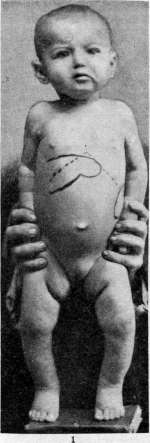
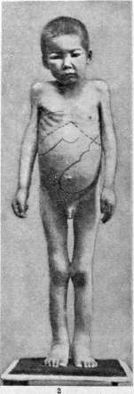
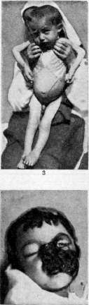
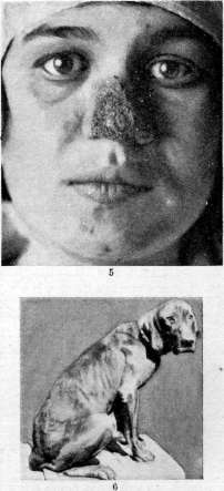
Figure 1. Kala-azar; case of a short, acute, severe course with a fatal outcome; exudatively-hemorrhagic diathesis. Fig. 2. Kala-azar; severe case with marked edema of the face and extremities; ascites. Figure 3. Kala-azar; severe case with a prolonged course. Figure 4. Kala-azar; case complicated by noma. Figure 5. Cutaneous leishmaniasis of the face. Figure 6. Typical habitus of a dog suffering from visceral leishmaniasis (ulceration of the eyelid margin, dried pus crusts around the eyelids due to conjunctivitis, ulcers on the lips and wings of the nose).
cases leishmanias can persist in the lymphatic glands, from which they enter the locus minoris resistentiae. Often near the ulcer it is possible to palpate thickened and painless lymphatic vessels in the form of a cord and along the course of the latter nodules ranging in size from a pea to a small cherry. In these nodules it was possible to detect leishmanias and obtain them from there in a pure culture. The classification of ulcers, most satisfactory, was proposed by Ledantec, but it is already outdated and requires changes. It is more correct to distinguish the following forms of this disease: 1) abortive, when the whole process is limited to the formation of a papule or nodule, which subsequently resolves; 2) desquamative, when leishmaniasis also proceeds without ulceration, in the form of a scaly plaque; 3) usual dry form of an ulcer covered with a crust; 4) suppurating ulcer as a result of secondary infection; 5) phagedenic form; 6) impetiginous form with a short course (about 2-4 months), resembling an impetiginous rash (secondary infection by pus-forming microbes); 7) keloid rash described by Balfour, when the whole process proceeds without ulceration, and the nodules consist mainly of dense connective tissue penetrated by foci of infiltrate; 8) papillomatous form; 9) leishmaniasis with predominant lesion of mucous membranes (American leishmaniasis). - Ulcers in a patient are rarely single; more often there are 3-5, and sometimes their number reaches 200 and 300 and can cause temporary disability. - Location of ulcers. As a rule, ulcers are located on open parts of the body; but they can also appear on covered parts of the body. On the face, their favorite place is the region of the zygomatic arch, the tip of the nose (see separate table, figure 5), the bridge of the nose, chin, lips, forehead, brow, edge of the ear. On the extremities, ulcers usually affect the dorsal surfaces of the joints. Sometimes ulcers are found on the genitals in men and women, which usually gives rise to confusing them with syphilis. - Complications. First of all, one has to reckon with the disfigurement that ulcers often cause. All other complications, such as phlegmons, erysipelas, gangrene, septicemia, etc., essentially represent in general complications of ulcers. Before the establishment of the true causative agent of the disease in the old literature, cases of the ulcerative process affecting muscles, cartilage (ear, nose), fascia, joints and bones, tooth loss, arrest of nail growth, abscesses in the liver, etc., were described. However, since the finding of leishmania was taken as the basis for the diagnosis of oriental sore, such complications have no longer been described by anyone, and undoubtedly they have nothing to do with leishmaniasis. Differential diagnosis. Cutaneous leishmaniasis is a granuloma, and it is often confused with similar processes in the skin, namely lupus, syphilis, blastomycosis, yaws, erythema multiforme and finally tropical ulcer. Cutaneous leishmaniasis differs from these diseases by the peculiar location of the ulcers, their course, and also their connection with the endemic focus of leishmaniasis. In addition, the finding of leishmania must be the basis of diagnosis. It is more difficult to distinguish leishmaniasis from tropical ulcer; the latter proceeds more acutely, and spirochetes and Vincent's bacilli are found in the juice of the ulcers. Treatment. There is perhaps not a single disease in which so many diverse methods of therapy are used. This is partly explained by the fact that if you start treating an ulcer with any preparation in the period when it passes into the last phase of healing, the effect is brilliant; conversely, in the treatment of the initial forms of the ulcer, almost all medicinal preparations usually turn out to be untenable. Cauterizing and tissue-destroying agents have always had especially wide application. Thus, ulcers are cauterized with a hot iron, thermocautery, and in the stage of papule and nodule this often gives positive results. All kinds of ointments with Cuprum sulfuricum, Stibio-Kalium tartaricum (20%), etc. are used. The latter ointment is successfully applied to the ulcer, and swelling and inflammation are obtained, infected granulations are destroyed, and in the future the ulcers begin to scar. To this group of preparations should also be attributed Kalium hypermanganicum, which is sprinkled in the form of a crushed powder onto the surface of the ulcer stripped of the crust. Cardamatis and Melissidis proposed using methylene blue in the form of an ointment 2-3 times a day, for 15-20 days. Such treatment can give relatively good results. Next should be mentioned the injection of 2% emetine around the ulcer and finally antimonial preparations (Stibenyl, Stibosan, Neostibosan, etc.), which are administered for 1-2 weeks, for 3 days in a row, with 5-7 day intervals and can to some extent be considered specific for this disease. Finally, recently X-rays, radium and ultraviolet rays have been used. The results from X-rays in the initial forms of the ulcer are insignificant, but in the flowering period, along with other therapy, it gives good results in terms of more rapid healing of ulcers. Regarding ultraviolet rays and radium, there is very little material that does not allow final conclusions to be made. In the initial stages, leishmaniasis gives good results by removing the affected area surgically. Change of climate has little effect on the course of the disease process. Prevention of cutaneous leishmaniasis and measures to combat leishmaniasis. The increased communication between the peoples of the USSR in recent years leads to the fact that many tropical diseases, including leishmaniasis, become ordinary even far beyond the borders of their endemic distribution. In connection with this, issues of prevention acquire great importance. The transmission of cutaneous leishmaniasis through insect bites dictates appropriate protective measures, namely: 1. In the foci of this disease, it is necessary to sleep under canopies dense enough so that phlebotomes cannot penetrate through them. It is desirable that the canopy be white, blue or red, because phlebotomes are almost invisible against a gray, yellow or even pale orange background. 2. Walls should also be painted in the specified colors. 3. If you do not use a canopy, it is necessary to close the windows by inserting nets into them, and sleep in a bright light, because phlebotomes are nocturnal insects and usually bite at night or in shaded rooms. 4. Since phlebotomes usually inhabit houses, in the south it is better to sleep outside the house - in the garden or on a flat roof, but obligatorily under a canopy. In general, in order to break the epidemiological chain, it is necessary to block the access of phlebotomes to the patient or, rather, to his skin lesions as sources of infection; for this purpose, as a therapeutic method, one should apply dusting the ulcers with Kalii hypermanganici powder or keep them under a bandage. The fight directed against phlebotomes presents great difficulties, because the biology of these insects has not yet been sufficiently studied. Phlebotomes breed near human habitation, and although the exact places of their breeding have not yet been studied, nevertheless it can be asserted that the life of these insects is connected with unsanitary conditions of the courtyard. Therefore, the maintenance of cleanliness of the yard, the dusting of latrines with lime, the timely removal of garbage, the cleaning of ditches (aryks), etc., in general, a thorough toilet of the soil surface of the populated area should be placed at the basis of measures to combat phlebotomes. Since the improvement of sanitary conditions of each individual courtyard largely depends on the improvement of general sanitary conditions, serious attention should also be paid to this side (water supply, sewerage). 5. Since there are a number of indications that the reservoir of the virus is sick dogs and relatively rarely other animals, it is necessary to properly organize the fight against dog leishmaniasis. For this, it is necessary to destroy sick dogs and issue appropriate mandatory regulations. 6. Preventive vaccination and vaccination with killed cultures. Martsinovsky, Shurenkova and others (see above) established that experimental oriental sores, as long as they exist for at least 3-4 months and heal independently, provide a fairly stable immunity, and repeated infections even after 5-7 years are not successful. Thus, by inoculating granulations from a patient onto the forearm or leg of a healthy person, we obtain the development of an ulcer at the site of inoculation, which guarantees against new infection through an insect. The practical inconvenience of this method is obvious, and recently various authors (Martsinovsky, Khodukin, Latyshev) have been making attempts to obtain vaccines from killed cultures of leishmanias. Such vaccines have been tested on a fairly large material, and although final conclusions about their action cannot yet be made, the impression is favorable. Forest American leishmaniasis (syn.: leishmaniosis Americana, uta, buha, forest yaw, espundia, leishmaniosis oro-pharyngealis). It is found in hot wooded areas, starting from Mexico and ending with the northern part of Argentina; it has existed in America for a long time, even before the arrival of Europeans there. In Peru on pottery dating back to the time of Inca rule, there are drawings depicting this type of leishmaniasis (uta). The disease is found mainly among workers engaged in rubber gathering and mat making in the virgin forests of America.
Migone notes a morbidity rate among workers in certain forests of Paraguay reaching 70-80% during the first months of their stay. Symptomatology: after an incubation period of two to three months, the disease begins with the appearance of one or several erythematous papules, sometimes itchy and resembling insect bites. After a few days, a small accumulation of pus-like fluid forms at the apex of the papule, and then upon scratching, the fluid protruding from it hardens, forming a blackish crust. Subsequently, tissue breakdown occurs beneath the crust, sometimes with the formation of pus, which emerges when the crust is pressed, emitting an unpleasant odor. Very often such ulcers are accompanied by lymphangitis, the appearance of nodes along the vessels, which in turn can turn into ulcers. Sometimes the appearance of ulcers is accompanied by fever, muscle, and periarticular pains. Approximately in 10-20% of cases, especially with a prolonged course of ulcers, involvement of the mucous membranes of the nose, mouth, and nasopharynx is noted. The process often spreads to the nasal septum, causing its destruction. Matta also points to ulcerative lesions of the vagina. With a favorable course, the disease ends in 8-12 months; but sometimes recovery drags on for 15-20 years. Many patients die from cachexia. The ulcers are located on hidden parts of the body, just as in cutaneous leishmaniasis. In approximately 30%, leishmaniasis affects the legs. Treatment with antimony preparations and intravenous infusions of emetine gives good results. There are indications of the beneficial effect of ultraviolet rays. Immunity in this disease is usually weakly expressed and is short-lived. The causative agent of American leishmaniasis, Leishmania brasiliensis, Vianna (1911) is morphologically similar to Leishmania tropica both in what is isolated from ulcers and in cultures. (Some authors describe flagellate forms in the ulcers.) Spontaneous diseases with this type of leishmaniasis have been noted in dogs; in other domestic animals it has not been observed yet. Townsend considers insects of the genus Forcipomyia (fam. Chironomidae) to be the transmitter of American leishmaniasis in Peru, which, however, is unlikely. Preventive measures and control of American leishmaniasis present great difficulties, since it must be assumed that some wild animals may also be the reservoir of the virus. Control aimed at transmitters also cannot yield much, since the biology of these insects has not yet been studied well enough. It has been established that with the felling of virgin forests and the establishment of plantations in their place, the disease disappears. As a preventive measure, it is recommended to set up settlements for workers away from the forest. Measures of individual prophylaxis, such as special clothing protecting against insect bites, mosquito nets, can play a certain protective role; finally, one should point out the necessity of keeping ulcers under a bandage or dusting them with potassium permanganate.
E. Martsinovsky. Kala-azar is a tropical infectious disease caused by the parasite Leishmania donovani. Synonyms: kala-azar, kala-jwar, kala-dukh, black sickness (all four names translate as "black disease"), tropical splenomegaly, fear of sir-caio, sahib, burdwan fever, dum-dum, Assam fever, anaemia splenica infantum, pseudoleukaemia infantum febrilis (Europe), ponos (Greece), haplopinakon (Cephalonia), marda tal biccia (Malta), malatha da mensa (Sicily), anaemia febrilis splenica (Fede), anaemia infantum a Leishmania (Pianese), leishmania-anaemia (Jemma, di Cristina), internal, visceral, general, Mediterranean, infantile leishmaniasis. The disease is characterized by persistent fever of various types, enlargement of the liver, spleen, lymph glands, marked emaciation, anemia, leukopenia, the presence of Leishmania donovani in the liver, spleen, and bone marrow, and the possibility of a wide variety of complications from which patients frequently die. It is observed in acute and chronic forms. It occurs in the form of separate foci, but can sometimes assume an epidemic spread. Kala-azar is predominantly a disease of childhood. The British first encountered kala-azar in 1849 during the occupation of the Garo Hills in India. In 1875, Rogers stated there that many families had died out from this disease. In 1882, the disease became widespread in the Brahmaputra River valley. In 1896, Rogers mistook kala-azar for a severe form of malaria. In 1902, Bentley mistook kala-azar for Malta fever. In 1903, Manson suggested that kala-azar is caused by a parasite from the trypanosome group. Several months later, Leishman gave a description of the true causative agent of kala-azar. In smears from the spleen of a patient who died of dum-dum fever, he discovered parasites, recognizing them as trypanosomes. Independently of Leishman, in the same year, Donovan reported finding similar bodies in the spleen of individuals who had suffered from dum-dum fever for a long time in Madras. Examining Donovan's preparations, Laveran and Mesnil recognized these bodies as trypanosomes. In 1904, Bentley in India finally established the unity of irregular fevers with splenomegaly, finding Leishman-Donovan bodies in all cases. Christophers provides an exhaustive description of the parasite's structure and its distribution in the host organism, and Rogers obtains its culture in citrate blood. In the USSR, the first case of kala-azar was discovered in 1909 by the Viennese physicians Sluka and Zarfl in a 9-year-old boy who fell ill in Tashkent; the 2nd case was described by Martsinovsky in the town of Klimovichi, Mogilev Governorate, in 1910; the third case belongs to M. N. Nikiforov and concerns a 6-year-old girl who fell ill in Tashkent in 1910; the fourth case was described by Kalashnikov in 1911 in a 37-year-old man from Tobolsk Governorate; the fifth case was described by N. V. Petrov in a 38-year-old woman who fell ill in Chardzhou in 1911; A. G. Gurko (1912) described 4 cases from Transcaucasia. In 1913, the expedition of V. L. Yakimov discovered 31 cases of kala-azar in Tashkent, Andijan, Samarkand, Bukhara, Termez, and Ashgabat. Since that time, a number of authors have published observations on human and canine kala-azar. Parasitology. The parasite causing kala-azar, Leishmania donovani, is morphologically indistinguishable from L. tropica (see above). In the search for a vector, attempts were made to infect head lice (Pediculus capitis), but these attempts proved fruitless; attempts to implicate fleas in the transmission of kala-azar were also unsuccessful: Pulex irritans, P. serraticeps, and Ctenocephalus canis. Suspicions regarding Anopheles maculipennis and Culex macrostoma (Franchini, Scordo) were not justified. In Cimex rotundatus and Cimex lectularius, Patton observed the complete development of leishmanias. Cornwall and La Frenais showed that from the intestine of a bedbug that had sucked the blood of a patient, a culture could be obtained 41 days after infection. Donovan was inclined to implicate Siphonvrinus rubro-fasciatus in the transmission of kala-azar. Brahmachari does not consider bedbugs to be vectors of kala-azar, since the distribution areas of the listed bedbugs and kala-azar do not coincide. Phlebotomus (Phi.) came under strong suspicion of having the ability to transmit kala-azar. Sinton pointed out in 1922 that the distribution area of kala-azar coincides with that of Phi. argentipes. However, Phi. argentipes has not been found in Central Asia. Knowles and Napier came to the conclusion that under favorable conditions of temperature and humidity in the body of Phi. argentipes, leishmanias can develop into flagellate forms. This was confirmed by Christophers and Shortt, who state that the reproduction of Flagellata in the body of Phlebotomus increases every day, and on the 5th day (according to Khodukin, on the 6th day) a strong infection of the intestine with flagellates can be observed. Shortt, Barraud, and Craighead found that from the 5th day after the first blood meal, flagellates are found in the pharynx. They are encountered in the oral cavity only after the second blood meal, so transmission is possible only at the third blood meal. Attempts by some authors to find Phlebotomus infected under natural conditions remained in vain; others found in an infected house Phi. argentipes, which turned out to be heavily infected with flagellates of the same type as artificially infected Phlebotomus. However, their extensive experiments on the transmission of kala-azar via the bite of Phlebotomus that had fed on infected peripheral blood of kala-azar patients gave negative results. Khodukin, Sofieva, and Shevchenko established that Phi. papatasi are readily infected with the virus of canine leishmaniasis, which allows the authors to suggest the role of these sandflies in the transmission of kala-azar. Geographical distribution (see map). Kala-azar occurs in Europe, Asia, and Africa between the equator and 40° N lat. and between 17° W and 12° E long. in the form of endemic, mostly small foci. Kala-azar reaches a strong spread in India in the valley of the Ganges and Brahmaputra rivers, in the provinces of Bihar, Bengal, and Assam; other places in India are affected less severely. Further east, foci of kala-azar are found in Indochina (Siam, Burma), on the island of Sumatra, the island of Formosa, in China (eastern coast and the valleys of the Yangtze and Yellow rivers). In the west: Arabia (Hejaz, Yemen), Asia Minor (Smyrna), Archipelago. In Europe: Greece, southern France, Spain, Portugal. In Africa: Cairo, Abyssinia, Egyptian Sudan, Tripoli, Tunisia, Algeria, Morocco, Guinea. In the USSR, kala-azar occurs in the form of small endemic foci in Transcaucasia (Erivan, Tiflis, Shulavery with the surrounding Borchaly uyezd) (Gurko, Vorobyov, Ushikov). In Central Asia, endemic foci of kala-azar are located along the line of the Central Asian railways, deviating from them deep into the country only around large centers (Samarkand, Tashkent, Kokand, and Andijan). Temperature and humidity conditions have a great influence on the spread of kala-azar. In India, for example, they are more favorable for the parasite and vector than in Central Asia. In accordance with this, morbidity in India is expressed in different figures than in Central Asia. The Director of Public Health in Calcutta defines the number of cases of kala-azar in Bengal in 1923 as 50,000, and in 1925 this number reaches 180,000 (Napier), whereas in Central Asia the annual number of detected cases is about 150. The age distribution of kala-azar is represented by the table (in % of the total). Assam 1924 Young, China 1923 Young, Spain 1925 Pittaluga, Central Asia 1923-28 Artamonov. Age: 0-1 year, 1-5 years, 5-10 years, 10-15 years, 15-20 years, 20-30 years, 30-40 years, 40 years and older. Total number of cases. In India and China, the highest percentage of cases falls on the age of 5-30 years, whereas in Spain and Central Asia, the age most susceptible to kala-azar is childhood up to 5 years. Social and domestic conditions. While in India kala-azar is distributed mainly among the rural population, in old villages with rich vegetation, in Central Asia kala-azar is found mainly in station areas and European parts of cities, and in kishlaks it occurs in single cases around large centers, with individual cases of kala-azar being discovered in homesteads devoid of vegetation.
In India, kala-azar is a disease of the poorest class. According to Brahmachari, kala-azar spreads slowly, advancing along trade routes at about 10 English miles per year. Capturing a new region, kala-azar disperses within it in small foci; in villages, it nests in certain houses. Brahmachari considers the conditions for the spread of kala-azar in cities to be the lack of paved streets, the immediate proximity of vegetation to housing, the presence of dirt floors in dwellings, and unhygienic living conditions. In Central Asia, foci of kala-azar are found in extremely overcrowded and densely built-up dwellings, near which dumping grounds are arranged, as well as in dwellings located on irrigation canals (aryks), which, in the absence of water supply and sewage systems, play the role of both. Sex and nationality apparently play no role in the epidemiology of kala-azar. Animal infection experiments. Besides humans, certain animals have also turned out to be susceptible to kala-azar. Donovan and later Patton infected dogs with a thick emulsion of infectious material from the liver, spleen, and bone marrow in amounts of 2.0-4.0 cm3. Experiments with the infection of monkeys of the genus Macacus, jackals, white mice, and rats also proved successful; rabbits and guinea pigs could be infected only with large doses of infectious material (Nicolle, Franchini). Sergent succeeded in causing a severe disease in a cat. Archibald succeeded in infecting monkeys by giving a large amount of virus per os. Hamsters turned out to be very susceptible to kala-azar. Pigeons, goats, chickens, calves, and pigs could not be infected. A. Artamonov. Pathological anatomy. The macroscopic pathologo-anatomical changes in kala-azar boil down to the following. The spleen is enlarged up to 17-26 cm, dense, fragile, cyanotic, sometimes withemic infarctions, less often hemorrhagic. Most lymph glands are medullary swollen, red. The bone marrow of the diaphyses is red, hyperplastic. Usually, the liver is enlarged. Anemia is sharply pronounced. The skin is characteristically waxy pale, sometimes pasto-oedematous (face, extremities), mucous membranes are pale. Muscles and adipose tissue atrophy in pronounced cases. Stunting of growth is noted in children. Various degenerative changes develop secondarily; catarrhal-ulcerative processes in the intestine (colitis), meningitis of various etiologies, peritonitis, pleuro- and bronchopneumonia often supervene secondarily; tuberculosis flourishes, noma develops, and various acute infections join in, giving the specific changes in organs inherent to them. - Microscopically, the most characteristic is the presence of leishmanias in the cells of the reticulo-endothelial system. The bone marrow is rich in large cells of the reticular type, laden with leishmanias [see separate plate (pp. 575-576), fig. 3], the parenchyma appearance is promyelocytic (according to Schilling). Spleen: sharp atrophy of follicles and pulp cells stuffed with erythrocytes. Reticulo-endothelial cells are numerous, sometimes completely packed with a huge amount of leishmanias [see separate pl. (pp. 575-576), fig. 2, 5, and 6]. Sinuses are dilated, endothelium is hyperplastic, partially desquamated into the lumen. Plasma cells are in the pulp. The reticular stroma coarsens and thickens. In lymph nodes, there is hyperplasia of reticulo-endothelial cells, leishmanias in them, transformation of sinus endothelium into "free macrophages"; coarsening of the stroma, atrophy of the parenchyma, erythrophagocytosis. Liver: abundant amount of leishmanias in Kupffer cells [see separate pl. (pp. 575-576), fig. 4], dilation of capillaries in hepatic cells or granular degeneration or fat droplets, more often along the periphery of the lobules. Both in the spleen and lymph nodes, and in the liver, there are foci of myeloid hemopoiesis. Digestive tract: frequent finds of leishmanias in the tonsils, leishmanias have been described in intestinal ulcers. Heart and blood vessels are mostly without specific changes, sometimes tigroid fatness; fatty degeneration of capillary endothelium, small hemorrhages in the skin, serous membranes. Kidneys: picture of nephrosis, sometimes nephritis. In the adrenal cortex, lipoid infiltration; usually early involution of the thymus gland. The remaining endocrine glands are without special changes. In the ganglion cells of sympathetic ganglia (solar plexus, Meissner's and Auerbach's), early pigmentation, shrinkage, pyknosis, vacuolation of pericellular spaces, neuronophagia (especially with complications). Brain and nerve fibers without special changes. The respiratory system is almost always involved by concomitant diseases. Death ensues from complicating factors (pneumonia, septic processes, infectious diseases, etc.) joining in the stage of exhaustion of the organism with already significant disability of systems (hematopoietic and reticulo-endothelial, liver, and kidneys). G. Terekhov. Clinic. The incubation period lasts from 10 days (Meneon) to 2 or more years (Brahmachari). In monkeys, it is equal to 50-60 days; in mice, 15-28 days (Yakimov). In the initial stage, the symptoms of the disease are little characteristic: indefinite febrile attacks with or without chills, at times irritability, in children loss of interest in games, slight enlargement of the liver and spleen, some May
June 1925. PALENESS OF THE INTEGUMENTS. Appetite
can remain normal or elevated. Sometimes the onset of January 1926. Date Temperature 3l| 1 { 2 j в | 4.j 5 | в | 7 ( S Days of illness 64 6 5[66 6 7|68 | | ^J Zl | rf з / [_ zr zr: t: f t _r i i ~t ' / ± it \ J f \ ir ±1 t tz t -м |Date 26 27 28 29 30 31 |temperature "1 п ' | \ 1 Ь \ Л / 1 / / / 1 M / л / Я / 35.5e 35- З-ч h3 ч <a -. -м----- м _й_ -Гп- Figure 1. Figure 2. kala-azar is masked by some other disease: malaria, influenza, measles, gastrointestinal disease, etc. If the diagnosis is not established at this time and treatment is not initiated, then after 3-4 weeks the symptoms begin to acquire a certain definiteness: unceasing fever of one type or another, more significant enlargement of the liver and spleen; paleness of the integuments progresses, the patient grows weaker; small children who shortly before this began to walk or stand on their feet stop walking and demand that they be constantly held in their arms; they sleep a lot; in the waking state, they are extremely apathetic and protest with a loud cry against any attempt to lead them out of this state. In children with exudative diathesis, puffiness of the face, swelling of the skin of the extremities are observed; in individuals of lymphatic constitution, tumors of the lymph glands catch the eye; in those who previously had tuberculosis, the process becomes aggravated or disseminated. Appetite decreases, emaciation begins. In the future, the symptoms may intensify further and become so characteristic that suspicions of kala-azar arise in the physician at first sight of the patient: sharpened facial features, skin that has lost its elasticity of a waxy color, musculature devoid of tone. Against the background of general sharp emaciation, a large abdomen catches the eye with dilated cutaneous veins, ascites, and greatly enlarged liver and spleen, occupying up to 2/3 of the entire abdominal cavity [see separate pl. (pp. 583-584), fig. 1, 2, and 3]. Urine is N or with traces of protein, sometimes lipuria, sometimes positive diazo reaction. The remaining organs are dependent on complications, very diverse in this stage, which is of varying duration. Next follows either a stage of recovery with the gradual disappearance of all morbid phenomena or a short stage of cachexia, sometimes with complications in the form of purpura, noma, sharply pronounced anemia, or leishmanial hemorrhagic colitis.
Figure 3. Kala-azar has a twofold course: short (1-2-3 months) and prolonged (up to 2 years and more). Many cases of the short course proceed in the form of severe acute febrile diseases and in the majority end fatally. These are patients with slightly enlarged liver and spleen, irregular fever, burdened with lymph- November 1927 December May 1929 1 Date 18 j 19 tempera- ture Days of illness 22 | 23 39.5° l / / [ / / / / / / / / / | ( / / / -l- 38.5! / / Chz-g- / Date 24(25 930|l|2|3J4 tempera-tu-ja Days of illness 1b]1 9J20 if / t : г г t j_ т ± t г г t IN" ^_ i ZZ& - O e z -e \zz m B i Pi =*' GTT z ~i April r. Date 2 122 2 3 24 25 26 2 7J2829 tempera-tu-ra Days of illness 6 6 67 6 70 717 2|73 74 3 F j ZJ m 1 1 ZI No. t =f _. 1 F - t II zr tit zr ± t.zt _ It ; ± t^ ±z. : iz'- uz. t- nz nu j t. tz1 ' ' rz u M tl tt 1 I tit 1 1 c 36- h 35.6- = 10L « 35^-5 & m Figure 4. Figure 5. ko-exudative or hemorrhagic diathesis; they come to a fatal end despite timely and adequate treatment. On the other hand, cases with small liver and spleen, moderate fever, ending in recovery within 1-2 months with relatively short treatment are observed. Part of the observed spontaneous cures from kala-azar pertains precisely to such cases. Cases with a prolonged course are more favorable with regard to quo ad vitam, although among them both mild ones, proceeding with moderate fever, without any complications, and severe ones, proceeding with debilitating irregular or hectic fever and accompanied by June u-----C=*ZB Date 12 13 14 1 5 16 2 . J 4 5 6 temperature, - - - i Z| Zl fl 1 Zl 1^1 zt ^fl Zl t -Г n t zlzn^ I± л I Zl it zzfl _zt fl ± / zu zt ± / ^ZL _zt iznz j_ it TZ /1 | ±Lt / r t: t^ ± iz t f ± i tz t / Г1 1: fl tz ( - t J I: / f | / tz j tz t 362- 35.5- l r and o f = 1 tr .f rf | » -is s~ й- s- I« n < ei >' _J4 jJ =0 a ^ a -° 7t -«1 G! n and i s : ь Figure 6. various complications, which are in many cases the cause of death of the patient. Cases with a prolonged course can also sometimes end in spontaneous recovery, but often the recovery turns out to be illusory, and is followed by a relapse. Fever at the beginning of the disease manifests itself insignificantly October________________________________November 1924 g. giving stadium decrementi (Fig. 4) and giving reason to confuse the disease with typhoid fever. In other cases, the fever takes the type of febris remittens, changing after a few days to another type or taking the character of febris remittens undulans (Figs. 5 and 6), in which the attacks of remittent fever continue for 10-12 days, separated
by 1-2-day afebrile intervals. The undulating type of fever sometimes takes the character of febris re-currens, resembling the temperature curve of Persian relapsing fever (Fig. 7). In some cases, severe febrile attacks are replaced by a longer or shorter afebrile period or subfebrile type of fever (Figs. 8 and 9), which may be followed by new outbreaks of the disease of considerable severity. Sometimes the fever takes a hectic type with a daily fluctuation of f
4-5° (Fig. 10). Increases can occur 1-2 times a day (Fig. 11), which should be considered specific for kala-azar. In some acutely proceeding cases, the fever takes an irregular type, continuing for many weeks (Figs. 12 and 13). The liver and spleen increase by 2-3 fingers. In the case of a prolonged course, the lower |Date 5 1 6 13 14 24 2 5 26 temperature - -|- - - / / U\ / tz\ \ \ \ 1 I \ / I | | л / | / \ n л \ й \ \ if \ i / / r\ \ | I I | /1 \ \ f K \ / tz \ / \ 1 l\ \ / ^ \ | it LI / \ \ \ / / / ^ / / \ | / \ | / / (1 | y / / \ _y \ \ \ / \ It ZZ1 \ \ / \ | \ i/ \ / / \ \ / \ [ KtJ- h \ ^ / Ts2 / \ \ and / and \ / t / \ \ / <* \ =-' /o1' 1 , ' 1 \ | G^ \ tzz m y ' $ o J2 « c if Ok > and Figure 7. edge of the liver can reach the navel. The spleen can occupy up to 2/3 of the abdominal cavity, causing a large belly together with the formed ascites. Dionisi established clinically and pathologo-anatomically that there may be no enlargement of the spleen. Enlargement of the liver is also inconstant: Rogers in India established that about 40% of cases proceed without enlargement of the liver. Red blood presents a picture of a gradually developing anemia, which by the end of the severe period reaches a very significant degree: the number of erythrocytes drops below 1 million. The amount of Hb drops (up to 10%) (Brahmachari). The resistance of erythrocytes decreases: 0.18-0.24. The sedimentation reaction in severe cases reaches 87 mm in 1 hour, according to Panchenko, but with the improvement of the general condition, the sedimentation rate decreases. Prognos- August
__September Date l temperature | Z1 i r, zr / t g | / / / /g / zr / za / / | / zt / / f f f f ± f / 3-3J f л / ( v si / T4 / I/1 /' / / k yz vn / \l y / f^ -i ' izhr / / fl / / / / f / h Vя / \ rt t \ \f / Hz \ / / and / rz Figure 8. tic significance this reaction apparently does not have. Coagulation is reduced. The number of leukocytes in the initial stage is increased to 12-14,000. As the painful phenomena develop, their number decreases and reaches 700 per 1 cm3, to rise again to the upper limit or slightly higher upon recovery and further normalize. Neutrophils show a sharp nuclear shift to the left, and their absolute number drops sharply. Lymphocytes in percentage terms remain within N; monocytes do not show characteristic changes. Eosinophils at the beginning of the disease are present in a reduced number, at the height of the disease are absent, and in the recovery period in some cases appear in an increased number. The pulse corresponds to temperature. A very frequent complication is protein-free edema of the face and extremities. Ascites is often added to them, especially in cases with a large liver and spleen. An absolutely bad prognosis is given by the addition of miliary
tuberculosis, purpura, pernicious anemia and leishmanial hemorrhagic colitis. Frequent causes of death are pneumonias and noma [see separate table (art. 583-584), Fig. 4]. Measles, pertussis and gastrointestinal diseases greatly worsen the prognosis, especially in small children. A rare complication is cutaneous kala-azar. In malaria-endemic areas, kala-azar is often combined with malaria. The prognosis in children is significantly worse than in adults. | Diagnosis. In addition to the signs listed above, certain reactions are also important for the diagnosis of kala-azar. Having noticed the increase in globulins in the serum of patients with kala-azar, Brahmachari proposed the following diagnostic reactions. 1. The patient's serum is diluted in a test tube with physiological saline 10-20 times, then a little distilled water is carefully added. With a positive reaction, a clear white ring is formed, as with a protein reaction. 2. If the patient's serum is diluted 1924 g.
diluted in a test tube with 2–3 parts of water, a white precipitate falls out. Napier proposed the formaldehyde test: 1.0 cm3 of clear test serum is poured into a test tube measuring 7.5 x 1.25 cm, a single drop of commercial formalin is added and shaken well; if after 1–2 minutes the serum becomes viscous, and after 3–20 minutes gelatinous and opaque, this points to kala-azar. All the mentioned reactions do not yield entirely reliable results. In differential diagnosis, it is necessary to keep in mind malaria, typhoid fever, Mediterranean fever, splenic echinococcus, Banti's disease (see), pernicious anemia, rickets, cirrhosis biliaris hypertrophica (Hanot), pseudo-leukaemia splenica infantum (Jaksch), splenomegalia (Gaucher). The most reliable and rapid diagnostic means is the microscope, the study of liver or spleen punctate. In the human organism Leishmania donovani were found, besides the liver, August g. september Da ga temperature i л \ / л л /1 л / / / 1} / / \ / л / г / '' « /' / / / | / ' / ¥ 1/ г / 1 /' л ч / / / I / / / f t i/ / ' ' / a / / |/ f < \ / / / / / y / / / / I " \ \ ¥ и ;г \т "П ~ю "o ^ < ^ "ST ^ ^ Угч Un й- »*i /Г /j /У1 Figure 9. of the spleen and bone marrow in the lymph glands, in the kidneys, in the muscles, in ulcerations and hemorrhagic spots of the skin, in intestinal ulcers, in the mammary gland, in the lungs, in the cutaneous papillae, in faeces. Treatment. Before the introduction into practice of the preparation of tartar emetic for the treatment of kala-azar, a whole arsenal of medicaments was tried that did not yield a positive result: Arsenophenylglycin, Atoxyl, mercury, aniline, Salvarsan, Neosalvarsan, Hectin, Electromercurol, Thiarsol, Natrium kakodylicum, X-rays. Antimony compounds were first recommended by Manson. At the present time, the following antimony preparations are used: Stibenyl, containing 33% 5-valent Sb, Stibosan, containing 31% 5-valent Sb, Antimosan, containing 12.5% February 1926 teXDa" [ra-tura. /1 п / / / 1 1 / 1 1 1 ft / д _ / / д / Zt. 1 I ±_ / 1 Л ±. \ \ tfc- \ 1 \ 1 гг. /1 \|| \ it и \ \ 4Ё /1 \ 1 / \ т^ \ 1ГТ \| V 1 ц y i _y- / 35.5- 35- / / / + f ~ei- *~ 4й Гц» °ч ^ 1 -° з га «2 j_ _._L-ffl_ Figure 10. 3-valent Sb. These patented preparations are tolerated by patients much easier than Stibio-Kalium tartaricum. They can even be administered subcutaneously and intramuscularly, whereas Stibium potassium tartrate can only be administered intravenously. Recently, the German firm Heyden has released a new preparation, Neostibosan, which holds promise of turning out to be a better remedy than those in use up to the present time. Stibenyl is used in a 1% solution, Stibosan in a 2% solution, Antimosan in a 5% solution. The minimum dose for infants is 0.1 into a vein or muscle, subsequent ones depending on the patient's tolerance. The onset of severe coughing or vomiting shortly after the administration of the medicament indicates that the dose must be reduced. Neostibosan is used in an initial dose for children aged up to 4 years—0.05, for children aged 5–14 years—0.1; subsequent doses are doubled. In addition to these "specific remedies," various authors recommend carrying out symptomatic treatment as well; Thyreocrin (resp. Thyreoidin) deserves mention upon the appearance of edema. It is also necessary to prescribe hygienic and dietetic treatment. The cessation of fever, a steady gain in weight, a progressive increase in hemoglobin, a slowing down of sedimentation, the approach of the leukocyte formula to normal can signal the end of treatment even in the presence of an enlarged liver and spleen, which in many cases return to normal only many months after the cessation of treatment. Prophylaxis and control measures. In the chain: human-parasite-vector, the last link is unknown, therefore the fight against kala-azar can currently be built only along the lines of combating the parasite. Treatment with antimony preparations in connection with dispensary registration and examination of areas endemic for kala-azar to identify and record cases is so far one of the main methods of combating kala-azar. Children, especially of younger age, who have ended up in the area of a leishmaniasis focus, should if possible be transferred to another region and placed under optimal conditions of hygiene and dietetics. The timely destruction of dogs sick with leishmaniasis must be considered a most serious preventive measure.
A. Artamonov. Canine leishmaniasis is an infectious disease analogous to human kala-azar, and in some cases to cutaneous leishmaniasis in humans. The parasite L. canis (Nicolle, 1908), the causative agent of canine leishmaniasis, is morphologically indistinguishable from L. donovani, the causative agent of human visceral leishmaniasis, and is cultivated on the same media. The question of the identity of the human and canine leishmaniasis viruses is still considered unresolved. Khodukin and Sofiev tend to believe that these two viruses are identical, because, in addition to epidemiological observations, serological studies of various strains of leishmania carried out by them indicate that L. canis is a synonym of L. donovani (Khodukin and Sofiev, 1928). The routes of spread of canine leishmaniasis are still not precisely known; Nicolle (1908) suggested that the vector of canine leishmaniasis is the flea (Ctenocephalus canis). However, subsequent observations established that fleas are not vectors of canine leishmaniasis. Figure 11. Leishmaniasis. These same studies proved that the bedbug is also not a vector of canine leishmaniasis. Following the publication of the reports of the Indian Kala-Azar Commission, Khodukin began investigating phlebotomine flies, and he succeeded in establishing that Phi. papatasi (Scop., 1786) becomes quite well infected with L. canis. Subsequent observations by Sofiev and Shevchenko established that 98% of Phi. papatasi become infected with L. canis. In the digestive tract of the phlebotomine fly, L. canis undergoes a cycle similar to the cycle in culture. Experiments involving the rubbing of infected phlebotomine flies into the skin of healthy dogs yielded negative results. Still, for now, Phi. papatasi must be considered the most probable vector, since in the stomach of this insect the leishmania remain fully viable for at least 2 weeks. Nicolle (1908) noted in some cases of leishmaniasis observed by him in children that they had contact with dogs exhibiting emaciation, eye lesions, and fever. Nicolle investigated the susceptibility of dogs to the virus of childhood leishmaniasis and found that dogs are readily infected by it. Following this, Nicolle undertook a systematic survey of dogs in Tunisia and succeeded in discovering 4 cases of spontaneous leishmaniasis (1.8%) among 222 dogs. The virus of canine leishmaniasis can infect white mice, monkeys, and hamsters (Cricetulus griseus). The cat did not become infected in the experiments of Alvarez and P. da Silva. To date, canine leishmaniasis has been established in North Africa, southern Europe (Italy, France, Spain, Portugal), and Central Asia (Yakimov and Shokhor, 1913–14). Within Central Asia, canine leishmaniasis is currently registered in the following locations: Toy-Tyube, Syr-Darya, Samarkand, Kokand, Andijan, Bukhara, Kagan, Termez, Merv, Ashkhabad; in Transcaucasia, leishmaniasis was established by Gurko, Dzhunkovsky, and Luss (1909). Cutaneous leishmaniasis in dogs has also been described in Persia and India. The epizootiology of canine leishmaniasis has been little studied. During the year, there are 2 rises in the morbidity curve: spring-summer and autumn-winter [Greece (Cardamatis), Tashkent (Khodukin)] (Fig. 14). In Central Asia, according to Khodukin's data, morbidity in various months ranges from 0.5% to 10%, with an annual average of 2% to 3%. With the exception of India, the distribution zone of canine leishmaniasis coincides with the distribution zone of kala-azar. In Tashkent, a close intertwining of foci of kala-azar and canine leishmaniasis has been established. Simultaneous illnesses in children and dogs in the same apartment were frequently observed. Over a period of 4 years (1926–29), the systematic elimination of leishmanial dogs apparently reduced the incidence of disease in children throughout the city. Thus, in 1924 there were 31 cases, in 1925—25, in 1926—24, in 1927—13, in 1928—14, and in 1929—11 cases. Consequently, the morbidity among children dropped sharply from the moment dogs sick with leishmaniasis were destroyed. In other Central Asian cities where control measures against canine leishmaniasis were not carried out, no decrease in morbidity is noted. The pathological anatomy of canine leishmaniasis has been poorly elaborated. The most characteristic feature is hyperplasia of the bone marrow (usually red in adult leishmanial dogs); an enlargement of the spleen by 2 to 3 times is also characteristic, and the spleen itself has an uneven, jagged, shrunken edge as a consequence of previous infarctions. Sometimes these infarctions are also discovered at autopsy. Microscopically in areas of alopecia...
Figure 13. Type of febris quotidiana. ...and <07
medicines often reveal leishmaniasis even in apparently healthy areas of skin, residing in endothelial cells; very often leishmaniasis is found in endothelial elements around hair follicles and sebaceous glands. In the spleen, hypertrophy of Malpighian corpuscles, infiltration of reticulo-endothelial cells by leishmanias. Leishmanias are found in lymph glands, the liver, the thyroid gland (rarely), the adrenal glands, in the epithelium of the intestines and urinary bladder. The clinical presentation of spontaneous leishmaniasis in dogs has been little studied. The incubation period is at least 3 months. Two forms are distinguished: acute and chronic. In the first form, there is a rapid disappearance of the subcutaneous fat layer, thinning of the hair coat, diarrhea sometimes appears, and irregular fever is noted. Da Silva observed (with artificial infection) purulent discharge from the eyes, patchy alopecia, and crust-covered excoriations. Lemaire, Ed. Sergent, and Lhéritier observed keratitis. Microscopically in these cases, interstitial keratitis with the presence of parasites was found. The disease lasts 4-6 months, the animals lose their characteristic mobility and usually die in a comatose state. Often before death, leishmanias can be detected in the peripheral blood. The chronic form, according to Yakimov, Laveran, and others, proceeds very slowly; according to these authors, clear visible signs are usually absent: some weight loss, reduction in the density of the hair coat, immobility. Yakimov considers sharp anemia and irregular fever to be one of the surest signs. Observations by Khodukin (1925-1930), carried out jointly with F. I. Shevchenko, showed that in only 2% (out of more than 4,000 autopsies of dogs) are visible manifestations of the disease absent in canine leishmaniasis in Central Asia. Among external signs, the most characteristic are blepharitis and conjunctivitis, encountered in 98% of all Khodukin's cases, followed by ulcerations on the eye, ear skin (86%), muzzle skin (60%), nose (20%), and ulcers on the toes, back, etc.—in 40% [see separate table (art. 583-584), fig. 6]. Very characteristic of general leishmaniasis in dogs is abundant shedding of the epidermis. Baldness is rare; it is more characteristic of the filarial syndrome. Fever in separate attacks (1-2 weeks) is especially frequent in dogs in the spring. The duration of the disease in most cases is difficult to establish. The chronic form of canine leishmaniasis can last for years. Along with general leishmaniasis, cutaneous leishmaniasis in dogs has been described by some authors. Cutaneous leishmaniasis in dogs, according to Yakimov, is caused by a special species of leishmania—L. canis var. cutanea—which, however, cannot be considered proven. Usually, cutaneous leishmaniasis of dogs is expressed in the presence of ulcers with a sluggish chronic course, but without parasites in the internal organs. Some authors (Khodukin and Shevchenko) believe that there is no independent skin disease (spontaneous) in dogs, at least within Central Asia. The most frequent localization of ulcers is on the bridge of the nose (in most cases), on the edge of the ear and toes; ulcers are rarely found on the back and eyelid. Like cutaneous leishmaniasis in humans, the disease begins with a nodule, painless and strongly infiltrated; subsequently, the flowering of the ulcer sets in, characterized by the breakdown of the nodule and the formation of a true ulcer. The ulcer has undermined, strongly infiltrated edges; the bottom is covered with sluggish, pale granulations. Spontaneous healing is frequent. The diagnosis of internal leishmaniasis of dogs in areas of its enzootic spread is quite easy based on the above symptoms: eye damage, emaciation, shedding of the epidermis, presence of characteristic ulcers. Of decisive importance is the examination of scrapings from the eyelid skin or from the bottom of ulcers. Eyelid skin scrapings, according to observations by Khodukin and Shevchenko, give a positive result in 98%. In case of a negative result when examining a scraping from the eyelid skin, trepanation (Yakimov), extraction of bone marrow and its subsequent examination (dry smear, Giemsa staining) can be recommended. Examination of the bone marrow must be carried out carefully, because there are cases when leishmanias are encountered in units. Post mortem examinations are performed on the bone marrow, spleen, and liver—in these organs it is easiest to find leishmanias. Treatment of internal leishmaniasis of dogs has not yet yielded satisfactory results. The only preventive measure at present can be considered the timely isolation of dogs suspected of leishmaniasis and the destruction of leishmaniotic dogs in areas of endemic spread of infantile and canine leishmaniasis.
N. Khodukin. Lit.: Artamonov A., Four cases of cure of leishmaniasis with inadequate therapy, Russ. zh. tropich. med., 1928, № 7; he same, Materials on the epidemiology of internal leishmaniasis in Central Asia, ibid., 1929, № 4; Borovsky P., On Sart sore, Voenno-med. zhurn., 1898, November (also Med. mysl Uzbekistana, 1927, № 2); Burova L., Rickenberg's reaction in leishmaniasis, Med. mysl Uzbek., 1928, № 1; Heydenreich L., Penda sore, SPB, 1888; Gershenovich R., On the question of R. Wassermann's [reaction] in leishmaniasis in children in Central Asia, Turk. med. zhurn., 1925, № 7; Lysenko V., Erythrocyte sedimentation rate in visceral leishmaniasis in children, ibid., № 4; Marcinovsky E., Etiology of Oriental sore, diss.—M., 1909; he same, Oriental sores and their etiology, Med. obozren., 1904, № 24; Marcinovsky E. and Bogrov S., On the question of the etiology of Oriental sore, ibid., № 3; Slonim M., On the question of the treatment of visceral leishmaniasis, Turkest. med. zhurn., 1922, № 1; Yakimov V., Leishmaniases (Trudy expeditsii po izucheniyu tropicheskikh b-ney lyudey i zhivotnykh Turkestanskogo kraya v 1913 g., t. I, P., 1915, lit.); Yakimov V. and Shokhor N., Leishmaniasis (Kala-azar) of humans in the Turkestan region, Med. obozr., 1914, № 18-19; Brahmachari U., Kala-azar (Hndb. d. Tropenkrankh., hrsg. v. C. Mense, B. IV, Lpz., 1926, lit.); Napier L., Kala-azar, L., 1927 (lit.). Canine Leishmaniasis.—Khodukin N., On the protozoa of the intestine of dog fleas in Tashkent and their role in the epidemiology of canine leishmaniasis, Med. mysl, 1927, № 3; he same, Attempt at infection of Phlebotomus lewisi (Nicolle), ibid.; he same, Basic problems of the epidemiology of Kala-azar in connection with the epidemiology of canine leishmaniasis in Central Asia, Tashkent, 1929 (lit.); Khodukin N. and Sofiev M., On the question of the identity of L. donovani and L. canis, Med. mysl Uzbekistana, 1928, № 1; Khodukin N. and Shevchenko F., On skin manifestations in canine leishmaniasis, ibid.; Yakimov V. and Shokhor N., Cutaneous leishmaniasis ("Oriental sore") in dogs in the Turkestan region, Vestnik obshch. veterin., 1914, № 6.
Related articles
Mentioned in
Cite this page
“Leishmaniases.” Soviet Medical Encyclopedia. English translation of Bolshaya Meditsinskaya Entsiklopediya, 1st ed. (Moscow, 1928–1936), ed. N. A. Semashko. https://sovietmedicalencyclopedia.pages.dev/article/leishmaniases/