Encephalography
Historical document, translated for reference. It reflects medical knowledge of the 1920s–30s and is not medical advice.
Summary
This 1930s encyclopedia article discusses encephalography (pneumoencephalography and ventriculography), detailing the historical development of replacing cerebrospinal fluid with air for X-ray visualization of the brain's ventricles and subarachnoid spaces. It covers the techniques of air introduction, equipment, physiological reactions, complications, and diagnostic interpretation based on early neurological experience.
Encyclopedia article (1928–1936)
ENCEPHALOGRAPHY (from the Greek encephalon — brain, and grapho — I write, depict). (Syn.: pneumocephalography, pneumoencephalography, encephaloventriculography). On ordinary roentgenograms, one can see cracks and fractures of various bones, ascertain the closure of sutures, expansion, deepening, and destruction of the sella turcica (pituitary and brain tumors), expansion of the internal auditory meatus (auditory nerve tumors), and sometimes detect a tumor if it contains calcareous deposits; in childhood, one can ascertain thinning of the cranial bones and divergence of the sutures (hydrocephalus); one can also ascertain bone erosions, expansion of the diploic veins, excessive proliferation of Pacchionian granulations, prominently expressed impressiones digitatae on the cranial bones, etc. Such a state of affairs did not satisfy clinicians, since the brain as such was almost never revealed on an X-ray image. Hence arose attempts to apply the method of contrast radiography to the skull and its contents. The first initiative in this direction belongs to the American neurosurgeon Dandy from Baltimore. He made a bold, but extremely simple in conception, attempt to replace cerebrospinal fluid with air. True, long before him, Schüller experimentally inflated the ventricles of dogs with air, but he did not publish the results of his experiments. In 1918, Dandy reported on 8 cases of intraspinal air injection, but the spinal method of air introduction did not satisfy him, and he soon replaced it with the direct introduction of air into the ventricles through trephine openings, calling this method ventriculography (syn.: pneumoventriculography). In 1920, Dandy already published 75 cases of brain tumors diagnosed and precisely localized with the aid of ventriculography. In the same year, the German neuropathologist Bingel in Brunswick and the Norwegian Widerøe in Kristiania, independently of each other and supposedly of Dandy, published the results they achieved with intraspinal air insufflation in diseases of the central nervous system and called this method encephalography. With the introduction of suboccipital puncture, air also began to be introduced through the posterior cistern of the brain. Thus, there are three methods of introducing air into the brain: direct introduction of air into the ventricles (ventriculography) and proper encephalography, for which either suboccipital puncture or, more frequently, lumbar puncture is used. Naturally, encephalography is technically simpler and causes less trauma, since the patient does not have to be trepanned and the brain is not traumatized by a puncture. The technique of encephalography is extremely simple: in a sitting position, the patient undergoes a lumbar puncture in the lower lumbar region between the LII–LIII or LIV–LV vertebrae, after which the fluid is allowed to flow out freely (under no circumstances should it be aspirated with a syringe), and for the first time, depending on the pressure, from 10 to 25 cm3 are released, after which 10 cm3 of air or another indifferent gas (oxygen, carbon dioxide, etc.) is injected, then 10 cm3 of fluid is released again and the same amount of air is injected, and so on, until a sufficient amount of air has been injected. For the technical performance of this operation, various more or less complex devices have been proposed by a number of authors: for gas filtration, its heating, measurement of cerebrospinal fluid pressure, etc., but one can easily dispense with them by using an ordinary Bier trocar or needle and a 10-gram Record syringe, and injecting unheated atmospheric air. Bingel exclusively uses the method he himself proposed of double puncture of two adjacent lumbar spaces, wherein he injects air into the upper trocar, while fluid flows out from the lower one; by this means, he brings fluctuations in cerebrospinal fluid pressure to a minimum, to which he attaches great importance, as he considers them very dangerous and capable of leading to a fatal outcome. A peculiar method of air introduction was proposed by Emdin. After puncture of the posterior brain cistern, the patient is asked to strain; this straining leads to an increase in intracranial pressure and the outflow of a certain amount of cerebrospinal fluid; upon cessation of straining, negative pressure is formed inside the skull, as a result of which an automatic suction of a certain amount of air occurs; by repeating this operation several times, it is possible in this way to introduce a sufficient amount of air to obtain clear encephalograms. Depending on the pathological process, the amount of air injected during encephalography varies within wide limits — from 40–50 cm3 to 125–150 cm3, and sometimes more. Usually, to obtain clear encephalograms in adults, it is sufficient to inject 75–80–100 cm3 of air. Air must be injected in an amount approximately 10 cm3 less than the fluid released, based on the consideration that the injected air, at room temperature, entering an environment with a significantly higher temperature (a difference of 15–20°), increases significantly in volume. Encephalography is based on the fact that air, having entered the subarachnoid spaces of the spinal cord and possessing a lower specific gravity, begins to rapidly rise upward through the subarachnoid spaces and partially through the foramina of Magendie and Luschka penetrates into the fourth ventricle and further through the cerebral aqueduct into the third ventricle and through the interventricular foramina into the lateral ventricles, displacing the cerebrospinal fluid from them, while partly, again displacing the cerebrospinal fluid, it spreads along the subarachnoid spaces and fills the cisterns of the brain. The introduction of a small amount of air can lead to false conclusions. This was proven by Kozhevnikov in an experiment performed on himself. By means of a lumbar puncture performed on him in a sitting position in front of the screen of an X-ray apparatus, fractional doses of air of 15 cm3 each were injected; after each injection of 15 cm3 of air, an image was taken, and on the first image, the right ventricle turned out to be significantly smaller than the left; with further air injection and repeated images, this difference gradually smoothed out, and after the injection of 90 cm3 of air, the ventricles became almost equal in size and configuration. Most authors who use encephalography also inject relatively large amounts of air, and only the Belgian neuropathologist Laruelle is a proponent of injecting small doses — 10–5 and even 2 cm3 of air. His method is hardly likely to find widespread use, given the above, but its appearance was caused by Laruelle's effort to make encephalography as harmless as possible. The following complications are observed during encephalography: headache, dizziness, nausea, tinnitus, profuse sweating, pulse changes, most frequently its slowing, less frequently acceleration, arrhythmias; after encephalography, an increase in temperature up to 39° and higher is usually noted, which typically lasts 1–2 days, less often longer. All the above complications usually pass quickly. In rare cases, collapse occurs. A number of authors have also described fatal cases: in most cases, patients with tumors of the posterior cranial fossa died; in these tumors, when fluid is released, due to elevated intracranial pressure, the cerebellum is pressed into the foramen magnum and compresses the medulla oblongata, which entails death. Recently, thanks to a more careful selection of encephalography cases and extreme caution during its performance, fatal outcomes have become relatively rare. Kozhevnikov has performed over 500 encephalographies and has not had a single fatal case. Sometimes encephalography produces a positive therapeutic effect; cases of cessation of stubborn, long-standing headaches have been noted; in epilepsy, seizures sometimes become less intense and rarer, and in some cases ceased for several years. Upon completion of air insufflation, X-ray imaging is commenced, which can be performed in both sitting and lying positions, but in no less than two directions — fronto-occipital and lateral; sometimes it is necessary to produce additional images in other directions. Khoroshko proposed taking 6 images: the first two in a sitting position (lateral and frontal), the second two in a lying position (on the forehead and on the abdomen; rays directed from above), and the third two in positions on the left and right sides (rays directed from the side — fronto-occipital); such a system of images makes it possible to obtain most clearly not only the general picture of the brain and ventricles, but also more detailed views of the frontal, occipital, and temporal lobes with the parts of the ventricles lying inside them. Due to the fact that air retains X-rays much less...
3.6
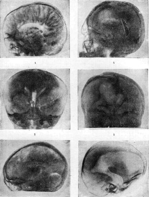
Figure 1. Profile of the brain of a hemiplegic patient; both ventricles are visible, one of which is larger than the other, there is a large subarachnoid accumulation of air, and the convolutions of the brain are clearly delineated. Figure 2. Brain of a child 1 year and 10 months old with oligophrenia; the lateral ventricles of the brain are small, the third ventricle is significantly enlarged and deformed. Figure 3. Brain of a 55-year-old woman suffering from cerebral syphilis; the upper parts of the lateral ventricles with the surface of the residual fluid are visible on it, and the cerebellum is outlined extremely distinctly. Figures 4 and 5. Brain of a 21-year-old patient suffering from traumatic epilepsy that developed in him in childhood after a blow from a horse's hoof to the head (in profile and in fronto-occipital direction); the lateral ventricles are visible, one of which is larger than the other and communicates with a cyst above which a bone defect is visible. Fig. 6. Encephalogram of a patient with hydrocephalus of the brain; huge, sharply deformed lateral ventricles are visible: the anterior, posterior, and inferior horns are clearly visible (the encephalograms are borrowed from the works of Kozhevnikov and Frenkel). 60S rays than cerebrospinal fluid, all parts of the brain are clearly visible on the images (see separate table), in which the fluid is replaced by air, such as the ventricles, mainly the lateral ones, which on the fronto-occipital image are projected in the form of a figure somewhat resembling a butterfly; downward from them along the midline, the III ventricle is visible in the form of a slit or narrow oval. On the lateral image, when the ventricles are uniform, they are projected one onto the other and have the shape of an arc running parallel to the cranial vault, from the rear end of which the inferior horn goes forward and slightly downward. Due to the fact that the fluid in the subarachnoid spaces is also replaced by air, the convolutions of the brain, as well as the cisterns of the brain, are visible more or less distinctly. This is how matters stand in the normal state; in pathological processes, depending on their nature, the picture changes sharply. In some cases, air does not pass into either the ventricles or the subarachnoid spaces, and then, naturally, one has to resort to ventriculography; in other cases, air penetrates only subarachnoidally, or only into the ventricles, or even into only one ventricle. Deformation of the ventricles and their displacement are frequently noted. With successful exposures on the lateral image, the III and IV ventricles are visible, and in rare cases the cerebral aqueduct as well. In atrophic processes of the cerebral cortex, the convolutions stand out extremely clearly, and limited accumulations of a large amount of air above the atrophied cortex are sometimes observed. Brain cysts can sometimes be detected. In Foerster's expression, encephalography is a intravital patho-anatomical study that makes it possible not only to diagnose brain tumors, but also to localize them precisely, as well as to clarify the anatomical basis of certain nervous and mental diseases, especially in relation to children, and thereby to make a correct prognosis. For diagnostic purposes, not only gases were used, but attempts were also made to introduce other contrast agents. Horoshko, for example, introduced ascending lipiodol using lumbar puncture (see Lipiodol and Myelography); being of a lower specific gravity than cerebrospinal fluid, it rises upward and penetrates into the ventricles and subarachnoid spaces. This method naturally did not become widespread because, on the one hand, it can lead to erroneous conclusions, and on the other hand, lipiodol is far from an indifferent substance, is absorbed extremely slowly, and can lead to inflammatory phenomena due to its prolonged stay in the cerebral ventricles and subarachnoid spaces. - For the X-ray imaging of the middle cerebral artery system, the Portuguese physician Egas Moniz proposed injecting from 4 cm3 to 6 cm3 of a 25% sodium iodide solution directly into the carotid artery. The patient is placed on the table for the image, an infusion is administered, and the image is taken immediately. The solution must be fresh, chemically pure, and absolutely sterile. By this method, called by Moniz radiodiaphoric or cerebral arteriography, it is possible to obtain fairly distinct images of the middle cerebral artery. This method sometimes makes it possible to localize tumors of the frontal lobes, in which the arterial system is displaced downward, and of the temporal lobes, in which displacement occurs upward. Moniz considers this method absolutely safe if all necessary precautions are observed, but nevertheless, fatalities have already been described in the literature. In addition to the above methods for determining the state of the ventricles, a method of direct examination of them was proposed, called encephaloscopy, or endoscopy, or ventriculoscopy by Volkmann: through a sufficiently large trepanation opening made, as in puncture of the corpus callosum, or in the frontal or occipital bone, or through the upper wall of the orbit (Hildebrandt), an encephaloscope (see figure) 22 cm long and the thickness of a pencil is introduced into the lateral ventricle of the brain through the brain substance. The instrument forms an angle of 150° with the shaft, and the rest of the device is the same as in cystoscopes (see Cystoscopy, cystoscope). To examine the ventricles with this instrument, the fluid in the ventricles must first be replaced with gas. This method could not catch on because, on the one hand, only a very limited part of the ventricles can be examined with its help, and on the other hand, very significant trauma is inflicted on the brain in the process. Ultimately, only encephalography and ventriculography have fully and deservedly won widespread use. Encephalography has made it possible to uncover previously completely unknown changes in the brain in a whole series of diseases (epilepsy, post-encephalitic parkinsonism, so-called traumatic neurosis, etc.).
Related articles
Mentioned in
Cite this page
“Encephalography.” Soviet Medical Encyclopedia. English translation of Bolshaya Meditsinskaya Entsiklopediya, 1st ed. (Moscow, 1928–1936), ed. N. A. Semashko. https://sovietmedicalencyclopedia.pages.dev/article/encephalography/