Vesiculitis
Historical document, translated for reference. It reflects medical knowledge of the 1920s–30s and is not medical advice.
Summary
Vesiculitis is inflammation of the seminal vesicles, first described by Morgagni in 1795. The article covers its causes, including gonococcal and tuberculosis infections, clinical manifestations, diagnosis, and treatment approaches from the 1930s Soviet medical perspective.
Encyclopedia article (1928–1936)
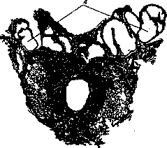
VESICULITIS (vesiculitis), inflammation of the seminal vesicles, was first described in 1795 by Morgagni. Due to the anatomically close, intimate contact (blood, lymphatic, nervous) with neighboring organs, the seminal vesicles reflect the condition of these organs. Figure 1. Normal seminal vesicles (A) of a 41-year-old man (natural size). (see Figure 1). Inflammation of neighboring organs can lead to vesiculitis by directly involving the seminal vesicles in the process. Hematogenous and lymphogenous routes of infection are also not uncommon. Among predisposing factors, frequent and prolonged congestion of pelvic organs as a result of masturbation and abnormal sexual intercourse, retention of secretion in the vesicles when the seminal duct is compressed, and diseases of the large intestine should be noted. These factors themselves can be the cause of catarrhal inflammation of the seminal vesicles. In bacterial vesiculitis, gonococcal and tuberculosis infections are most common; less frequently, vesiculitis can be caused by staphylococcus, streptococcus, E. coli, etc. The frequency of involvement of the seminal vesicles in the gonorrheal process ranges from 4% (Peterson) to 50% and even 80% (Joung, Marton) of all prolonged cases of urethral gonorrhea. Tuberculosis of the seminal vesicles, as a primary disease, is rare, but often occurs (in up to half of all cases, Teutschlander) in combination with other manifestations of genitourinary tuberculosis. Morphologically, single or multiple nodules are found, which merge into foci with caseous decay. Patho-anatomically, in the course of acute gonorrheal V., one can distinguish forms 1) superficial, 2) deep, and 3) empyema. In the first form, the mucous membrane is infiltrated in its superficial layers, the epithelial covering is desquamated, thickened in places, the vessels are markedly dilated, and the submucosal tissue is almost unchanged. Subsequently, the submucosal and muscle layers of the walls appear infiltrated, in places penetrated by connective tissue strands. Sometimes one can trace how the connective tissue strands pass into the surrounding cellular tissue (second form). The third form is characterized by the formation of small abscesses in individual convolutions, diverticula of the seminal vesicles, which often merge into one common purulent sac-empyema. The last two forms can involve the surrounding cellular tissue in the inflammatory process, and then we are dealing with paravesiculitis. A prolonged gonorrheal process in the seminal vesicles is patho-anatomically characterized by diffuse infiltration of the walls, their subsequent thickening or sclerosis, which in the final stage turns into atrophic cirrhosis. In other cases, the seminal vesicles are cystically expanded (see Figure 2)

Figure 2. Cystically expanded lumens (A) of the seminal vesicles of a 59-year-old man (natural size).
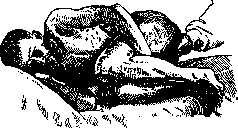
and filled with serous content, pus, decay, or granulations. The seminal vesicles are examined through the rectum in the standing position, making the patient sit on the doctor's index finger, or in the patient's lateral position (see Figure 3) with the lower limbs drawn to the abdomen (Fronshtein). According to clinical course, V. can be divided into acute and chronic. The younger the age of the subject, the more acute the course of the process. Inflammation of the seminal vesicles of sac-like shape proceeds most favorably and subjectively bothers the patient little. In the presence of a large number of diverticulum-like expansions, long additional passages, high and dense villiform-like epithelial growths, the process is clinically manifested by disturbances in the function of the sexual apparatus. Heaviness and pain in the rectal area, intensifying during urination and defecation to tenesmus, increased frequency of pollutions, sometimes with purulent and bloody content. There is general weakness, temperature rise to 39° and above. In these cases, the diagnosis does not present
Figure 3. Position of the patient for examining the seminal vesicles by the Fronshtein method. particular difficulties. Anamnesis, enlargement, doughy tension of the seminal vesicle, pain on pressure on it are the most characteristic objective signs of the acute process. The secret obtained by massage is characterized by the presence of a large number of leukocytes, amorphous decay, epithelium, blood or hematoidin crystals, as well as the presence of deformed immotile spermatozoa. The acute process usually proceeds violently for 5-7 days and resolves either by reverse development or transition to a chronic form or formation of empyema. In the latter cases, the abscess may rupture into the urethra, which is the most favorable outcome, less often into the surrounding cellular tissue, then into the ampulla of the large intestine, bladder, abdominal cavity. V. can from the very beginning proceed sluggishly, chronically; in cases of involvement of a small area of the mucosa or after the acute phenomena have subsided, the process can pass into a latent state and exist in this way for many years. In the chronic stage, symptoms of a neuro-sexual nature come to the forefront: frequent pollutions, painful erections, increased sexual desire, weak orgasm, sluggish and rapid ejaculation of semen, sometimes with an admixture of blood. Three forms of chronic gonorrheal vesiculitis should be distinguished - Catarrhal inflammation: on palpation, nothing pathological, except for slight tenderness on pressure, can be detected; in the secretion of the seminal vesicle, leukocytes, an increased number of epithelial cells and spermatozoa. - The fibrous form is caused by sclerotic changes in the entire thickness of the mucosa and the formation of connective tissue strands in the surrounding cellular tissue. On palpation: a dense elongated seminal vesicle, painful on pressure; in the secretion obtained by massage, a significant amount of pus cells and epithelium. Spermatozoa may be absent, as pathological products of secretion subject them to cytolysis. - In empyema, the most common form, with virus in the secretion, the seminal vesicle S95 is palpated either as a single, cherry-sized formation or as a collection of bulges in the shape of a bunch of grapes. - In suspected chronic gonorrheal V., in each individual case, it is necessary to differentiate a number of other diseases, which can be accompanied by similar symptoms and on palpation simulate the seminal vesicle. Knowledge of the topographic location of the seminal vesicles, taking into account the condition of the prostate gland, their deeper position, clarifies the true nature of the diseases.
Treatment of gonorrheal V. should be directed first of all against the primary focus of infection (see Gonorrhea). Treatment of the acute stage of the disease should focus on increasing the body's protective strength through immunotherapy. In the treatment of chronic forms, in addition to general treatment, direct effect on the seminal vesicles is also necessary, which consists in systematic massage, warming them by diathermy or hot microclysters, which usually gives good results. In case of failure, in catarrhal forms, surgical treatment is indicated - vasotomy, vasopuncture (see Vasostomy, vasotomy), for fibrous V. and empyemas, vesiculotomy and vesiculectomy are indicated. Therapy for tuberculous V. consists in improving the general condition of the body, and in advanced cases - in surgical excision of the seminal vesicles. Satisfactory results are obtained from the quartz lamp.
Related articles
Mentioned in
Cite this page
“Vesiculitis.” Soviet Medical Encyclopedia. English translation of Bolshaya Meditsinskaya Entsiklopediya, 1st ed. (Moscow, 1928–1936), ed. N. A. Semashko. https://sovietmedicalencyclopedia.pages.dev/article/vesiculitis/