Promorphology
Historical document, translated for reference. It reflects medical knowledge of the 1920s–30s and is not medical advice.
Summary
This historical article from the 1928-1936 Soviet Medical Encyclopedia explores promorphology, the study of the structure of germ cells (primarily eggs) and its significance for embryonic development, tracing the evolution of scientific thought from preformation and epigenesis to modern concepts of egg polarity, symmetry, and cytoplasmic localization.
Encyclopedia article (1928–1936)
PROMORPHOLOGY, the doctrine of the structure of sex cells, mainly the egg, and of the significance of this structure for development. The end of the 17th century is characterized by the emergence of biological theories that attempted, to a significant extent speculatively, to find in the egg and embryo at early stages of its development parts corresponding to the features of the formed organism. These theories, which are grouped under the name of preformation, or evolution, were contrasted with the theories of epigenesis, or the doctrine of new formation, Fig. 1. Egg of the hemipteran Metapodius femoratus: 1—from the ventral side; 2—from the right; A and P—anterior and posterior poles; D and V—dorsal and ventral sides. The naive view of early preformationists, based on metaphysical notions of organismal development as the unfolding (evolution, literally, unfolding) of pre-existing parts of the organism within the sex cell, was replaced by the doctrine of the absolute homogeneity of the egg, of the absence of any structure whatsoever within it. Everything that arises in the egg during development is formed entirely anew, by unknown pathways. The modern view of the structure of the egg and its development is reconcilable neither with the view of the preformationists nor with the doctrine of the early epigenetists. Direct observation of the course of development and the study of the egg's structure lead to the conviction that the egg, prior to the onset of cleavage and sometimes prior to fertilization, possesses a definite structure, and this latter is by no means indifferent to the direction of the embryo's development. In a number of cases, the phenomena of polarity, bilateral symmetry, and sexual dimorphism can be ascertained in the egg (Fig. 1). The arrangement of cytoplasmic inclusions determines the unequal specific gravity of various parts of the egg, which is of essential importance for the eggs of aquatic animals, influencing the position of the egg floating in water. As early as Baer (1834), von Baer distinguished two hemispheres in the frog's egg, differing in the arrangement of pigment, and gave them the names of the animal and vegetative layers (or poles), of which the former gives rise to the epidermis, nervous system, and sense organs, and the latter to the intestine and its appendages. Subsequently, these poles were also called formative or protoplasmic and nutritive or deutoplasmic. In telolecithal eggs of a number of vertebrates, the heavier yolk usually accumulates on the
vegetative pole, and such eggs float with the animal pole uppermost (cartilaginous fishes, amphibians). Yolk arrangement of the same kind is also observed in the eggs of some terrestrial vertebrates (reptiles and birds). Sometimes the vegetative pole of the egg is provided with drops of fat, making it lighter, and such eggs, for example in annelids and some fishes, float with the vegetative pole uppermost. Frequently, however, substances having different specific gravities are distributed more or less evenly in the egg, and then the polarity of the egg can be judged not by its position in the water, but by the localization of the micropyle (an opening in the membrane for the entry of the spermatozoon). Van Beneden already pointed out that in
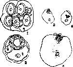
bilaterally symmetrical animals, the eggs also possess bilateral symmetry. Excellent examples of this are the eggs of insects and cephalopod mollusks. Driesch and Boveri attempted to explain the polarity and bilateral symmetry of the egg by the hypothesis of the "polar-bilateral orientation" of the minute protoplasmic particles forming the "finest structure" of the egg. This hypothesis is fundamentally incorrect, since it seeks to mechanistically reduce the structural features of the egg to the spatial arrangement of protoplasm particles, coinciding in this sense with Nägeli's idea on the micellar structure of living matter. Sexual dimorphism in uncleaved and even unfertilized eggs has been noted repeatedly. It can pertain to both the features of nuclear morphology and the cytoplasmic characteristics of the egg. Sexual dimorphism in the structure of eggs is observed particularly vividly in rotifers, aphids, and the worm Dinophilus. In the latter (according to Korschelt) (Fig. 2), eggs producing females are incomparably larger in size than eggs producing males. A similar phenomenon of sexual dimegaly was described by Reuter in mites and Montgomery in spiders. Riddle showed that in pigeons, eggs producing males are smaller, contain more water and less fat and phosphorus than eggs producing females. Depending on the arrangement of cytoplasmic inclusions, which are important for the further development of the egg, Pflüger (1883) suggested speaking of anisotropic and isotropic eggs. The former, on the initiative of Heider (1900), received the name of mosaic eggs, because experiments separating blastomeres at early stages of development led to the formation of defective larvae, while the latter were called regulative eggs due to the fact that isolated blastomeres gave rise to full-fledged embryos differing only in correspondingly smaller sizes. Düsberg Figure 2. Sexual dimegaly of eggs in Dinophilus apatris: 1—egg capsule containing large (female-producing) and small (male-producing) eggs; 2—cross section of a female, showing two kinds of eggs in the ovary; 3 and 4—small eggs producing a male; two stages of fertilization; 5—analogous stage of a large egg producing a female. (After Korschelt and Nachtsheim.) (Düsberg, 1926) classifies the eggs of ascidians, ctenophores, mollusks, the polychaete worm Lanice, the roundworm Ascaris, and insect eggs as anisotropic, and the eggs of jellyfish, echinoderms, the nemertean Cerebratulus, the earthworm, amphioxus, and fishes as isotropic eggs. The results of studying anisotropic eggs gave rise to the so-called "mosaic theory," whose originator is considered to be W. His (1874), who proceeded from the proposition that every point in the embryonic region of the blastoderm (of the chicken) must subsequently be an organ or part of an organ, and that every organ developing from the blastoderm has its preformed primordium. His gave this viewpoint the name of the "principle of organ-forming regions of the embryo." Later, Ray Lankester (1887) and Rabl (1879) joined His's views, with Rabl showing that in the mollusk Planorbis, the internal mesoblast originates from 12 cells arranged in three quartets. A number of facts obtained on various objects led to the conviction that the pattern of cleavage must be considered as mosaic work ("Mosaik-arbeit"). The most distinct distribution of various substances in the uncleaved egg has been described for ascidians. In Styela partita, Conklin distinguishes special regions of the egg that subsequently give rise to particular organ systems. Thus, the fine-grained plasma of the animal hemisphere of the egg and a region of light plasma at its equator form the ectoderm of the embryo, the yellow plasma located in the form of a crescent in the vegetative hemisphere gives rise to the mesoderm, the coarse-grained plasma of the vegetative hemisphere forms the entoderm, and finally the white plasma, which becomes noticeable on the anterior part of the egg at the very first cleavage, gives rise to the notochord and nervous system. In ctenophores (type of coelenterates), the promorphology of the egg was studied by Yatsu. Parts of the cut egg of Beroë yielded a normal larva, regardless of the plane of the cut, if the cut
was performed prior to the discharge of reduction bodies. If, however, the operation took place after maturation, the cleavage of the fragments proceeded normally, but the larvae showed defective development. Yatsu comes to the conclusion that the specialization of egg parts sets in after the formation of reduction bodies. At the same time, the form of cleavage does not depend on the distribution of cytoplasmic inclusions. Mollusks provide rich material on the promorphology of the egg. In the gastropods Dentalium and Ilyanassa, in the unfertilized egg, the yolk is located in the middle part of the egg, while on the upper and especially the lower pole there are regions of light plasma. During cleavage, the vegetative pole departs into one of the first two blastomeres, and then the animal blastoblast divides earlier than the vegetative one, forming the so-called trefoil stage (Fig. 3). The significance of this distribution of ooplasmic material
MORPHOLOGY
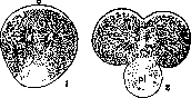
clarified in the experiments of Crampton and Wilson (see below). Hallez (1886) drew attention to the promorphology of insect eggs. He established the remarkable fact that the eggs are arranged in the oviduct in such a way that the head end of the future embryos is directed toward the head end of the mother. Hegner (1909) noted that in the egg of the beetle Leptinotarsa decemlineata, by the time of maturation, certain areas of the peripheral protoplasm have different prospective significance. Kasper described in the mosquito Chironomus plumosus at the vegetative pole of the egg, while still in the ovary, an accumulation of pigment grains; later, germ cells are formed from this area of the egg. Similar phenomena were established by Buchner (1910) in the worm Sagitta, Haecker (1847) in the crustacean Cyclops, and Kühn (1911) in the crustacean Polyphemus. In a number of cases, this pigment spot, which is related to the formation of the germ cells of the future embryo, is formed due to the penetration into the egg during its maturation of one or several cells of the germinal epithelium. The place of penetration of these cells into the egg indicates the localization of materials for the future germinal primordium. It is possible that the remainder of these cells takes part in the formation of the germ cells of the embryo. Dusberg classifies, for example, the eggs of echinoderms, particularly the sea urchin, in the category of isotropic eggs. Boveri (1901) found that in the mature egg of Strongylocentrotus the orange pigment is located in the vegetative hemisphere, forming a belt slightly below the equator. The first two cleavage furrows run meridionally, dividing the egg into parts with quite evenly distributed cytoplasmic inclusions. The third furrow is located in the plane of the equator, so that the quartet of animal blastomeres receives much less pigment than the quartet of vegetative ones. However, all 8 blastomeres contain both pigmented and uncolored plasma. A similar phenomenon of the uniform distribution of ooplasmic material up to the 3rd cleavage was discovered in the nemertean Cerebratulus, in the lancelet Amphioxus lanceolatus, and in fishes (lamprey according to Bataillon's data and Fundulus according to the studies of Morgan et al.). The isotropy of the egg manifests itself even more clearly in hydromedusae. The pigmented granular plasma in Clytia is located in the central part of the egg, while the peripheral zone is occupied by transparent plasma. The first 4 cleavages proceed in such a way that the egg is divided radially and thereby each blastosome is supplied with a section of pigmented and colorless plasma. Only the 5th cleavage leads to the formation of 16 internal blastomeres containing granularity and pigment, and 16 external ones filled with non-pigmented and more homogenous plasma.
The significance of the promorphology of the egg for its further development was established both by direct observation of certain structural elements by applying color marks (in vivo marking) to the studied parts of the egg or embryo, and by active experimental intervention in the process of development. In the latter case, cutting the yet uncleaved egg into parts or isolating individual blastomeres was most often used. From experiments of the latter kind, it is necessary to note the removal of the vegetative polar lobe at the "trefoil" stage (see above) in the eggs of Ilyanassa and Dentalium. An egg that has lost its polar lobe gives a deformed larva devoid of a part of the organs. Similarly, underdeveloped larvae were obtained from isolated blastomeres in ctenophores, nemerteans, etc. Depending on the behavior of isolated blastomeres, it became customary to distinguish so-called "mosaic" and "regulative" eggs. "Mosaic" eggs are called eggs with an early distribution of various formative substances between individual blastomeres, as a result of which isolated blastomeres develop into a part of the embryo. "Regulative" eggs, on the contrary, retain isotropy for a long time, which is why an entire embryo of a correspondingly smaller size can develop from isolated blastomeres. Wilson tries
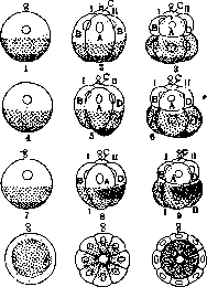
Figure 4. Diagram of the primary stratification of the eggs of a sea urchin (1-3), an annelid or gastropod mollusk (4-6), an ascidian (7-9), and a hydromedusa (10-12). White zone—ectoblast; dotted zone—endoblast; hatched zone—mesoblast; I and II—the first two cleavage planes. In the sea urchin, all zones are distributed evenly among the blastomeres. In worms and mollusks, the lower zone falls only into quadrant D. In the ascidian, the mesoblastic zone falls into quadrants A and B. In the medusa, horizontal stratification is absent. attempts to eliminate the contradiction in the cited classification of eggs of different animals by pointing to the absence of fundamental differences between the "mosaic" and "regulative" types of development. He suggests that in "regulative" eggs, the first cleavages are quantitative, distributing the ooplasmic material evenly. The cleavage leading to the uneven distribution of formative material is called qualitative, and, starting from this moment, the blastomeres lose equipotentiality. Such qualitative cleavage in the eggs of the hydromedusa is the 5th cleavage, i.e., the 32-blastomere stage, and in the sea urchin, the 3rd (8-blastomere stage). The arrangement of ooplasmic material in the ascidian, annelids, and gastropods is such that already the 2nd and even the 1st cleavage (4-2 blastomere stage) is qualitative (Fig. 4). Wilson's view reflects the desire to free oneself from the chains of metaphysics, but the path chosen by him suffers from eclecticism. Indeed, the preformationist point of view of the proponents of the mosaic theory is characterized by the metaphysical notion of the unfolding of pre-existing diversity or, at best, the notion of the "transition of invisible diversity into visible" (Roux). This concept of development, understood "as decrease and increase, as repetition," is-"dead, poor, dry" (Lenin). The epigenetic point of view bears a no less metaphysical character. Proponents of the theory of new formation place the driving force, the motive of development, outside the material system of the organism. K. F. Wolff, one of the founders of the theory of epigenetics in biology, took a vitalistic position, attributing the source of new formation during the development of the embryo to an entelechial "forming aspiration" (vis essentialis). Not a single consistently developed epigenetic theory is conceivable without a vitalistic superstructure. Of course, the attempt to reconcile the epigenetic and preformationist points of view with compromise formulations—such as the recognition that the egg already contains preformed parts of the embryo, but not all and not fully developed, while further development by new formation creates the missing parts and completes development—will not lead to any fruitful results. There is no doubt that the egg is neither a homogeneous drop nor a miniature model of a formed organism. The egg is a stage of development of the organism; it possesses a definite species-specific structure and a definite set of functions. The structural and functional features of the egg, being mutually conditioned, at the same time reflect the historical character of the organism, which represents the result of the development of a vast number of preceding phylogenetic forms. Thus, the egg is not a simple mixture of chemical substances, but a complex material system characterized by the main sign of the living—metabolism. At the same time, the egg is immeasurably simpler than the organism that will develop from it. The development of an organism from an egg is a process of new formation, a process of complication, the emergence of diversities not existing in the undeveloped egg. A consistently materialistic point of view on development has as its task to uncover the nature of the emergence of the new, the emergence of diversity.
"The condition for the knowledge of all processes of the world in their 'self-movement,' in their spontaneous development, in their living life, is the knowledge of them as a unity of opposites. Development is the struggle of opposites" (Lenin). The task of Marxist-Leninist biology is to uncover at each stage of the organism's development that contradictory relation which is leading, determining, that relation which, arising as a result of the "splitting of the single," leads to the struggle of opposites and to the spasmodic "destruction of the old and emergence of the new." To solve this problem, a comprehensive study of the development process itself is necessary, since each subsequent stage of development has the prerequisites of its emergence in the previous stage; the subsequent stage arises only when all conditions for the transition to the next stage have matured in the previous stage (see Developmental mechanics, Organism, Preformation, Epigenesis).
Related articles
Mentioned in
Cite this page
“Promorphology.” Soviet Medical Encyclopedia. English translation of Bolshaya Meditsinskaya Entsiklopediya, 1st ed. (Moscow, 1928–1936), ed. N. A. Semashko. https://sovietmedicalencyclopedia.pages.dev/article/promorphology/