Neurotomy
Historical document, translated for reference. It reflects medical knowledge of the 1920s–30s and is not medical advice.
Summary
Neurotomy is the complete transverse section of a nerve trunk, initially used for treating trigeminal neuralgia and later expanded for various conditions including severe spastic paralysis and certain ulcers. The article details the surgical technique, indications, and specific applications for different body regions.
Encyclopedia article (1928–1936)
NEUROTOMY, neurotomia (from Greek neuron - nerve and tome - section), complete transverse section of a nerve trunk. Neurotomy is the most radical method of interrupting nerve conductivity. Initially, neurotomy was proposed by Schlichting (1748) as a method for treating trigeminal neuralgia. Somewhat later, Quenu introduced neurotomy for relieving pain in spontaneous gangrene. Neurotomy gained wider application since Stoffel (1911). The latter proposed, to reduce or even completely eliminate severe spastic paralysis of the limbs (upper and lower), the section of motor nerve branches going to the affected muscles. In the last 8-10 years, indications for N. have been significantly expanded. This has been and is being facilitated by the desire to use N. as a method of treatment (Moltokov) for certain diseases, the connection of which with the nervous system has either been disputed (chronic non-healing ulcers of the leg, spontaneous gangrene, etc.) or was not even suspected (tuberculous ulcers of the tongue, etc.). However, in practical work, it is premature to extend indications for N. so widely. The considerable clinical material already available on this issue shows that along with brilliant therapeutic results after N., very severe complications are also observed in the form of persistent paresthesia, edema, pains, trophic disorders, etc. Moreover, in some cases, it creates the impression that N. somehow activates the pathological process for which they were performed. Therefore, the scope of their application is currently more advisable to limit to those cases where other methods of treatment, both therapeutic and surgical, prove powerless. Thus, direct indications for N. at the present time should be considered persistent disorders in the area of sensory, motor, vasomotor, secretory and "trophic" functions of the nervous system that do not respond to other forms of therapy, and the described operation is permissible in most cases only in the area of those spinal nerves whose motor function does not have essential significance (for example, n. peronaeus prof. in the lower third of the leg, intercostal nerves, etc.). Of course, cases with impairment of the motor sphere are excluded from this, where surgical intervention is undertaken precisely on the nerve fibers. Technique of N. The operation N. consists of two moments: exposure of the nerve and its section. The first moment is carried out according to general surgical rules. The location of the incision, its direction and length are determined by the data of topographic anatomy and the rules of operative surgery. The external appearance of the nerve usually allows it to be distinguished without much difficulty from the surrounding tissues. More critical is the second moment - isolation of the nerve from the surrounding tissues and its section, requiring certain special techniques. Isolation of the nerve should be performed on the smallest possible extent. Usually 1/3-2 cm is quite sufficient to perform neurotomy. Anatomical preparation of the nerve trunk for the purpose of exposing it over a large extent should be considered absolutely contraindicated. This technique leads to excessive trauma to the nerve trunk, disruption of its nutrition and formation of extensive scars with all the resulting consequences (paresthesia, edema, neuromas, etc.). When isolating the nerve from the surrounding cellular tissue, it is necessary to avoid grasping it with forceps: for fixation, it is better to use a small blunt hook (metal or glass), on which the nerve is lifted from its bed. After isolation is complete, a gauze tampon is placed under the nerve, after which its section is undertaken. The best instrument for this purpose is a razor or a razor-sharp scalpel. Scissors should not be used, as they crush the nerve fibers. The section should be performed in one pass as much as possible, in order to obtain a smooth, flat surface of the nerve's cross-section. One of the main moments of this part of the operation is the careful stopping of bleeding from the severed nerve, since hematomas forming on this basis create a significant obstacle for the growth of axial cylinders. The usual hemostasis technique - applying a forceps with subsequent ligation of the vessel - is not applicable here due to the possible compression of individual nerve fibers, which may subsequently cause a series of painful symptoms. The most advisable method is light tamponage of the bleeding ends of the nerve with a gauze ball for 5-10 minutes. After completion of wound toilet, the ends of the severed nerve are brought almost into contact with each other by two silk sutures applied on the epineurium (see Nervous suture) [it is recommended to leave a gap of 2-3 mm between the ends, which creates the most favorable conditions for subsequent regeneration (Nageotte)]. Where the caliber of the nerve is very small (n. interosseus dorsalis), or the divergence of its ends does not exceed 3-4 mm, the application of sutures is unnecessary. In these cases, the proximal and distal segments of the nerve are carefully laid end-to-end, and the connective tissue sheath in which the nerve was located is closed with 1-2 sutures. The operation ends with the layer-by-layer closure of the tissues tightly. Almost all N. are performed under local anesthesia (1% solution of novocaine). Only in rare cases (for example - operations on Gasser's ganglion) is anesthesia given. Special cases of neurotomy for various diseases (by regions). Head. Most often, neurotomy is performed for trigeminal neuralgia. At the beginning of the disease, usually the affected branches are sectioned in the peripheral parts, namely - at the site of their emergence on the face (see Trigeminus nerve).-Neuralgia n. glossopharyngei. Excellent results according to American and Russian authors are obtained by section of the glossopharyngeal nerve. It is better to refrain from the intracranial approach to this nerve proposed by Dandy, as this route is technically quite difficult. The same results are obtained by sectioning the nerve in the neck, which is much simpler and safer. Approach - an incision 8-10 cm from the tip of the mastoid process toward the middle of the hyoid bone, one transverse finger posteriorly and below the angle of the lower jaw. The parotid gland is pulled upward, m. sternocleidomastoideus and m. stylohyoideus are displaced laterally, deep under the lower edge of m. stylopharyngei, the nerve is found on the external carotid artery. N. of the glossopharyngeal nerve also gives positive results in terms of eliminating pain and in inoperable cancers of the tongue and tuberculous ulcers of it. In the latter cases, healing of the ulcers is even noted. - Neck. Neuralgia cervico-occipitalis. A good therapeutic result is obtained by section of n. occipitalis major. Approach - a transverse incision 6-7 cm long posteriorly, at the level of the spinous process of the 2nd cervical vertebra, starting from the midline. The m. cucullaris, m. splenius capitis, m. semispinalis are sectioned layer by layer. Deep, on m. obliquus capitis int., the occipital nerve is located. - Pain in tuberculous ulcers in the area of the larynx; a positive result is obtained by section of the superior laryngeal nerve (n. laryngeus sup.). Approach - an incision 5-6 cm along the anterior edge of the sternocleidomastoid muscle, starting from the angle of the lower jaw and downward. The muscle is pulled outward, the vascular bundle (carotid artery and jugular vein) - inward. Along the posterior surface of the latter runs n. vagus, and medially, in the upper angle of the wound, n. laryngeus is located (Figure 1).-Hyperhidrosis of the face. In many cases, it is possible to achieve cessation of sweating by section of n. auricularis magni. The latter is exposed along the posterior edge of the sternocleidomastoid muscle over its middle third. - Phrenicotomy - section of n. phrenici. Chest. Neuralgia intercostalis (mainly localized in the V-IX intercostal nerves and sometimes accompanied by a rash in the form of herpes zoster). In severe cases, section of the affected nerves is indicated. Approach - parallel to the lower edge of the corresponding rib, 0.5 cm below it, an incision (6-7 cm) is made, starting four fingers to the side of the line of spinous processes. Layer by layer, the external intercostal muscle is reached, which is sectioned at the site of its attachment to the rib. When it is pulled downward, the nerve is exposed. In addition to the neuralgias mentioned, section of intercostal nerves (in combination with avulsion of the central and peripheral ends) is sometimes used in tabetic crises (Leriche and others).-Upper extremities.
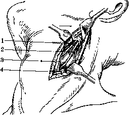
Figure 1. Surgical approach to n. laryngeus sup.: 1 - site of section of the laryngeal nerve; 2 - n. vagus; 3 - a. carotis int.; 4 - m. sternocleidomastoideus.
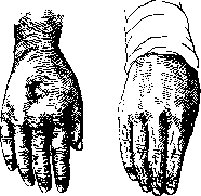
Figure 2.
Figure 3.
Figure 2. Hard edema of the back of the hand before neurotomy. Pit from finger pressure. Figure 3. The same after neurotomy. Hard edema of the back of the hand, usually accompanied by reflex contracture of the finger extensors. Excellent results are obtained from the incision of the n. interossei dors. (ram. n. radialis) (figures 2 and 3). Approach - incision on the dorsal side of the forearm in its middle third along the line connecting the head of the radius with its styloid process. The nerve lies on the membrana interossea between m. extensor pollicis longus and brevis. A positive result is obtained from the severing of this same nerve in non-healing ulcers on the back of the hand. - Disease Raynaud, x-ray examination, eczema. Experience in their treatment by N. so far does not give definite results. Usually they sever in the lower third of the forearm n. medianus - with localization of the process on the palmar side of the distal phalanges of the 1st-3rd fingers, n. ulnaris (ram. superfic.) - with lesions of the terminal phalanges of the 4th-5th fingers, ram. superficialis n. radialis - with the presence of the process on the back of the 1st-3rd fingers. In dry forms of eczema on the palmar surface of the forearm, the incision of n. cut. antibrachii med. in the lower third of the arm gives a positive result in a number of cases.
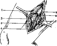
Figure 4. Surgical approach to n. saphenus: 1 - lamina vasto-adductoria; 2 - m. sartorius; 3 - n. saphenus; 4 - ramus saphenus a. femoralis supremae; 5 - m. gracilis; 6 - n. obturatorius; 7 - anastomosis.
s n. saphenus. Approach - incision (8-10 cm) is made along the posterior edge of the sartorius muscle (m. sartorius) 4 fingers above the medial condyle of the femur. The muscle is pulled forward and under it in the fatty tissue the n. saphenus is exposed (figure 4). With another, rarer localization of ulcers, namely - on the same side, but in the upper third of the leg, a good effect is obtained from the incision of n. obturatorius (see technique of exposure below). With the position of ulcers on the posterior and lateral surface of the leg, one can attempt to sever n. cut. surae med. et lat., but in most cases these N. are little effective. Statistics of results after N. for ulcers give 70-72% of immediate success. Stable results (from 1 to 3 years) are observed in 20-25% (fig. 5 and 6). Within the nearest half year, 18-20% recur.
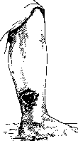
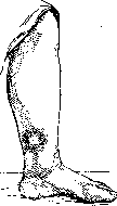
Figure 5.
Figure 6.
Figure 5. Non-healing ulcer of the leg (duration 1 year) before neurotomy. Figure 6. The same after neurotomy. Smooth scar at the site of the former ulcer. Duration of observation 4 years.
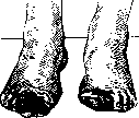
Spontaneous gangrene. The highest percentage of success 1& is obtained from the severing of n. peronaei profundi. This should be considered equally correct for both initial forms and forms with developed gangrenous foci. In this case, the location of the foci (on the foot or toes) does not play a special role. The nerve is exposed by an incision (6-7 cm long) in the middle third of the leg, a finger's breadth outward from the crista tibiae. By separating m. extensor hallucis longus and m. tibialis anterior, the nerve is found in depth between them on the interosseous septum. If the therapeutic effect of the indicated N. proves insufficient, then to it is added the severing of n. cut. surae medialis in the popliteal fossa with localization of gangrene on the little toe, or the severing of the corresponding branch of n. peronaei superficialis in the presence of gangrenous ulcers on other toes of the foot. It is better to refrain from severing such large nerve trunks as, for example, n. ischiadicus and even n. tibialis and n. peronaeus comm. (in the popliteal fossa). The therapist, the value of such N. is more than problematic, and the motor disorders caused by them unfavorably affect the function of the limb. Immediate success after neurotomy for spontaneous gangrene
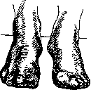
gangrene of both legs before neurotomy.
Figure 8. The same after neurotomy of n. peronaei profundi. At the site of ulcers smooth scars. Duration of observation 2V2 years. Within the nearest half year recurrences are noted in 10-15%. Duration of therapeutic effect in individual cases has been traced up to four years (figures 7 and 8). In addition to spontaneous gangrene, the incision of n. peronaei profundi gives good results in hard edema of the back of the foot, in Kehler's disease, as well as in Morton's disease.
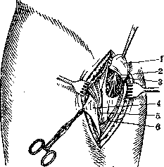
Disorders of the motor sphere. Little's disease, spastic paralyses in injuries of the spinal cord and hemiplegias. A relatively good functional result is obtained from the severing of n. obturatorii. Approach - vertical incision (10-12 cm long), beginning at the border of the middle and inner thirds of the Poupart ligament. The interval between m. pectineus and m. adductor longus is exposed layer by layer. When m. pectinei is pulled outward in the depth of the wound, all branches of n. obturatorii become visible under the fascia (fig. 9). - There are still some forms of diseases which are treated by neurotomy. First of all, the so-called reflex epilepsies should be mentioned here, individual cases of treatment of which by N. gave positive results. As for the rather large group of malignant neoplasms, the attempt to treat them by N. must so far be recognized as unsuccessful. N. for neuromas - see Neuroma.
Figure 9. Surgical approach to n. obturatorius: 1 - fascia m. obturatoris ext.; 2 - n. obturatorius; 3 - a. obturatoria; 4 - fascia pectinea; 5 - m. pectineus; 6 - m. adductor longus.
Related articles
Mentioned in
Cite this page
“Neurotomy.” Soviet Medical Encyclopedia. English translation of Bolshaya Meditsinskaya Entsiklopediya, 1st ed. (Moscow, 1928–1936), ed. N. A. Semashko. https://sovietmedicalencyclopedia.pages.dev/article/neurotomy/