Hemophilia
Historical document, translated for reference. It reflects medical knowledge of the 1920s–30s and is not medical advice.
Summary
This article from the first edition of the Great Medical Encyclopedia (1928–1936) provides a historical overview of hemophilia, tracing its recognition from the Talmud to the 20th century. It details the evolution of scientific understanding regarding its hereditary nature, specifically focusing on the transition from the 'Lossen rule' to modern genetic theories of sex-linked recessive inheritance.
Encyclopedia article (1928–1936)
HEMOPHILIA (from the Greek haima—blood and philia—inclination), a term introduced at the beginning of the 19th century by Schönlein for a disease known much earlier. Hemophilia is a hereditary (according to peculiar laws) hemorrhagic diathesis (see), in which a decrease in blood coagulability represents the primary deviation from the norm. Indirect references regarding hemophilia are encountered as early as the Talmud. More definite information about it exists in the works of the 11th-century Arab physician Albucasis. The first data on the hereditary nature of this disease were reported by Fordyce in 1784. In the 19th century, the problem of hemophilia was developed very widely (works of Nasse, Virchow, Grandidier, Lossen, and others). However, only in the 20th century did most of the controversial questions of hemophilia receive their scientific resolution (works of Sahli on blood coagulability in hemophilia, and of Bauer and Schloessmann on the heredity of hemophilia). Although it had long been known that hemophilia is a hereditary, genotypic disease, the question of the nature of hemophilia inheritance was, until recently, extremely confused. Most authors held the viewpoint of the 'Lossen rule,' empirically derived by the latter in 1876 and 1905 based on the study of the famous Mampel family (see Figure 1). This rule stated: 'Only men suffer from hemophilia; they cannot transmit the disease to the next generation (provided they marry a woman from a healthy family); inheritance occurs through women, who, however, do not suffer from it themselves' ('conductors'). Soon, however, facts began to accumulate proving that if the Lossen rule is suitable as a generalization of the course of inheritance in the Mampel family, it still cannot be recognized as universal, and that in some cases, the old Nasse rule (1820), which allowed for the inheritance of hemophilia from a sick father through a healthy daughter to a grandson, should be recognized as more plausible. Only in recent years has this question been successfully resolved, thanks to the works of K. Bauer and Schloessmann.
Bauer put forward the hypothesis that hemophilia is a sex-linked recessive trait. Accepting the difference in the chromosomal apparatus of men and women in the form of a paired sex chromosome (XX) for the latter and an unpaired (XY) for the former, one obtains the following two usual combinations: [Diagram omitted]. Designations: ♂—healthy man; ♂—sick man; ♀—healthy woman; [Diagram omitted]. Such cases should be encountered extremely rarely. However, such a case is known, for example, in the Mampel family (see Figure 3). Theoretically, from the latter marriage, three sick daughters should have resulted, but in reality, all 6 turned out to be healthy. Based on the analysis of such cases, Bauer put forward a hypothesis about the lethal nature of the hemophilia gene in the ho[mozygous state].
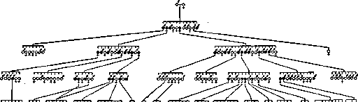
Figure 1. o - woman-heterozygote ("conductor"); xt - sex chromosome carrying the hemophilia gene. In other words, from the marriage of an affected man and a healthy woman, the entire generation will be outwardly healthy, but while all sons will be truly healthy (both themselves and their descendants), all daughters will be heterozygotes (see Homo-heterozygotes), and from their marriage with healthy men, half of the male offspring will be affected, and half of the female offspring will again be heterozygotes (the other half of both male and female offspring will be truly healthy). Thus, Nasse's law is incorrect: a hemophiliac can transmit the disease through an outwardly healthy daughter to his grandson. And indeed, all known facts of hemophilia inheritance fit perfectly into this scheme. The following pedigree tree can serve as an illustration of what has been said (for the case of Levit, see Figure 2). No less confusing in the literature until recently was the question of hemophilia in women. Some authors denied it, others, on the contrary, recognized it (admittedly, as a relatively rare phenomenon). The view of hemophilia as a recessive, sex-linked pathological trait seems to clarify this question as well. Indeed, hemophilia in women becomes possible only with the following combination: Figure 2. XtXt - designations are the same as above; f - affected woman. In other words, only in the marriage of an affected man and a heterozygote woman should half of the daughters theoretically turn out to be affected (homozygotes with respect to the hemophilia gene). Already proceeding from these theoretical data, it is not difficult to understand that in the homozygous state, i.e., about the inability of the zygote (see) xtxt to further development. Regarding this hypothesis, it can be said that it is quite probable, since, indeed, it has not yet been possible to observe hemophilia in a woman. Schloessmann, it is true, described mild phenomena of bleeding in "conductor" women, as well as a slight decrease in blood coagulability. Theoretically, this is possible, since sometimes a recessive trait manifests itself in a significantly weakened degree in heterozygotes as well. Nevertheless, Schloessmann's data cannot be considered indisputable, since a number of authors (Opitz and others) could not confirm them. In connection with the problem of the heredity of hemophilia, there is the question of "sporadic," or non-hereditary cases of it. Recently, this question has been covered in detail by Schloessmann. In many cases of "sporadic" hemophilia, it is possible to prove the heredity of the affliction with a more thorough anamnesis. This can definitely be said in relation to those cases where several patients appear in the same family. But still, there are cases where the hereditary factor cannot be proven in any way and where, consequently, one has to admit the emergence of a new mutation (see). No clinical or hematological differences between "sporadic" and hereditary hemophilia can be proven. It is necessary, however, to note that there is still no data to suggest that such a sporadic case has been the source individual of a new hemophilic family. Finally, regarding the heredity of hemophilia, Schloessmann's observations are of great interest, speaking of a specific type of disease in individual families; the latter can differ from each other: by the character of the bleeding, the age at which the clinical manifestations of the disease begin, and the degree of decrease in blood coagulability. Such familial differences can, perhaps, be interpreted as multiple allelomorphism (see Allelomorphs). Hemophilia belongs to those hereditary afflictions that do not require special exogenous moments for their manifestation. Its main clinical symptom—bleeding—appears very early, in most cases in the first years of life, sometimes already at the tying of the umbilical cord, but no later than the 8th-12th year of life. A particularly predisposing moment for bleeding is the period of the first teething. The intensity of bleeding is not the same in all cases: in some it is expressed very insignificantly, while in others the bleeding is so profuse that death occurs within a few hours. Traumatic hemorrhages are characteristic of hemophilia, primarily. The possibility of spontaneous hemorrhages is disputed by some authors. It is hardly correct, however, especially if one takes into account the frequency of hemorrhages into internal organs (kidneys, lungs, brain) in hemophilia. On the skin, there are isolated bruises and hematomas of different sizes, forming at the site of trauma. Multiple petechiae, so characteristic of Werlhof's disease, are not noted in hemophilia. Bleeding from mucous membranes is not uncommon, with the nasal mucosa taking first place in frequency, followed by the gums and the gastrointestinal tract. Surgical intervention, even minor (tooth extraction, ritual circumcision), is often the cause of fatal bleeding. The same can happen with such relatively innocent manipulations as bougienage, probing, etc. Very typical for hemophiliacs are hemorrhages into joints, and it is not always possible to prove the presence of trauma. These hemorrhages are extremely painful and, by their symptoms (pain, swelling, temperature), can resemble attacks of acute articular rheumatism, and in a later stage, the joint lesion resembles that in tuberculosis (tumor albus). The knee and elbow joints are most frequently affected. Hemorrhages resorb relatively quickly, but due to their frequent relapses, partial limitations of mobility, deformities, and ankyloses appear. The X-ray picture of the affected joints is quite characteristic: in the early stage, a patchy veiling of the joint space is noted, and later—deformation and destruction of the articular ends. Sometimes a picture of deforming arthritis is obtained. Most hemophiliacs die from bleeding in their early years; with age, on the contrary, the phenomena of bleeding decrease, and there are cases where hemophilia patients lived to be 70 years old and older. The cases of so-called local hemophilia described by old authors (Senator and others) (bleeding, for example, from only one kidney without anatomical changes in it) cannot currently be considered as such, as they are not evidence for the existence of this form. The pathological anatomy of hemophilia does not present anything characteristic; there are only phenomena of secondary anemia. Occasionally, a moderate enlargement of the spleen is observed. In the works of old authors, a special thinness of the vessel walls is noted, and Virchow in one case found a narrow aorta; it was with these changes that they sought to link the pathogenesis of hemophilia. However, in the overwhelming majority of autopsies, no special changes were found in the vessels. Nothing specific is noted regarding blood morphology either. Its regeneration after bleeding proceeds relatively quickly. The number of Bizzozero's platelets is normal or even increased. Clot retractility is well expressed. The bleeding time (see) is normal or only slightly prolonged. The Rumpel-Leede symptom (see Rumpel-Leede symptom) is usually negative. One of the most important symptoms of hemophilia is the decrease in blood coagulability, a circumstance known to old authors but first well studied by Sahli, which opened a new era in the understanding of the pathogenesis of hemophilia. The degree of decrease varies in different cases, but the very fact of slowed blood coagulation can always be noted. However, this sometimes requires long-term observation of the patient and repeated examination, since towards the end of the bleeding, normal blood coagulation is sometimes noted. Less significant differences in the duration of coagulation are also observed in the intervals between paroxysms of bleeding. There are great disagreements in the literature about the causes of slowed blood coagulation in hemophilia, as, indeed, about the mechanism of coagulation in general. Nevertheless, on some issues, complete unity of opinion has been reached in this direction: this applies to fibrinogen, calcium, and antithrombin (anticoagulin), the normal content of which in the blood of hemophiliacs is recognized by everyone. Also, there is almost no doubt that the serum of hemophiliacs contains a sufficient amount of thrombin (fibrin-ferment). Thus, one should look for deviations from the norm not so much in the quantity of the latter as in the slowed process of its formation. Sahli already expressed the opinion that in hemophilia, the disorder of coagulation depends on a lack of thrombokinase, which originates from the formed elements of the blood and, perhaps, from all the cells of the body. This thought was confirmed by the studies of a number of authors. Some proof that there is a deficiency of all cells of the organism here is the circumstance that aqueous extracts of various organs of a normal person (kidneys, liver, testicles) promote the rapid coagulation of the blood of hemophiliacs. The idea of a lack of thrombokinase (or, according to the more plausible view of other authors, of a disorder in its release by the body's cells) as the cause of the decrease in blood coagulability of hemophiliacs follows, naturally, from the enzymatic theory of Schmidt (A. Schmidt).
It is interesting that the founder of the colloid-chemical theory of coagulation, Nolf, sees the cause of the coagulation disorder in hemophilia as a deficiency of thrombozyme, which is essentially an analogue of thrombokinase. Representatives of other blood coagulation theories consider the cause of the latter's disorder in hemophilia to be a deficiency of prothrombin (thrombogen, serozyme). In general, the question of the slowing of coagulation in hemophilia cannot be considered finally resolved. One thing is clear: no matter what point of view one holds regarding the mechanism of blood coagulation, one must admit that in hemophilia there is a disorder in the process of thrombin (fibrin-ferment) formation. Most recently, Stuber and Lang have conducted an interesting study that sheds light on the question of decreased blood coagulation in hemophilia from a completely new point of view. According to these authors, the processes of coagulation and glycolysis occur in the blood in parallel: in hemophiliacs (as well as in geese, in which blood coagulation is also sharply reduced), the process of glycolysis in the blood is sharply slowed. The authors reject the existence of a specific coagulation ferment. In their opinion, the cause of the first phase of coagulation is lactic acid, or its breakdown products, formed during glycolysis. In the blood of two hemophiliacs, a fluorine content 10 times higher than normal was found; a high content of it was also found in the blood of geese. The authors assume a possible inhibitory effect of fluorine on glycolysis. Since the discovery of decreased coagulation in hemophilia, various authors began to explain all the phenomena of bleeding observed here by this phenomenon alone. At the present time, such a solution to the question cannot be considered correct, and one must again return to the view of the old authors who attached great importance to the vascular factor: this is supported by both spontaneous hemorrhages and hemorrhages after minor injuries that do not cause any hemorrhages in a normal person. What is new is that these changes are no longer thought of as gross-anatomical: such cannot be detected. One must therefore think about more subtle, perhaps physico-chemical, changes in the vascular endothelium, or the connecting substance (Kittsubstanz) between them. - The diagnosis of hemophilia, taking into account the above-mentioned genetic, clinical, and hematological data, is comparatively easy. Most often, it is necessary to differentiate it from Werlhof's disease (see; differential diagnosis there). - Prophylaxis of hemophilia can for now be thought of only along eugenic lines: sterilization of women from hemophilic families; healthy men from the aforementioned families, however, may marry without hindrance. Prophylaxis of bleeding in hemophilia should consist in protecting the hemophiliac from all injuries that could cause bleeding. Special attention should be paid to the timely treatment of dental diseases. - Therapy is possible only symptomatically (stopping the bleeding). Parenteral administration of various protein preparations (serum, etc.) is used. More effective is blood transfusion, which in most cases gives a good result (see also Bleeding).
Related articles
Mentioned in
Cite this page
“Hemophilia.” Soviet Medical Encyclopedia. English translation of Bolshaya Meditsinskaya Entsiklopediya, 1st ed. (Moscow, 1928–1936), ed. N. A. Semashko. https://sovietmedicalencyclopedia.pages.dev/article/hemophilia/