Craniometry
Historical document, translated for reference. It reflects medical knowledge of the 1920s–30s and is not medical advice.
Summary
Craniometry is the measurement of the dimensions of the skull and head. The article describes various instruments used for this purpose, including sliding calipers and more complex devices like the cephalometer and dioptragraph.
Encyclopedia article (1928–1936)
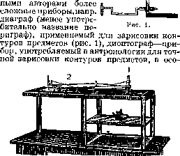
CRANIOMETRY (from Greek cranion - skull and metron - measure), measurement of the dimensions of the skull and head. The simplest and most commonly used craniometer is the sliding caliper, whose legs, when folded, touch each other tightly only at the midpoint of the instrument, after which they diverge to the sides, bending in an arc, and converge again on the midline. The legs of the caliper are connected by a hinge. The curved parts are rounded and end in button-like thickenings. From the hinge extends a crossbar attached to one of the legs by a pin that allows it to move freely. In the corresponding place on the opposite leg is also attached a metal box rotating on a pin, the front wall of which is replaced by a plate with an edge beveled toward the zero division, serving for reading. The crossbar is marked with a scale with divisions exactly corresponding to the divergence of the caliper legs. The number of divisions on the crossbar is 30; the zero point is at the fixed end of the crossbar. An exact correspondence of the divisions on the crossbar to the divergence of the caliper legs, good fitting of the sleeve and screws is required. This description is standard, according to which craniometric calipers are prepared in the USSR. For the simultaneous measurement of several dimensions of the skull, more complex* instruments are used, for example, the Kreilein cephalometer apparatus, consisting of a system of elastic plates used for measuring arcs on the heads of living people; (see Craniocerebral topography). For the simultaneous measurement and graphic recording of the contours of a human skull in different positions (relative to objects), the dioptragraph is used (Fig. 2). There are also other devices used for the same purposes: craniophor, Kocher's craniostat, etc., which do not have wide practical application.
Рисунок 2. Диоптограф: 1- трубка c визирным крестом; 2-пантограф c графитом, вычерчивающим контур предмета.
The dioptragraph: 1 - tube with a sighting cross; 2 - pantograph with graphite, drawing the contour of the object.
Related articles
Mentioned in
Cite this page
“Craniometry.” Soviet Medical Encyclopedia. English translation of Bolshaya Meditsinskaya Entsiklopediya, 1st ed. (Moscow, 1928–1936), ed. N. A. Semashko. https://sovietmedicalencyclopedia.pages.dev/article/craniometry/