Epistropheus
Historical document, translated for reference. It reflects medical knowledge of the 1920s–30s and is not medical advice.
Summary
The Epistropheus is the second cervical vertebra, also known as the axis. It features a tooth-like projection called the dens epistrophei, which forms a pivot joint with the atlas vertebra above it.
Encyclopedia entry (1928–1936)
EPISTROPHEUS (from Greek epistrepho - I turn), the second cervical vertebra. Older anatomists called it the axis, axis (also achop) or vertebra dentata, while the epistropheus was sometimes called the atlas. The E. is constructed according to the type of cervical vertebrae and differs from them in that its body upward continues into the so-called dens epistrophei (dens epistrophei) (figs. 1 and 2), of cylindrical form, somewhat thickened upward and provided
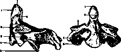
Figure 1.
Figure 2.
Figure 1. Epistropheus from the left side: 1 and 2 - fac. articul. ant. et sup.; 3 - for. transvers.; 4 - proc. transv.; 5 - fac. artic. inf.; 6 - fac. articul. post.; 7 - dens. Figure 2. Epistropheus from the front: 1 - dens; 2, 3 and 5 - fac. articular, ant., sup., inf.; 4 - proc. transversus. (From Rauber.) with articular surfaces on its anterior and posterior surfaces for the anterior arch of the transverse ligament of the atlas. On the sides of the dens are slightly convex articular surfaces on which the lateral masses of the atlas rest. In a mechanical sense, the listed articular surfaces form one conical rotational joint. The dens represents the separated body of the first vertebra. Even at the cartilage stage, the body of the atlas fuses with the cartilaginous body of the epistropheus, forming the dens, which has its own center of ossification and finally fuses with the bony body of the E. at 3-5 years of age. Differentiation of the E. begins in reptiles, but only in mammals does the dens fuse with the body of the second vertebra. - Pathology of the E. mainly concerns fractures of it, which are fatal in most cases. They are observed very rarely, more often in the form of pathological fractures (tuberculosis); in this case, there is a fracture of the dens and its displacement into the spinal canal. Several cases of dislocation of the epistropheus have been described.
Related articles
Mentioned in
Cite this page
“Epistropheus.” Soviet Medical Encyclopedia. English translation of Bolshaya Meditsinskaya Entsiklopediya, 1st ed. (Moscow, 1928–1936), ed. N. A. Semashko. https://sovietmedicalencyclopedia.pages.dev/article/epistropheus/