Plasteins
Historical document, translated for reference. It reflects medical knowledge of the 1920s–30s and is not medical advice.
Summary
Plasteins are protein precipitates formed during the action of rennet on concentrated solutions of peptic digestion products. They are insoluble in water but dissolve in dilute acids and alkalis, and their formation involves a decrease in free amino groups, suggesting they may be products of protein resynthesis rather than further hydrolysis.
Encyclopedia article (1928–1936)
PLASTEINS, the name given by Zavyalov to precipitates obtained in an incubator when chymosin acts on concentrated solutions of products of peptic digestion of proteins. The phenomenon itself was first described by A. Danilevsky. The formation of P. also occurs when all proteolytic enzymes and extracts of fresh and autolyzed organs act. Substances of a similar nature, formed under the action of papain, were called coaguleases by Kuraev. P. are also obtained from products of tryptic digestion and acid or alkaline hydrolysis of proteins. P. are insoluble in water, easily dissolve in dilute acids and alkalis; their solutions in alkalis, taken not in excess, coagulate into a transparent mass upon heating. Most authors considered P. to be products of the reverse synthesis of proteins, although some attributed the formation of P. to the further course of protein hydrolysis. Henriques and Gjaldbaek showed that in the formation of P. there is a decrease in the amount of free NH2 groups titrated by Sorensen, which speaks in favor of the first of the two views; Glagolev came to the same result later. It has not been clarified whether all fractions of albumoses and peptones are necessary for the formation of P., or whether some of them may be absent. In the formation of P., an equilibrium is established between the products of protein hydrolysis remaining in solution and precipitating as sediment. Most P. give the color reactions characteristic of proteins.
PLASTIC SURGERY
82в
absent. In the formation of P., an equilibrium is established between the remaining in solution and precipitating as sediment products of protein hydrolysis. Most P. give the color reactions characteristic of proteins.
i
PLASTIC SURGERY
.
gS» of the surface (mainly with thin flaps). After engraftment, they are more mobile and resistant to external irritations. The skin transplant is taken 0.5-1.0 cm wider than the size of the defect; the shape is outlined with a knife according to a gauze pattern; at the same time, care is taken so that the main direction of skin contractility of the transplant corresponds to the contractility of the skin at the edges of the defect. After the beginning of detachment, the assistant stretches the flap with two forceps so that it is convenient to remove a layer of equal thickness only of the skin, without subcutaneous fat. The edges of the defect are detached for 1 cm along the periphery. The transplant is placed in the correct position, the edges of the defect and flap are turned back and sutured in such a way as to achieve close contact of the edges. After applying the main sutures (with hair), a forceps is inserted under the flap to lift it from the bottom of the defect, and very small holes are made with a thin sharp scalpel. Then a continuous suture with thin hair is applied. The flap should be evenly stretched. The dressing consists of several layers of gauze with uniform pressure from cotton or sponge; a fine rubber foam sponge (Rauer) can be used. Limberg uses a thin rubber balloon, which is placed under the dressing and connected by a rubber tube to a mercury manometer. By pumping air into the balloon through a side tube with a bellows, pressure is maintained for several days at about 32 mm Hg. Elimination of scars! To eliminate superficial and deeper scars on the face and other parts of the body, formed from mechanical injuries, burns, inflammatory processes, in addition to therapeutic measures (massage, ionization, d'Arsonval currents, injections of fibrinolysin, etc.), excision of scar tissue with subsequent closure of skin defects by linear suture with mobilization of wound edges, sometimes with the addition of relaxing incisions, or with the formation of skin flaps on a short or long pedicle (see below) is used. The same procedure is followed after excision of birthmarks, spots from the introduction of coloring substances (tattooing), skin horny growths, superficial tumors, etc. When excising scars, rules must be observed that ensure subsequent cosmetic closure of the defect. Incisions outlining the scars should be made strictly perpendicular to the surface of healthy skin, through its entire thickness; scar tissue (with narrow to wide scars) is not excised in a wedge shape, but is cut parallel to the skin surface at the level of the subcutaneous tissue; the edges of the skin are mobilized to full mobility at the same level and advanced onto the surface of the cut scars. The needle entry at a distance of 3-4 mm is made perpendicular to the skin through its entire thickness. Only under such conditions are not depressed scars obtained after suturing (Fig. 1 a and b"). a
Figure 1.
Deep, contractile scars that fix soft tissues to bones are mobilized at their point of attachment. The defect after excision to prevent depression is filled by moving adjacent, deeper tissues (adipose tissue, muscles) or by performing a free fat graft simultaneously with excision or after wound healing. Fat is best taken from the anterior surface of the thigh over the fascia lata. Most skin defects resulting from excision of superficial tumors and scars have the form of simple geometric figures: triangle, rectangle, rhombus, oval. In Fig. 2, the shapes of the defects and the direction of the movable edges are shown schematically. After healing of flap wounds (machine-gun wounds, gunshot wounds at close range), irregularly shaped linear and star-shaped scars remain, sometimes with defects in normal skin, which sharply displace parts of the face: lips, corners of the mouth, wings of the nose, eyelids, etc. Before deciding on the amount of missing skin and cutting new flaps to close the defect, linear and star-shaped scars must be excised, the skin edges mobilized, and the resulting flaps moved to their normal position—in other words, used for the rational closure of the resulting defect—and only in case of insufficient skin should one resort to forming new flaps or closing the defect with epithelial transplantation according to Thiersch or Krause.
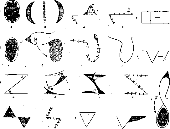
Through-and-through contractile scars in the mouth area, scars that contract the jaw, are excised until complete exposure of the jaw is achieved, after which all exposed surfaces are covered with healthy skin flaps on a pedicle to prevent formation of new scars. From the oral cavity side, it is sometimes advisable to cover the exposed surfaces with epithelium, which is applied with the cut surface upward on a previously taken impression with the mouth open from dental stent material; the impression is reinserted into the oral cavity from the outside of the teeth for 10-14 days, after which all epithelium takes (Esser's method).
In treating contractile scars on the flexor surfaces of the extremities, many authors do not excise flat scars but move them in the form of a triangular flap above or below the site of contraction and cover the exposed surface with a counter triangular flap of healthy skin taken from nearby (Joseph, Limberg, etc.), or they use a transverse zigzag incision of the contractile skin scar, after which the lateral prepared flaps are moved without tension (Morestin). Epithelial transplantation according to Thiersch onto the exposed surface in these cases does not always succeed due to the difficulty of applying an immobile pressure dressing, for example in the axillary region.
For contractile scars in the axillary region, on the anterior surface of the elbow flexure, in the popliteal fossa, between the thumb and index finger, Rauer recommends the following method. The flat contractile scar is outlined with an oval incision from the peripheral side outside the flexure: in the axillary area, on the forearm below the flexure, on the leg below the popliteal fossa; all scar tissue is dissected off until complete straightening of the limb is achieved, with all the dissected skin being moved to the site of the flexure, and the resulting (crescent-shaped) skin defect outside the flexure, if it cannot be closed with mobilized flaps from the sides of the defect, is covered with epithelium applied to an impression taken from stent material previously from the surface of the wound. Dense, protruding scars can be excised after wound healing and mobilization of the limb.
Large through-and-through defects on the face with loss of soft tissues, bones result from gunshot wounds from modern small-caliber jacketed bullets at close range (up to 500 m), as well as from wounds from fragments of artillery shells, airplane bombs, etc. These defects are characterized after healing by extensive contractile scars. In addition, extensive through-and-through defects of the mouth area are observed after destructive pathological processes, primarily in gangrene of the lips, cheeks, nose after typhus and relapsing fever. A characteristic feature of these defects is smooth, as if stamped edges, penetrating through the entire thickness of the cheek or lip, nose, sometimes with a defect of the alveolar border of the upper and lower jaw. Defects of the lips, cheeks are also observed after noma, but since here they result from a prolonged inflammatory process starting from the mucous membrane, they are accompanied by extensive contractive scars of the surrounding tissues. In lupus, where the process begins from the surface of the skin, flat scars are observed on thinned skin, defects with thinned edges (obliterated, as if melted, worn-away parts of the face). After syphilis, where gummatous processes begin primarily from bones, from the cartilage of the nose, hard palate, the defects are characterized by depression of soft tissues and retracted edges. After tuberculosis, removal of malignant tumors, less characteristic defects remain.
Through-and-through defects of the face, mainly of the mouth area (lips, cheeks, chin), can be restored by various techniques depending on the nature of the injury, its extent, availability of sufficient plastic material near the defect, the patient's endurance, and the surgeon's experience and ingenuity. Defects can be closed: by mobilization of adjacent tissues with freshening and approximation of the defect edges; by moving skin flaps taken from nearby, in one layer or in the full thickness of the cheek (Dieffenbach, Bruns, Morestin, Lexer, etc.). They can also be closed with flaps on a long pedicle, taken from nearby, for example from the neck, chest, or from distant sites: from the arm, abdomen, in the form of tubed, duplicated, or migrating flaps. Rauer considers it possible to recommend following the following general rules when choosing the operation method. Median and lateral slit-like defects of the face, lips, chin (from saber cuts, from bullet wounds and from wounds from fragments of shells, etc.), as well as transverse facial defects when the defect depends less on lack of tissue than on divergence of the contracted wound edges—are closed after excision of scars and mobilization of the mucous membrane and skin by pulling the tissues, with the mucous membrane (with submucous catgut sutures from the outside of the wound), muscular layer, and skin sutured separately.
Through-and-through defects of the mouth area with significant loss of soft tissue, sometimes with partial or complete loss of the nose, are most conveniently closed with the help of flaps on a pedicle from more distant areas—from the forehead, neck, from the arm—or with migrating tubed flaps. The most important requirement in restoring defects of the natural orifice area and nose should be the restoration of both the internal and external layers of the wall replacing the defect. The internal layer is formed from a skin flap on a long or short pedicle, taken from nearby or from a more distant place and turned with its surface inward; functionally it can completely replace the mucous membrane: this surface does not undergo maceration and inflammatory processes; it becomes only thin and tender, although it does not metaplasize into mucous membrane; hair continues to grow, glands are preserved. Skin flaps on a pedicle for closing the external layer can be formed from the forehead (Indian method), from the hairy part of the head for restoring the chin and upper lip in men (Lexer), from the cheek, especially in the nasolabial fold, from the supraclavicular area, neck, shoulder (Italian method) or in the form of tubed flaps from distant sites (Fig. 3, diagram of flap location).
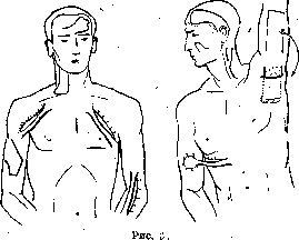
The site for raising the flap is dictated by the location of the defect, its size and shape, the availability of intact skin, i.e., absence of scars at the site where the flap will be formed, cosmetic considerations, sometimes the patient's reluctance to have new scars on the face or neck. When forming single-layer flaps on a pedicle, certain general rules must be observed to ensure their nutrition and survival: first, the contractility of the skin at the sites where the flap is raised must be taken into account (for example, on the neck the flap contracts almost by half in length and width). The length of the flap should not exceed more than 3-3½ times its width, the pedicle should not be narrower than the end of the flap except for flaps where large blood vessels pass through the pedicle
large blood vessels, e.g., the temporal artery, the inferior maxillary artery, etc. When raising the flap, parallelism between the outer and inner surfaces of the flap must be maintained, sometimes guided by the layer of fascia. On the neck, it is better to take the flap with the platysma muscle. When applied, the flap should cover the defect freely, with almost no tension. The pedicle should lie freely (in an arc), without twisting or bending at a sharp angle. To close a through-and-through defect, the edges of the defect are first freshened, i.e., scars are excised, and the mucous membrane and skin are mobilized at the edges; then two flaps are outlined: one is sutured to the edges of the mucous membrane with the epithelial side inward, the other is placed on top and sutured to the edges of the skin, with the upper layer fitting tightly against the lower one, with no clots from accumulating blood between them. Sometimes, to replace the inner layer, it is advantageous to outline a wide flap with its base at the edge of the defect (without freshening the edge at this point) and turn it inward; sometimes instead of one flap, two shorter flaps can be taken from both sides of the defect and sutured along the midline. A through-and-through defect can also be closed at once with a single flap bent at the posterior edge of the cheek defect (Almazov), where the end of the flap forms the inner wall, and its middle part forms the outer wall; at the posterior edge, at the bend, a temporary slit-like opening remains. Eiselberg first sutured one end of the flap with the skin inward, and after it took, he covered the granulating defect with the remaining part of the flap. To close extensive through-and-through defects, it is convenient to use pre-doubled flaps. For example, a long flap from the hairy part of the head with its base in the temporal region (Lexer) is lowered and connected with a hairless flap on the lateral surface of the neck, and thus the fused, doubled flap is transferred to close a defect of the upper or lower lip (in men), sometimes the cheek. A double flap can be used, formed from a flap on the inner surface of the arm with its base in the upper third at the border of the axillary fossa, and from a bridge flap formed on the lateral surface of the chest below the axillary fossa by two horizontal parallel incisions. After the skin is undermined on the chest, the arm flap is inserted into the resulting pocket, like into a pedicle, and the exposed surfaces are sutured together. After fusion, the two pedicles on the chest are severed, and thus the doubled flap with the arm is transferred to the face, where it is sutured into a defect two-thirds of the circumference; after 2-3 weeks, the pedicle is severed from the arm, and the defect is finally closed (fig. 3).
In addition to simple and doubled flaps, tubed flaps, introduced by Filatov, are now widely used in the USSR. A tubed flap is formed from a simple bridge flap with two pedicles, created by two parallel incisions; after the skin is undermined, the edges of the flap are carefully sutured, and thus a closed, continuous tube is formed, remaining connected to the base at two pedicles. The width of the flap is usually taken as 5-9 cm, the length from 10 to 25 cm; in individual cases, the tube can be shorter and correspondingly thinner. Due to the central vascular trunk formed in the tube and the absence of exposed surfaces, the tubed flap has greater stability in terms of nutrition and is less prone to secondary infection. Sometimes the tube serves as a stalk for a flap-platform formed at one end, which may be more or less wide (the dimensions of the platform should not significantly exceed the width of the unfolded flap, and the platform can be slightly longer than the width), or the tube itself or part of it, when unfolded, serves to close the defect. Sometimes after the "maturation" of the flap, it is advantageous to almost completely detach the platform formed at one end, and then resuture it in the same place, and after 8-10 days, use it for plastic surgery. An important advantage of tubed flaps is that they can be formed on the most distant parts of the body and, by sequential movement of the pedicles, brought closer to the site of the defect; thus, wandering-migratory caterpillar tubed flaps are formed. The duration of "maturation" of the flap is 2-3 weeks at each stage. To reduce time, it is better to transfer one pedicle of the flap to the arm or forearm (preferably to the middle of the upper arm) on the inner side, and together with the arm, after severing the pedicle from the chest or abdomen, transfer it to the face. For fixation of the arm, a figure-of-eight bandage of soft bandage is used, firmly fixing the shoulder to the head, in front of the ear; the bandage can be secured on top with a starched or plaster bandage. Sometimes it is more convenient to fix the arm in front of the chest in a bent position with the hand resting on the opposite shoulder. A tubed flap can be complicated by preliminary implantation of cartilage or bone into it, or by forming elements of a future organ on it, e.g., a nose. The only inconvenience of using tubed flaps is the longer treatment time with a large number of stages before final plastic surgery. Although a tubed flap for facial plastic surgery can be formed anywhere, there are still places most convenient for flap formation, e.g., the neck (one pedicle in the mandibular area, the other in the supraclavicular region); the outer surface of the elbow joint (Rauer) (one pedicle on the lower third of the arm, the other at the border of the upper and middle thirds of the forearm; Danelidze proposed making a tube on the flexor surface of the shoulder joint); the anterior surface of the shoulder joint (beginning - on the upper third of the arm, end - on the chest over the mammary gland); the chest from the axillary fossa to the clavicle; the abdomen and lateral surface of the chest in a transverse direction (fig. 3). The introduction of the tubed flap into practice created an era in the development of Russian plastic surgery.
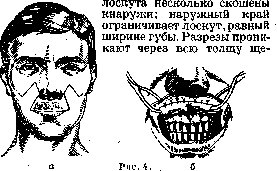
Plastic surgery of the lips (cheiloplasty). On the upper and lower lips, due to the significant mobility and contractility of the tissues, wounds with considerable divergence of edges are observed even in the absence of tissue loss. With contusions and falls, the lip is often cut by the sharp edge of the adjacent teeth. In fresh cases, relatively smooth edges of the wound are sutured without any tension with a good cosmetic effect, even if there is a slight loss of tissue. When the edges of the wound are crushed, they are freshened, the suture is applied from the side of the mucous membrane and from the side of the skin, with the muscular layer also being included in the suture. Healed triangular defects of the upper and lower lip, after freshening of the edges, are sutured by the method of Mirault and others, as in cleft lip. More or less significant and complete defects of the lip most often remain after extensive gunshot wounds, as well as after destructive pathological processes (noma, lupus, etc.). Restoration of the upper and lower lip is achieved primarily by forming flaps from tissues located in direct proximity to the defect, if the condition of the surrounding tissues is favorable, or flaps are taken from more distant places (neck, arm, hairy part of the head, etc.) in the form of simple, doubled, or tubed flaps. With any method of lip formation, one must not forget the general rule - when freshening the defect, preserve the remnants of the red border of the lip in the form of a flap on a pedicle, or outline a flap from the mucous membrane at the edge of the defect, in order to then use it to form the edge of the lip; due to the remarkable ability of the mucous membrane to take, even long and narrow mucous flaps take well. A tube consisting of three layers of muscles and mucous membrane must essentially be restored from the corresponding layers of surrounding tissues. These conditions are best satisfied by flaps taken from the cheek in its entire thickness. Along this type, the well-known operations of older authors (Brans, Langenbeck, Sedillot, Ombredanne, Morsten, Lexer, Dieffenbach, Esser, etc.) are constructed. To form the upper lip with a complete defect, two quadrangular flaps are outlined in the area of the nasolabial fold; the inner edges of the flaps limit the lateral edges of the defect and extend slightly above the wings of the nose, the upper edges. Turned at their base, these flaps are sutured with their short sides in the middle of the defect (fig. 4a). The red border, separated during freshening of the defect, on a pedicle is sutured to the edge of the formed lip. The remaining lateral defects are sutured over the mucous membrane and from the side of the skin (fig. 4b). The blood supply of the through flap (in its entire length) in the nasolabial fold is provided by the angular artery. Esser uses the course of this artery to form a wedge-shaped flap on each side; these flaps are sutured one on top of the other to form the lip. A defect of the upper lip can also be restored by a tongue-shaped flap from the arm (brachial method) in the case
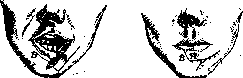
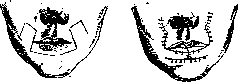
838. In cases where the mucous membrane can be formed from the remaining skin of the lip turned inward or mobilized for this purpose from the mucous membrane of the cheek, in men a flap from the hairy part of the head can be taken, doubled with a flap from the lateral surface of the cheek, and then transferred to form the lip. Additionally, a tubed flap transferred from the arm to the face serves as convenient material for forming the upper and lower lip. Usually only the end of the flap is sutured to the edge of the defect on the freshened skin surface or simultaneously to the mucous membrane and skin, and then the entire tube is used to form the lip, being incised along the suture line and first sutured with one edge to the remaining mucous membrane of the upper vermilion border or to the skin turned inward, and with the other edge sutured to the edge of the freshened defect at the base of the nose. If the red border is preserved during freshening of the defect, it is sewn into the new incision along the edge of the sutured flap. The final shaping of the lip (flattening, excision of fat, connection with the muscular layer of the cheek) is postponed until complete take of the flap. When forming the upper and lower lip "from tubed flaps for shaping the corners of the mouth," "Fig. a," "b," tongue-shaped or triangular flaps can be cut from the upper edge of the lower flap on each side with the base inward and sewn into horizontal incisions on the upper flap. Partial and complete defects of the lower lip, which is more mobile and active than the upper, are accompanied by more significant functional disorders, especially during food intake, constant drooling, etc. Relatively small triangular defects after injuries, tumor removal can easily be drawn together after freshening of the edges with sutures through the skin and muscular layer and separately for the mucous membrane. Large defects with complete absence of the lower lip, observed after gunshot wounds, often in connection with complete avulsion of the chin, "Fig. 6," "b'," as well as defects after removal of malignant tumors, are closed by plastic surgery. Defects of the lower lip from gangrene after typhus and other destructive processes are less common. In choosing the method of operations for plastic surgery of the lower lip, the same considerations are followed as in restoring the upper lip. In the absence of scars and defects of healthy tissue on the cheek, flaps are cut from the full thickness of the cheek; otherwise, plastic surgery with simple or tubed doubled flaps is used. With a simultaneous defect of soft tissues of the chin, plastic surgery of the lip is performed together with restoration of the chin defect. For restoration of half of the lower lip, the Moresten method is convenient. A flap is cut in the nasolabial fold on the corresponding side-through the full thickness of the cheek, with the inner edge of the flap limiting the outer edge of the lower lip defect during formation. The shape of the flap and its position after suturing are shown in fig. 5 a, b.-When preserved mucous membrane or the possibility of mobilizing it from the cheek mucosa exists, a single-layer flap can be formed at the border of the chin and cheek, with the inner edge of the flap starting from the corner of the mouth, limiting the defect on its outer side (fig. 6 a, b). For complete restoration of the lower lip, operations with formation of symmetrical flaps through the full thickness of the cheek are often used. Operations of this type include the Bruns method with formation of flaps from the cheek in the direction of the nasolabial folds (fig. 7).
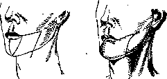
After cancer surgery of the lower lip, the Differenbach method in Bergman's modification is often used. From each side of the cheek a flap is formed, bounded above by a line, "Fig. 8," extending from the corner of the mouth to the auditory canal and not reaching 2-2.5 cm to the auricle; from here the incision line is directed at an acute angle along the edge of the m. masseter and descends below the lower jaw border. The inner line of the flap is parallel to the outer one and limits the outer edge of the defect; incisions penetrate through the full thickness of the cheek, sparing the fascia of the parotid gland above; the reflected flap on most of its inner upper surface is covered by mucous membrane. The flaps are moved to the midline and sutured, along the edge of the formed lip the skin is sutured to the mucous membrane (fig. 8). Lexer suggests closing the lower lip defect with a flap with the base directed toward the cheek and bounded by an arcuate line extending beyond the midline of the chin. The flap is displaced upward, with the preserved mucous membrane of the oral vestibule serving to form the mucous membrane of the lip. When it is possible to successfully close defects of the lips and cheeks with various flaps taken from distant sites, operations with cutting flaps from the cheek hardly have an advantage, all the more so that when cutting these flaps the 513» and feeding nerves and muscles are severed, the oral vestibule is reduced, and depressed scars remain.-When removing malignant tumors of the perioral area, it is necessary to try to expand the area of operation as much as possible, operating in healthy tissues; the resulting defects are left unclosed until the wound heals, and only in the absence of recurrence after a more or less long time are they closed with the help of tubed or doubled flaps. A gaping oral cavity is temporarily closed with a thin rubber plate-shield, preventing saliva leakage and drying of the tongue. As mentioned above, extensive defects of the chin area are restored simultaneously with plastic surgery of the upper lip. A good method for this purpose in men is formation of a flap from the hairy part of the head according to Lexer for the outer layer, with formation of a bib-like hairless flap from the neck for the inner layer.-The flap from the head has the shape of a pistol grip about 4 or more transverse fingers wide, the rounded end extends beyond the midline of the forehead, the stalk is formed at the upper base of the ear, with the anterior incision being conducted in front of the ear and temporal artery, the posterior one behind the auricle; both incisions end at the level of the zygomatic arch; the flap is easily separated from the galea aponeurotica, and when separating the base it is necessary to be careful not to injure the feeding temporal artery. To obtain the correct shape of the soft tissues of the chin, the remaining fragments of the lower jaw are connected with a wire arch fixed on the preserved teeth; on this arch a convex shield is fixed to protect the tissues from depression and wrinkling. To form the red border of the lip, a strip of mucous membrane is cut from each edge of the defect, which is sutured to the upper edge of the mucous membrane with skin. With deficiencies of the mucous membrane and significant scar changes of the corners of the mouth, scars are excised, and the defect is replaced by transplantation of healthy skin on a stalk in the form of a tubed or simple flap, from which after take the corners of the mouth are formed.-With shortening of the upper lip and its strong depression in profile, e.g. after suturing triangular defects, the upper lip can be lengthened by the Abbe method by insertion of a triangular flap from the lower lip with a stalk on the mucous membrane of the red border, the flap being turned 180° (fig. 10). The principle of cosmetic operations undertaken to eliminate the so-called sorrow folds, wrinkles, and sagging skin of the cheeks, chin, comes down to tension, excision, and fixation of skin in certain directions, whereby folds are smoothed and straightened. To ensure sufficient tension of the skin cover, excisions of varying sizes of skin areas (oval, semilunar or spindle-shaped) are made on less noticeable
"Fig. 9."
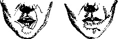
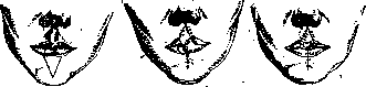
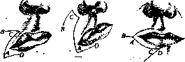
of given flaps. In women, to form a chin, a doubled flap can be taken from the inner side of the arm and chest, as described above. Scar changes in the position of the lower lip in the form of eversion outward are eliminated by freeing it from below from deep scar changes and replacing the resulting defect with skin from the arm, neck, or by free skin grafting according to Krause. Drooping of one corner is eliminated by moving triangular flaps, bounded by an incision in the shape of the letter Z at the corner of the mouth—after rearrangement, the incision line has the shape of a Z reflected in a mirror (fig. 9 and 2, 3). Scar narrowing of the oral fissure and widening of the mouth are corrected by wedge-shaped excision of tissues at the corners of the mouth, followed by submucosal excision of scars and suturing of the mucosa. Figure 10. On certain parts of the face—at the temples on the border of the hairy part of the head, forehead, behind the ear—with subsequent tight suturing of the skin defects. Noel (S. Noel) proposes for greater precision of incisions of corresponding shape to use cutouts from celluloid plates, which are applied to specific places, measured with a centimeter tape like a craniometer. Lexer excises S-shaped areas of skin on the hairy part of the temples above the ear and a similarly shaped area of skin located under the earlobe and behind the ear. Suturing of these defects, sometimes with undermining of the skin edge towards the face, gives a reliable effect of smoothing wrinkles. For stronger fixation, when suturing the edges of the wound on the temples, Lexer captures some sutures the underlying fascia, and when tensioning the lower flap behind the ear, fixes it with sutures to the periosteum of the occipital bone. In some cases, for greater tension of the skin cover, a greater or lesser undermining of the skin towards the middle of the face is applied, where the skin is not excised in the form of platforms of a specific shape, but is separated in small sections with significant tension at the edge of the defect. Noel uses the following technique for this operation: the incision begins on the hairy part of the temple, at a greater or lesser height, with an arc-shaped line curved backward, passes along the anterior edge of attachment of the helix and descends in front, at the very attachment of the auricle, to the middle of the earlobe (the incision is made with pointed scissors, advancing the lower branch of the scissors under the skin). Then the inner edge of the skin is separated to the fascia for 2-2.5 cm towards the face. Strong forceps (e.g., Duayen's trident) grasp the edge of the skin, starting from below, and with strong tension upward and towards the parietal bone, the edge of the skin is cut off with short cuts of scissors at the level of the posterior edge of the wound; after each cut, a suture is applied, so that the facial skin with the cheek is gradually pulled upward. If necessary to smooth folds on the chin or neck at the same time, the mentioned incision is continued around the earlobe behind the ear and from there directed to the occiput. Here also the skin is undermined to a greater or lesser extent, and the wound is sutured. When pulling the chin skin, it is recommended to pull the subcutaneous fat separately, suturing it with several catgut sutures in the lower parts of the wound. To remove wrinkles on the forehead, an incision is made at the border of the hairy part of the head and forehead; the skin is undermined almost to the eyebrows and excised, the remaining spindle-shaped skin defect is sutured. Vertical folds between the eyebrows are excised along the direction of the folds. Sagging bags under the eyes in the absence of edema are removed under the edge of the lower eyelid after careful verification of the size of the flap to be removed; excessive removal of skin can cause an unpleasant eversion of the eyelid edge. Disfigurement of the auricle is observed in various forms. First of all, congenital anomalies are encountered, starting from complete absence of the auricle, with complete atresia, underdevelopment of the auditory canal and auditory organ; then rudimentary rudiments of the auricle are encountered in the form of small soft hanging tumors or the auricle or earlobe is underdeveloped. The operation to restore the auricle has not yet given satisfactory results by any author, since the ear formed from flaps from the arm or from the back of the neck, in its shape, could not satisfy either the surgeon or the patient. It is of course possible with the help of tubular flaps with cartilage transplantation to create with great difficulty a more or less similar ear, but in view of the fact that the auricle is not functionally important and besides its deficiency can be hidden in the hair, it is hardly necessary to undertake the difficult operation to restore a whole ear, especially since an ear made of soft rubber of natural skin color can completely replace the natural ear. This cosmetic prosthesis can be easily fixed in place with the help of live loops formed from skin folds at the site of the ear. More often requests are made to reduce overly large ears or to correct wide protruding ears. Reduction of the auricle can be achieved by excising the upper edge of the antihelix in the shape of a semilune, where the upper skin flap and cartilage are removed. The posterior skin flap is preserved. Then from the helix and the remaining posterior skin flap, a triangle is excised with the apex at the lower concave edge of the excision. The helix is sutured and attached to the remaining antihelix—(Lexer's method, figure 11). Another method consists in excising part of the helix in front—at the upper semilunar part of the antihelix while preserving the posterior skin flap, where from the remaining cartilaginous part of the antihelix in the middle, a triangle of corresponding size is excised so that when sutured all parts fit well (Colle's method, fig. 12). To eliminate protruding ears, it is sufficient to make an oval-shaped skin incision in the longitudinal direction along the ear, placing it so that one half goes behind the auricle and the other lies at its base. With a wide convex cartilage backward, after skin undermining, an oval-shaped piece of cartilage is excised in the longitudinal direction. The cartilage is sutured with separate sutures, the excess undermined skin is excised, and its edge is sutured to the scalp. Organ transplantation—see Transplantation.
Related articles
Mentioned in
Cite this page
“Plasteins.” Soviet Medical Encyclopedia. English translation of Bolshaya Meditsinskaya Entsiklopediya, 1st ed. (Moscow, 1928–1936), ed. N. A. Semashko. https://sovietmedicalencyclopedia.pages.dev/article/plasteins/