Hemofuscin
Historical document, translated for reference. It reflects medical knowledge of the 1920s–30s and is not medical advice.
Summary
Hemofuscin is a golden-brown pigment found in old hemorrhages and certain diseases, particularly hemochromatosis. The article discusses its properties, relationship to hemochromatosis, and the historical confusion with lipofuscin.
Encyclopedia article (1928–1936)
HEMOFUSCIN, a golden-brown pigment, found intracellularly around old hemorrhages and in certain pathological processes (mainly hemochromatosis) in the smooth muscle of blood and lymphatic vessels and the intestine. Insoluble in acids and alkalis. Does not contain iron, even when bound. The corresponding microchemical reaction (for iron) is negative. H. is considered a blood pigment. To date, however, it has not been definitely established what substance is the basis of its origin: hemoglobin and its derivatives or other protein substances and lipoids that make up the stroma of the blood corpuscle. Hueck identifies H. with the lipofuscin pigment of non-blood origin. It can be added that in former times the term H. was applied to denote the pigment which is now called lipofuscin, since it was previously incorrectly thought that the latter had a blood origin. HEMOCHROMATOSIS (from Greek haima-blood and chroma-color), a disease whose essence is a disturbance in the body's iron metabolism. This term was introduced by Recklinghausen and indicates the abnormal (brown) coloration of the skin and organs, which is essentially of hematogenous origin. H. is often combined with hypertrophic cirrhosis of the liver and glycosuria. Hence its former designations: "cirrhose hypertrophique pigmentaire dans le diabete sucre" (Hanot, Chauffard) or "diabete bronze" (Marie). In the past, bronze diabetes was by some authors isolated as an independent nosological unit. This finds its justification in that the phenomena of diabetes-polydipsia, polyphagia, glycosuria, and exhaustion-in the picture of H. sometimes play a dominant role. The accompanying
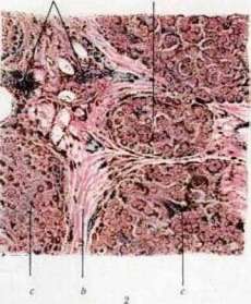
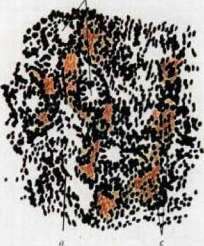
Fig. 1. Liver in hemochromatosis. a - cut surface. b - microscopic section of liver tissue. Hypertrophic cirrhosis of the liver; dense, brownish-red tissue (tr) replacing parenchyma with separate islands of liver cells (lc). Stained by Heidenhain's method. Fig. 2. Sympathetic ganglion and nerve; yellowish degeneration of nerve cells and nerve fibers (rf). Stained by Nissl's method. Staining by Weigert's method
melanoderma completes the picture. At present it is well known that "bronze diabetes" is only one of the symptoms of hemochromatosis. Etiology. Despite the obvious independence of H. from the disturbance of hemoglobin metabolism, its cause was still recently considered to be the slow intravascular breakdown of blood, caused by this or that accompanying chronic diseases (tuberculosis, cancer, etc.) or the absorption of harmful substances from the intestine. Special mention should be made of the opinion that H. is caused by erythrophagocytosis by liver cells. At present, the view is becoming more firmly established that pathological blood breakdown plays no role in the pathogenesis of H. In other words, it is now believed that H. is not a "maximum hemosiderosis" but a disturbance of the general or local iron metabolism, in which the iron molecule, brought into the cell in the usual amount, is not assimilated by it but is deposited and accumulates. Whether this happens because the cells are incapable of assimilating Fe due to their toxic damage, or the iron is offered to them in an unassimilable form-is an open question. Alcoholism is indicated as an important etiological factor. Pathological anatomy. At autopsy, it is found that all internal organs are colored rusty-brown or chocolate-colored, with large hemorrhages that could cause such extensive "pigment metastases" being described only as exceptions. The liver, pancreas, and intra-abdominal lymph glands are particularly strongly pigmented. The weight of the liver reaches 2,500-3,000 g, its appearance corresponds to the picture of hypertrophic cirrhosis (see separate table, figs. 1 and 2). The pancreas is dense, difficult to cut. In the trunk of the brain, small hemorrhages may be found, and in the putamen-foci of softening. In cases of the initial stage of H., who died from intercurrent diseases, only the 1st symptom of the disease-pigmentation-is clearly expressed; at this time, cirrhotic processes may be completely absent or weakly expressed. Microscopic examination reveals that the rusty coloration of internal organs depends on the extensive deposition of a golden-brown pigment in them, and the latter is always of two kinds. One pigment, which does not give a reaction for iron, is located in the skin and in the smooth muscle of the digestive tract and vessels. It is interpreted as hemofuscin or, according to the latest views, as lipofuscin, and in the skin-as melanin. The other, iron-containing pigment-hemosiderin-is deposited mainly in all secreting organs (liver, pancreas, salivary glands, thyroid, pituitary, choroid plexus of the brain and many others), in the heart and lymph nodes. To a lesser extent-in the spleen, bone marrow and kidneys. In the latter-it may be completely absent. Particularly abundant deposition of pigment is noted in the parenchyma and proliferated stroma of the liver and pancreas, then-in periportal and retroperitoneal glands. In the latter, the deposition of iron can be so great that it leads to the disappearance of lymphoid tissue; iron deposition is also observed in the reticular stroma. The further from the main focus of the lesion, the poorer in pigment the lymphatic glands become. The amount of iron deposited in this way in organs can exceed the norm 100 times; e.g., in the dry residue of the liver, with a normal iron content of 0.08%, there can be 7.62%, in the pancreas-5%, and in the lymph nodes-14.69%. The iron found in the brain cannot in any way be considered physiological, because its location (perivascular and in the walls of capillaries) does not correspond to the centers where, according to Spatz, physiological iron is normally located. Symptomatology. The most striking symptom-bronze (or smoky) coloration of the skin, most sharply expressed on open places-face, neck, hands (except palms), on places normally more pigmented (genitalia, nipples), and finally, in areas subjected to pressure or irritation (armpits, inner surface of thighs). Like Addison's disease, on the mucous membranes (on the gums, on the inner side of the cheeks, on the tongue) smoky-brown spots are sometimes observed. The coloration of the skin begins insidiously and sometimes reaches strong degrees. Sometimes it may be completely absent. Often accompanied by early loss of hair. Pigmentary cirrhosis is expressed by the symptoms of ordinary cirrhosis of the liver: digestive disorders, pressure in the right hypochondrium, later-signs of portal hyperemia (hemorrhoids, nosebleeds). Jaundice is rare; ascites is observed in the later stages and is usually small. The liver is always enlarged, its edge is sharp, the surface is smooth. The spleen is almost always enlarged. Diabetes is accompanied by the usual symptoms: polydipsia, polyphagia, fatigue, polyuria, and more or less pronounced glycosuria; acetonemia-a rare phenomenon. The condition of the blood and in particular the amount of Hb-is within normal limits. Occasionally a small anemia is described. The osmotic resistance of erythrocytes is normal. Urobilin is absent in the urine; hemoglobinuria is observed only in individual, far advanced cases. It is now established that pigmentation is the very first symptom. It is followed by cirrhosis of the liver and other organs. Diabetes is caused by damage to the pancreas and is an inconstant and terminal complication. H. is an extremely rare disease. Only about 60 cases have been described. It occurs in adults, mainly in men; in women only 6 cases have been observed. The diagnosis of H. presents no difficulties if the typical symptom complex is expressed: pigmentation of the skin, cirrhosis of the liver, and glycosuria. If the first is absent, which is by no means rare, one must be guided by the combination of hypertrophic cirrhosis with glycosuria in the absence of jaundice. The prognosis of H. is unfavorable, especially when diabetes is present. Death may be preceded by a long coma. Treatment of H. is purely symptomatic. Cholesterol was administered orally up to 1.0 pro die (with the aim of increasing the resistance of erythrocytes) and splenectomy.
E. Gertsenberg.
Related articles
Cite this page
“Hemofuscin.” Soviet Medical Encyclopedia. English translation of Bolshaya Meditsinskaya Entsiklopediya, 1st ed. (Moscow, 1928–1936), ed. N. A. Semashko. https://sovietmedicalencyclopedia.pages.dev/article/hemofuscin/