Vuzin and Lice
Historical document, translated for reference. It reflects medical knowledge of the 1920s–30s and is not medical advice.
Summary
This article from the 1928-1936 Great Medical Encyclopedia discusses vuzin, an iso-octylhydrocupreine alkaloid used historically as a disinfectant and antiseptic in wound care, as well as the biology, habits, and medical significance of human lice (Pediculidae).
Encyclopedia article (1928–1936)
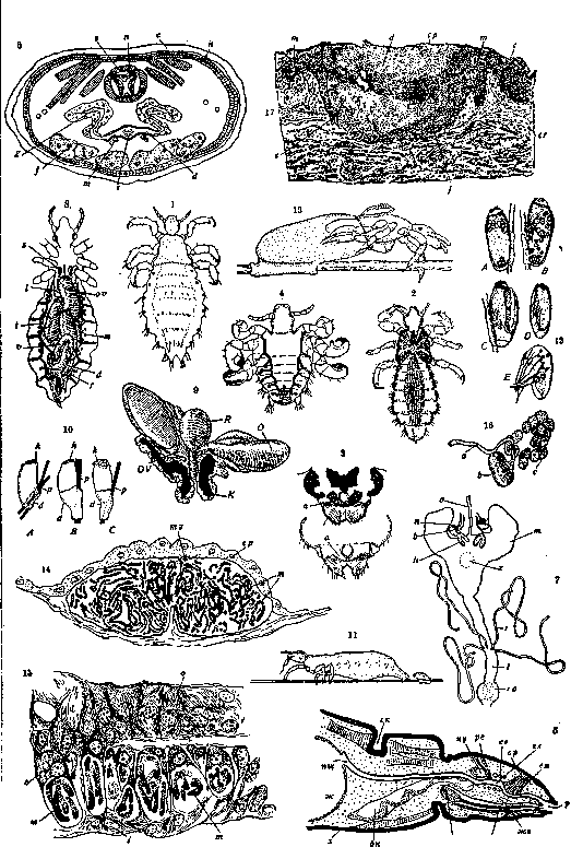
VUZIN, Vuzin, iso-octylhydrocupreine, belongs to the quinine series of alkaloids. Its dihydrochloride C19H23(C8H17)N2O2·2HCl + 2H2O forms colorless, fine needles with a slightly acidic taste and an acid reaction; it causes a weak but prolonged numbness on the tongue. Vuzin is soluble in hot water, alcohol, and chloroform; sparingly soluble in ether, gasoline, and HCl. An aqueous solution of Vuzin gives an emerald-green coloration with chlorine water and an excess of NH3 (the thalleioquin test). Vuzin possesses an elective potency against staphylococci and streptococci; it is used for wound disinfection by irrigation and for antiseptic injections into surgically prepared wounds. The disinfecting action of Vuzin is little diminished by tissue fluids; wounds of soft parts and joints, as well as gas gangrene, show an equally favorable course. For antiseptic infusions, solutions of 1:10,000 to 1:500 are used. To eliminate pain upon injection, 0.6 g of NaCl, 0.5 g of novocaine, and 0.002 g of suprarenin bitartrate (Suprareninum bitartaricum) are added per 100 cubic centimeters of solution. Vuzin solutions should not be prepared in stock for more than 4 days. Side effects upon injection into tissue include severe reaction, skin swelling, blister formation, fever, exanthema, and headache. Intravenous and intraperitoneal injections are cautioned against. Following intralumbar administration of Vuzin, a weakening of the reflexes of the lumbar and sacral parts of the spinal cord is observed. Vuzin produces a precipitate with blood serum and exhibits a hemolytic and phagocytosis-limiting effect. For mice and cats, the lethal dose is 0.2 per kg of body weight; with slow intravenous injection in cats, the lethal dose is 0.015 per kg of body weight. Large doses lead to a fatal outcome, accompanied by a strong slowing of the pulse, a severe drop in blood pressure, and convulsions. LICE, false-beaked insects (Pseudorhynchota) parasitizing exclusively on mammals, upon whose blood they feed. Various species of lice are strictly adapted to feeding on specific hosts and do not transfer to other hosts. Thus, the dog louse (in contrast to fleas) does not occur on humans, and vice versa. Therefore, from the standpoint of contemporary medical knowledge, only human lice belonging to three species of the family Pediculidae have medical significance: Pediculus capitis—head louse (see Figure 2), Ped. vestimenti—body louse (see Figure 1), and Phthirius pubis—pubic louse, or crab louse (see Figure 4). The first two species of lice are considered by some investigators to be races of the same species—Ped. humanus. The Russian names of lice are given according to the place on the human body where these parasites live. However, determining the species of a louse based solely on the place of its capture is impossible, as cases of unusual localization of lice are not uncommon; for example, crab lice are found on the beard and eyelashes, head lice on body hair, etc. The crab louse is most characteristic and easily distinguishable. The body and head lice are not always easily distinguished from each other. Usually, the head louse is sharply pigmented on the sides of the body, whereas the body louse is of a uniform gray color. The genital falciform appendages (located in females on the underside of the body, just before the end of the abdomen) are broader and blunter in Ped. capitis, and narrower and more pointed in Ped. vestimenti (see Figure 3). Prehensile legs serve for the locomotion of lice; they lack wings. A temperature of 25–27° causes the greatest mobility of lice; at temperatures from +5° to -6°, the latter cease moving. Along a vertical plane, lice can crawl up to a height of up to 1 m. In a minute, a louse crawls no more than 30–35 cm. In all stages of transformation, all human lice are blood-sucking. The piercing proboscis of the louse is hidden at rest in a special sheath in the head and is pushed outward during the act of sucking, at which point it plunges into the skin and pierces blood vessels. Blood is sucked by the louse through the action of sucking muscles located in the head (see Figures 5, 6, and 7). Normally, the body louse drinks blood 2–3 times a day. Lice fast for a short time—a maximum of 10 days (at temperatures of 10–20°). At a temperature of 40°, lice perish within 12 hours. Humidity affects the duration of starvation in lice. If it is desired in practice to exterminate lice by starvation, clothing and other items are left in a dry room in a tightly closed box for 3 weeks. Lice can survive under water at a temperature of 15–17° for two days. They possess a sense of smell and distinguish human skin at a distance of 1–2 cm. There are people who are immune to lice and do not become infected by them even if they live in a lice-infested environment. Lice react differently to odorous substances. The state of satiety also influences the nature of the reaction. Sated lice avoid substances that repel them with their odor, whereas hungry lice will suck human skin rubbed with substances toxic to them. The optimal temperature for the life of a louse is about +30°. Lice are dioecious, but hermaphrodites are also found among hybrids between the head and body louse (see Figure 8). Mating can occur at any time; in the body louse, the store of spermatozoa received by the female during copulation lasts for 7–12 days. Louse eggs are called nits. During egg-laying, a drop of glue gland secretion is squeezed out from the genital opening (see Figure 9); it instantly solidifies and firmly glues the nit to the substrate (see Figure 11), which consists of hairs (pubic louse, head louse) or the intersection of fabric threads or hair (body louse). Egg-laying on unusual substrates (buttons, buckles, belts, etc.) also occurs. A well-fed female can lay eggs even without fertilization, but they do not develop. Not all eggs laid by a fertilized female yield larvae. The percentage of viable eggs fluctuates between 70% and 97%. Egg-laying occurs at temperatures between 20° and 37°, with an optimum temperature of about 32°. The intensity of laying drops sharply with a decrease in temperature. The body louse lays 0–14 nits per day, and no more than 295 over its entire life; the head louse yields no more than 4 per day, and up to 141 nits in total. The nits of crab lice (see Figure 10) are very characteristic due to their pear-shaped form and dome-shaped lid with high cells (nit length 0.65–0.67 mm). Body and head lice in their extreme forms of nits also differ from each other (see Figure 10). In the former, the nit has a flat lid and very low cells shifted to the side (length 0.9–1 mm); in the latter, the egg is of a more oval shape, with a weakly convex lid of moderate height (length 0.75–0.8 mm). The duration of embryonic development (see Figure 12) of embryos in nits is determined by the external temperature. The minimum development period is 4 days at 36–37°; however, at this same temperature, other nits yield larvae after 8 days. At 35°, development lasts 6–8 days; at 30°, from 7 to 14 days (most often 11–13 days); at 25°, 16 days. Alternating cooling and heating of nits slows down the development of lice to 5–6 weeks (e.g., periodic removal and putting on of the same clothing). In permanently worn clothing, nits yield larvae after 7–10 days. At temperatures above 40–45° and below 22°, hatching of larvae does not occur. The optimum development is observed at 30–31°. Humidity at the optimal temperature accelerates the embryonic development of lice. The resistance of nits to external influences is determined by the protective properties of their shell. At temperatures from +1° to -3°, they survive a week; a temperature of +98° kills them in 1/2 minute, and a temperature of +54°... [Captions to the table omitted in text OCR]
Figure 12. Various stages (A-E) of development of the louse embryo in nits. Figure 13. Hatching of the Pediculus larva from the nit. Figure 14. Cross section of the stomach of Pediculus in the region of the mycetome; ep—stomach epithelium; mz—mycetome cell; m—symbiont fungi. Figure 15. Symbiont fungi (m) in the wall of the oviduct (o, g) of Pediculus; z—cell with symbionts. Figure 16. Bean-shaped salivary gland of Pediculus (b); a—excretory duct; c—nephrocytes. Figure 17. Section of a human skin papule at the site of injection of an extract from the bean-shaped glands of the body louse: m—unchanged epidermis; c—stratum corneum; d—focus of epidermal necrosis; cp—stratum corneum of the epidermis above it; e—extravasate at the base of the papule; f—fibrin and infiltrate at the base of the papule; cr—corium of the louse
em
ej
in 35 minutes; a 2.5% solution of carbolic acid kills them in 10 minutes, and a 2% lysol solution in 5 minutes. Upon hatching from the nit, the louse larva pushes off the operculum and emerges to the outside under the pressure of air pumped by the larva through the digestive canal into the main part of the nit shell. The hatched larva is almost transparent (see figure 13); it is noticeable only by two black dots on the sides of the head, i.e., the eyes. The larva resembles an adult louse, but has a disproportionately small abdomen and three-jointed antennae. The larva is able to suck blood as early as 3.5 hours after hatching from the nit. The larva molts three times and after the third shedding of the skin turns into an adult louse. Metamorphosis in the louse is therefore incomplete, since the pupal stage is lacking. The duration of the metamorphosis phases varies depending on the intensity of feeding, temperature fluctuations, humidity, and individual characteristics of the insect. On average, about 12 days are spent on the entire larval period. The complete life cycle of the louse of the genus Pediculus consists of the following periods: a) embryonic development—from 4 days to 6 weeks; b) larva of the 1st phase of development—3-5 days; c) larva of the 2nd phase of development—4-5 days; d) larva of the 3rd phase of development—3-4 days; e) sexually mature form—maximum life for the female body louse is 46 days, for the male—32 days; for the female head louse—up to 38 days, for the male—up to 27 days. The life cycle of the body louse from the moment of egg laying to the beginning of egg laying by a female hatched from this egg, when kept on the human body, lasts 16 days. The body louse lives in total up to 2 months, while the head louse lives about 4 weeks. By the end of its life, a female body louse can have 4,160 descendants (of the first, second, and other generations). - Among the features of the organization of lice, the presence of symbiont fungi in their body should be noted, living in the so-called mycetome—a cellular organ of the abdominal wall of the stomach (see figure 14). These fungi also populate the cells of the inner lining of the oviduct (see figure 15), from where they pass in groups into the adjacent chambers of the egg tubes, where they settle in the yolk of the maturing egg. Thus, the symbiont fungi of lice pass from generation to generation of their hosts via intrauterine infection of the eggs. The effect of lice on man manifests itself in two ways. - Lice permanently harm man as blood-sucking ectoparasites; in addition, under certain circumstances, lice are vectors of the pathogens of typhus and relapsing fever. When sucking blood, lice inject into the human body saliva secreted by the bean-shaped and horseshoe-shaped glands (see figure 16). The function of the former was elucidated by the experiments of Pavlovsky and Stein. When an emulsion of the bean-shaped glands of Pediculus in a physiological solution is injected into the papillary layer of human skin, a dense bluish papule appears at the injection site after 8-10 hours with a sensation of itching and burning. In 3-4 days it flattens, pales, and disappears. Sections through the papule (see figure 17) show that in its area only the stratum corneum remains of the epidermis, while the rest of the epidermis is degenerated and necrotic. Here, and deeper in the corium, there are hemorrhages; in the thickness of the deposited fibrin, there are dense accumulations of polynuclear cells, polyblasts, lymphocytes, and eosinophils. The papule is rather sharply demarcated from normal tissue also in its dermal part. These changes depend on local inflammation caused by the active principle of the bean-shaped salivary glands, since emulsions from other organs of the body of the louse do not possess such properties. The bluish color of the papule depends on local hemorrhage and inflammatory vascular dilatation. The density of the papule is explained by the formation of an infiltrate; upon the regression of the latter, a pigmented spot remains. These pathological moments explain the characteristic symptoms of pediculosis: itching (irritation of the nerve endings of the skin by louse saliva), coarsening of the skin in vagabond's disease (see) (chronic summation of the effect of saliva on the dermis), and melanoderma (destruction of hemoglobin in tissue hemorrhages). In similar experiments, an emulsion from the bean-shaped salivary glands of the pubic louse causes the formation of "grey spots" on the skin—taches bleues, or maculae caeruleae, in the area of which no pathological changes can be seen microscopically. Vectors of infectious diseases are only lice of the genus Pediculus. Both the body louse and the head louse transmit typhus and relapsing fever, as well as trench fever (febris quintana). Typhus is transmitted by the louse when sucking man on the 4th-5th-7th day after the louse had previously drunk the blood of a sick person. Relapsing fever is not transmitted by the louse's sucking (C. Nicolle), since Obermeier's spirochetes have no natural exit from the body of the infected louse and can only come out upon their crushing. The louse remains infectious between the 5th and 12th days after its blood meal on a relapsing fever patient. For the transmission of relapsing fever, the louse must be crushed on the human body and the spirochetes introduced into his organism either through scratches or through mucous membranes. The role of lice in the transmission of pyogenic and fungal microorganisms (staphylococci, diplococci, Achorion, etc.) is purely mechanical and accidental. See Pediculosis, Relapsing fever, Typhus, Trench fever, Disinsection.
Related articles
Cite this page
“Vuzin and Lice.” Soviet Medical Encyclopedia. English translation of Bolshaya Meditsinskaya Entsiklopediya, 1st ed. (Moscow, 1928–1936), ed. N. A. Semashko. https://sovietmedicalencyclopedia.pages.dev/article/vuzin-and-lice/